
Recommended
More Related Content
What's hot
What's hot (20)
Similar to 9. smooth muscle lecture 4
Similar to 9. smooth muscle lecture 4 (20)
More from Sam Phiri
More from Sam Phiri (13)
Recently uploaded
Recently uploaded (20)
9. smooth muscle lecture 4
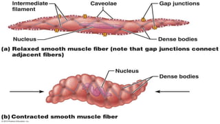
- 1. Smooth Muscle • Involuntary in function • Cells are not striated • Fibers smaller than those in skeletal muscle • Spindle-shaped; single, central nucleus • More actin than myosin • No sarcomeres – Not arranged as symmetrically as in skeletal muscle, thus NO striations. • Caveolae: indentations in sarcolemma; – May act like T tubules • Dense bodies instead of Z disks – Have noncontractile intermediate filaments • Slow, wave-like contractions
- 2. Smooth Muscle • Grouped into sheets in walls of hollow organs • Longitudinal layer – muscle fibers run parallel to organ’s long axis • Circular layer – muscle fibers run around circumference of the organ • Both layers participate in peristalsis
- 3. Smooth Muscle • Is innervated by autonomic nervous system (ANS) • Visceral or unitary smooth muscle – Only a few muscle fibers innervated in each group – Impulse spreads through gap junctions – Who sheet contracts as a unit – Often autorhythmic • Multiunit: – Cells or groups of cells act as independent units – Arrector pili of skin and iris of eye
- 4. Properties of Single-Unit Smooth Muscle –Gap junctions –Pacemaker cells with spontaneous depolarizations –Innervation to few cells –Tone = level of contraction without stimulation –Increases/decreases in tension –Graded Contractions • No recruitment • Vary intracellular calcium –Stretch Reflex • Relaxation in response to sudden or prolonged stretch
- 5. Chemical Basis for Smooth Muscle Contraction • Smooth muscle contains actin filaments contractile machinery are predominantly composed of α- and γ-actin • myosin filaments, having chemical characteristics similar to those in skeletal muscle. • Smooth muscle does not contain the troponin complex that is required in the control of skeletal muscle contraction • Calmodulin - takes on the regulatory role in smooth muscle
- 6. Physical Basis for Smooth Muscle Contraction • Large numbers of actin filaments are attached to dense bodies. Dense bodies are rich in α-actinin • Some of dense bodies are attached to the cell membrane. Others are dispersed inside the cell. • The dense bodies of smooth muscle serve the same role as the Z discs in skeletal muscle.
- 7. Calcium sources •Smooth muscle has two sources of calcium to initiate contraction; •Calcium stored in SR of the cells •Extracellular calcium that can enter muscle via Ca2+ channels on membrane of smooth muscle •One source does not control the other and a combination makes longer lasting contractions
- 8. Ca2+ from the ECF • Ca2+ enters through VG Ca2+ channels stimulated by depolarising wave • Ca2+ ligand gated channels respond to ligands such as epi and other hormones allowing an influx of Ca2+ Ca2+ from the ICF • Smooth muscle has a second messenger system used to open the RYR on the SR instead of depolarisation as in the skeletal muscle • Receptors of ligands (epi, ACH) are noted as transmembrane proteins that are G protein coupled • When activated, they stimulate the G protein to undergo a cascade of processes that lead to production of inositol triphosphate (IP3) • This inositol triphosphate stimulates the RYR channels on SR allowing Ca2+ to enter sarcoplasm from the SR
- 9. G Proteins coupled Receptor
- 10. Cont
- 11. Cont
- 12. Cont
- 13. G Protein Coupled Receptor Regulation
- 14. SECOND MESSENGER INOSITOL TRIPHOSPHATE & DIACYLGLYCEROL
- 15. Inositol Triphosphate & Diacylglycerol
- 16. Smooth Muscle Contraction: Mechanism
- 17. MOLECULAR BASIS OF CONTRACTION • The following sequence of events occurs after a rise in cytosolic calcium in a smooth muscle fiber : 1. Calcium binds to calmodulin, 2. Calcium-calmodulin complex binds to another cytosolic protein, myosin light-chain kinase, thereby activating the enzyme 3. Active myosin light-chain kinase uses ATP to phosphorylate myosin light chains in the globular head of myosin. 4. The phosphorylated cross-bridge binds to actin. • The smooth muscle myosin isoform has a very low maximal rate of ATPase activity, on the order of 10 to 100 times less than that of skeletal muscle myosin. • Since the rate of ATP splitting determines the rate of cross-bridge cycling and thus shortening velocity,
- 18. RELAXATION • To relax a contracted smooth muscle, • Activated Myosin must be dephosphorylated because dephosphorylated myosin is unable to bind to actin. • Dephosphorylation is mediated by myosin light-chain phosphatase, which is continuously active in smooth muscle during periods of rest and contraction. • When cytosolic calcium rises, the rate of myosin phosphorylation by the activated kinase exceeds the rate of dephosphorylation by the phosphatase, and the amount of phosphorylated myosin in the cell increases, producing a rise in tension. • When the cytosolic calcium concentration decreases, the rate of dephosphorylation exceeds the rate of phosphorylation, and the amount of phosphorylated myosin decreases, producing relaxation.
- 19. Latch Mechanism for Prolonged Holding of Contractions of Smooth Muscle. • Once smooth muscle has developed full contraction, the amount of continuing excitation usually can be reduced to far less than the initial level, yet the muscle maintains its full force of contraction. • Further, the energy consumed to maintain contraction is often minimal, economic utilization of ATP. • The importance of the latch mechanism is that it can maintain prolonged tonic contraction in smooth muscle for hours with little use of energy.
- 20. Comparisons Among Skeletal, Smooth, and Cardiac Muscle
- 21. THANK YOU Dr. Phiri S B