
Recommended
More Related Content
What's hot
What's hot (20)
Similar to CRANIAL NERVES- ANATOMY
Similar to CRANIAL NERVES- ANATOMY (20)
Recently uploaded
Recently uploaded (20)
CRANIAL NERVES- ANATOMY
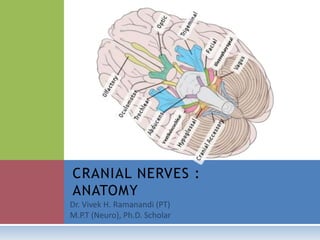
- 2. INTRODUCTION Twelve pairs Pass through or into the cranial bones (thus cranial nerves) Numbered I to XII roughly in order from top (rostral) to bottom (caudal) Some cranial nerves contain only sensory fibres, some contain only motor fibres, and some contain both.
- 3. Some cranial nerves convey parasympathetic fibres, some convey taste fibres, some convey both, and some neither Olfactory and optic nerves are not “proper” nerves I and II are attached to the cerebral hemispheres III to XII to are attached to the brain stem (midbrain, pons and medulla)
- 4. CRANIAL NERVES NAME TYPE PRNCIPAL FUNCTION I Olfactory Sensory Smell II Optic Sensory Vision III Oculomotor Motor Movt of eyeballs IV Trochlear Motor Movt of eyeballs
- 5. NAME TYPE PRNCIPAL FUNCTION V Trigeminal Va: Ophthalmic Vb: Maxillary Vc: Mandibular Va: Sensory Vb: Sensory Vc: Mixed Va: Sensation from eyeball, anterior scalp, upper face Vb: Sensation from nasal cavity and sinuses, palate, mid face, maxillary teeth Vc: Muscles of mastication, tensor tympani Sensation from chin, temple, oral cavity, tongue, TM Joint, mandibular teeth, ear, proprioception from muscles of mastication VI Abducens Motor Movements of eyeball: lateral rectus muscle VII Facial Mixed Muscles of facial expression, stapedius (middle ear) (parasympathetic: lacrimal,
- 6. NAME TYPE PRNCIPAL FUNCTION IX Glossopharyngeal Mixed Sensation from oropharynx, posterior tongue, carotid body and sinus (taste: posterior tongue) (muscle: stylopharyngeus) (parasympathetic: parotid gland) X Vagus Mixed Muscles of larynx, pharynx (phonation, swallowing) Sensation from larynx, hypopharynx, heart, lungs, abdominal viscera (taste: epiglottic region, hypopharynx) (parasympathetic: cardiac muscle; muscles and glands of foregut and midgut: intestinal activity) XI Accessory Motor Muscles: sternocleidomastoid, trapezius
- 7. DEVELOPMENT Forebrain: 1. Telencephalon (the cerebral hemispheres) and 2. Diencephalon (the thalamic structures surrounding the third ventricle) Midbrain, or mesencephalon Hindbrain: pons, cerebellum and medulla
- 9. ATTACHMENTS OF CRANIAL NERVES Forebrain Cranial n. I Telencephalon: limbic system Cribriform plate (ethmoid) Cranial n. II Diencephalon: lateral geniculate body Optic canal (sphenoid)
- 10. Midbrain Cranial n. III Upper midbrain (ventral): Interpeduncular fossa Superior orbital fissure (sphenoid) Cranial n. IV Lower midbrain (dorsal): below inferior colliculi Superior orbital fissure (sphenoid)
- 11. Hindbrain Cranial n. V Pons (lateral aspect) Va: superior orbital fissure (sphenoid) Vb: foramen rotundum (sphenoid) Vc: foramen ovale (sphenoid) Cranial n. VI Pontomedullary junction (Near midline) Superior orbital fissure (sphenoid)
- 12. Cranial n. VII Pontomedullary junction (Cerebellopontine angle ) Internal acoustic meatus, facial canal, stylomastoid foramen (temporal) Cranial n. VIII Pontomedullary junction (Cerebellopontine angle ) Internal acoustic meatus (Temporal)
- 13. Cranial n. IX, X, XI Medulla (Rootlets, lateral to inferior olive, extending down to cervical cord) Jugular foramen (between occipital and temporal bones) Cranial n. XII Medulla ( Rootlets between pyramid and olive) Hypoglossal canal (occipital)
- 16. TYPES OF NERVE FIBRE WITHIN CRANIAL NERVES Motor nerves: voluntary/involuntary Motor nerves: somatic/visceral Sensory nerves: somatic/visceral
- 17. GANGLION AND NUCLEUS Easily confused Both contain nerve cell bodies Some cranial nerves are associated with both a ganglion and a nucleus with the same name Example, the trigeminal nerve (V) is associated with the trigeminal ganglion and several trigeminal nuclei
- 18. In a nerve, ganglion means a swelling on the nerve. Caused by a collection of nerve cell bodies on a peripheral nerve A nucleus is an aggregation of cell bodies in the CNS Exception: basal ganglia of the brain Ganglia are peripheral; nuclei are central
- 19. CRANIAL NERVE NUCLEI A nucleus is a collection in the central grey matter of cell bodies of neurons serving similar functions Motor and sensory nuclei In the developing neural tube, motor components are in the ventral portion (basal lamina) and sensory components in the dorsal portion (alar lamina) separated by the sulcus limitans
- 20. Visceral elements develop near the sulcus Somatic elements towards the dorsal and ventral margins Ventral to dorsal arrangement: Somatic motor, visceral motor, visceral sensory and somatic sensory.
- 22. In the brain stem: dorsal aspects of the brain stem were forcibly parted by the enlarging central canal which becomes the fourth ventricle. The sequence of somatic motor, visceral motor, visceral sensory, somatic sensory is medial to lateral