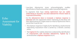
Stress
Echocardiography
By
Dr. Gunjan Shah
DM Resident, UNMICRC
Definition
 To detect the presence and extent of coronary artery disease by
provoking regional ischemia with resulting wall motion
abnormalities.
 “stress induced regional wall motion abnormalities”
 addition of exercise doppler permits evaluation of exercise-
induced changes in valvular function
 eg, severity of mitral regurgitation/mitral stenosis gradient,
pulmonary artery systolic pressure, left ventricular outflow tract
gradient, and global left ventricular systolic and diastolic function.
Pathophysiol
ogy
 Stress induced demand-supply mismatch causing perfusion defects
 wall motion abnormality on echo  ECG changes  Angina
Indications
 Evaluation of patients with known or suspected coronary
artery disease (particularly in patient with preexiting ST-T
changes that precludes interpretations of electrocardiogram
TMT)
 Assessment of myocardial viability.
 Evaluation of dyspnea of possible cardiac origin.
 Evaluation for pulmonary artery hypertension (pulmonary
artery systolic pressure can be estimated at rest and post-
exercise)
Indications
 Evaluation of mitral valve disease, including MS gradient and MR.
 Evaluation of aortic stenosis (assess severity of low gradientAS)
 Evaluation of LVOT gradients, MR, and pulmonary hypertension in
patients with HCM.
Pretest
Probability for
evaluation of
Angina
Stress
techniques
 Exercise stress
 Pharmacological stress
 Pacing
 Handgrip exercise
Pharmacologic
al Stress Echo
 patients who are unable to perform exercise testing
 may be preferable to exercise testing for preoperative risk
and myocardial viability assessment
Agents for
pharmacologic
al stress
 Dobutamine (with atropine as needed to achieve HR)
 Adenosine
 Dipyridamole
 Regadenoson
 ASE recommend dobutamine as preferable to vasodilators.
(there is greater experience using dobutamine for SE in )
 dobutamine and vasodilators are equally potent ischemic
stressors for inducing wall abnormalities in the presence of a
critical CAD.
 dipyridamole is a popular pharmacologic regimen for SE in
Europe.
DSE
(Assessment of
obstructive
CAD)
 Graded dobutamine infusion in five three-minute stages
starting at 5 mcg/kg/minute, followed by 10, 20, 30, and 40
mcg/kg/minute
 Atropine in divided doses of 0.5 mg each to a total of 2 mg,
as needed.
 Atropine increases the Sn of SE in patients receiving beta-
blockers and in single-vessel disease.
 Use of a sustained isometric hand grip or a low-level
dynamic foot exercise (with or without atropine) in the late
stages of the dobutamine protocol as a supplemental
maneuver to achieve peak heart rate also described.
DSE (Viability
assessment)
 Dobutamine dosing for viability assessment starts at a lower
initial dose than for assessment of cad.
 Continuous dobutamine infusion initiated at 2.5
mcg/kg/minute, and increasing at five-minute intervals in
2.5 mcg/kg/minute increments until a contractile response is
noted, up to a maximum dose of 10 mcg/kg/minute.
End points for
DSE
 Exceeding target heart rate of 85% age-predicted maximum
 Development of significant angina
 Recognition of a new wall motion abnormality
 A decrease in systolic blood pressure >20 mm Hg from
baseline
 Arrhythmias such as atrial fibrillation or nonsustained
ventricular tachycardia
 Limiting side effects or symptoms
Effects of Beta
blockers
 Use of beta-blockers may attenuate HR response and
evidence of myocardial ischemia during DSE.
 The effect of beta-blockers on HR response to dobutamine
can be overcome by the administration of atropine.
 Moreover, performing the SE while the patient stays on all
medications, including beta-blockers, may be useful for
assessment of treatment efficacy.
Vasodilator stress
echocardiography
 Dipyridamole (dose): up to 0.84 mg/kg in two separate
infusions: 0.56 mg/kg over 4 minutes ("standard dose"),
followed by four minutes of no dose, then an additional 0.28
mg/kg is given over two minutes if no HR achieved. If no
endpoint is reached following total of 0.84 mg/kg,
then atropine (doses of 0.25 mg, up to a maximum of 1 mg).
 Adenosine (dose): max dose of 140 mcg/kg/minute over six
minutes Imaging is performed prior to and after starting
adenosine infusion.
 Adenosine is a shorter-acting agent employed for MPI and,
to a lesser extent, to detect stress-induced changes in wall
motion, but this method has not been in wide clinical use.
 Vasodilator stress is better suited for perfusion than wall
motion analysis. (as they redistribute coronary blood flow–
steal phenomena)
 Both these C/I in reactive airway obstruction and significant
conduction defects
 Dobutamine  Inducible ischemia &myocardial viability
 Adenosine, dipyridamole  myocardial perfusion imaging
Pacing
 In selected patients with a PPM, increasing the pacing rate
to facilitate achievement of target heart rate may be used
 Can also be done via trans esophageal pacing wire
 combined with dobutamine infusion, starting with dobu
infusion following the usual protocol and then holding the
infusion at 20 mcg.
 At this dose of dobutamine, we start a stepwise increase of
the HR by increase pacing rate until achievement of the
THR.
 During recovery the pacing rate is progressively reduced at
one-minute intervals.
Handgrip
exercise
 Handgrip may be used as an adjunct to exercise or DSE.
 During the last stage of exercise or dobutamine infusion and
30 seconds before acquiring the peak exercise images,
patients are asked to exert a sustained grip on a tennis ball.
 The hand grip response reliably raises BP at least 10 mmHg
and usually also increases HR; superimposed on the
maximum achieved levels, this augmentation results in a
"bonus" increment in rate pressure product.  increasing
sensitivity of method
Complications
and risks of SE
 Risk of complications is very low
 In the largest survey to date, the overall rate of life-
threatening events was 1 per 1000 examinations (0.015% for
exercise and 0.18% for dobutamine)
 MC minor complications include PVCs and atrial arrhythmias
 Most frequent major complications were acute MI or
ventricular tachycardia or fibrillation.
Safety of DSE
 No absolute contraindications to DSE.
 Unstable patients as in with ADHF or unstable angina,
should rarely be subjected to stress testing of any kind.
 Can be been safely performed in patients with recent MI,
extensive left ventricular dysfunction, abdominal aortic
aneurysm, syncope, aortic stenosis, hypertrophic
cardiomyopathy, history of ventricular tachycardia, and
aborted sudden death. But to be performed according to
risk benefit ratio.
 Unlike dipyridamole/adenosine, dobutamine is safer in
bronchospastic lung disease.
Echo
assessment for
CAD
 When SE is performed to evaluate CAD, the main goal is to
side-by-side comparison of images for assessment of global
and regional LV systolic function at rest and immediately
after stress.
 Images are also compared with pre-peak and peak stress,
during pharmacologic stress.
 With stress, the normal LV becomes hypercontractile, cavity
size is reduced, and EF increases.
 Function in each segment is graded (normal, hyperdynamic,
hypokinetic, akinetic, dyskinetic, or aneurysmal) at rest and
with stress.
 Imp to detect RWMA wall thickening and endocardial
excursion
Interpretation
 The presence of baseline RWMA that remain “fixed”
(unchanged) with stress  a previous infarct.
 The development of a new or worsening RWMA  a flow-
limiting stenosis in the coronary artery supplying the
abnormal segment or segments.
 A large ischemic territory—such as left main or multivessel
CAD  diminished global LVEF and chamber dilation with
stress (i.e., transient ischemic LV dilatation).
Rest Stress Interpretation
Normal Hyperkinetic Normal
Normal Hypokinetic / akinetic Ischemia
Akinetic Akinetic Infarction (previous)
Hypokinetic Akinetic / dyskinetic Infarction and/or ischemia
Hypokinetic / akinetic Normal Viable
Response to
stress and
interpretation
Echo
Assessment for
Viability
 Low-dose dobutamine stress echocardiography enables
identification of dysfunctional but viable myocardial segments.
 In segments that have resting dysfunction but are viable
(hibernating or stunned), low-dose dobutamine infusion leads to
improved contractility.
 As the dobutamine dose is increased, a biphasic response is
detected with the segmental motion improving at low-dose and
then deteriorating at a higher dose to a level of dysfunction that is
worse than it was at rest.
• This biphasic response is highly Sp for identifying a dysfunctional
hibernating myocardium that is served by a stenotic coronary
artery.
• The highest Sn for viability detection is obtained by identification
of improvement with low-dose dobutamine; highest Sp is
achieved when a biphasic response occurs.
Interpretation
of response
(viability)
 Monophasic  contractility increase at low dose and also
increase at high dose  viable (most sensitive)
 Biphasic  increase at low dose and worsens at high dose
 viable (most specific)
 Non phasic  no improvement at any dose  non viable
scarred (thinned out <5mm)
Doppler
assessment
 Mitral valve – Changes in mitral stenosis gradient or mitral
regurgitation following stress
 Aortic valve – Evaluation of aortic valvular gradients in
suspected low-flow, low-gradient aortic stenosis
 Tricuspid valve – Assessment of tricuspid regurgitation
velocity for estimation of pulmonary artery systolic pressure
in patients with suspected pulmonary hypertension
 Left ventricular outflow tract (LVOT) – Evaluation for
inducible or worsening LVOT gradient in suspected or
known HCM
SE after MI
 Particularly helpful in case of multivessel disease (post
revascularization of IRA)  if stenosis in other vessels is
significant or not (to identify need of revasc)  identify
ischemia at distance  new onset RWMA compare to
previous one.
Choosing b/w
stress
modalities
Clinical situation treadmill bicycle dobutamine
Chest pain evaluation ++ ++ +
Postmyocardial
infarction risk
++ ++ ++
Viability -- -- ++
Evaluation of
dyspnea/fatigue*
+ ++ +
Preoperative risk
assessment
+ + ++
Severity of valve
disease*
-- ++ --
Pulmonary
hypertension*
-- ++ --
*Inclusion of Doppler parameters is recommended
Prognostic
value of SE
 Higher WMSI = worse prognosis
 Extent of ischemia  more segments involved = worse
prognosis
 Magnitude of hypokinesia in involved segments  more
severe = bad
 Low stress EF = bad
 Ischemic dilatation of LV
Comparison
with stress
nuclear
imaging
 The accuracy of SE (stress echocardiography) is similar to
that of stress radionuclide perfusion imaging.
 From meta-analyses, as well as from comparisons of the
accuracy of SE and nuclear imaging in the same patient
population, Sn 88% and Sp 83% for significant CAD
(generally defined as >50% coronary artery stenosis by
angiography).
 The specificity of SE appears to be higher than that of
nuclear imaging for left main and triple-vessel CAD.
 Time and cost efficient compare to nuclear.
Limitations of
SE
 Potential for rapid resolution of ischemia prior to image
acquisition post exercise
 Hypertensive response to stress  greater likelihood for
false +ve test
 Suboptimal image quality (can be tackled by contrast)
 May be exacerbated by respiratory motion d/t heavy
breathing post exercise
False results in
SE
THANK YOU

Stress echo.pptx

  • 1.
  • 2.
    Definition  To detectthe presence and extent of coronary artery disease by provoking regional ischemia with resulting wall motion abnormalities.  “stress induced regional wall motion abnormalities”  addition of exercise doppler permits evaluation of exercise- induced changes in valvular function  eg, severity of mitral regurgitation/mitral stenosis gradient, pulmonary artery systolic pressure, left ventricular outflow tract gradient, and global left ventricular systolic and diastolic function.
  • 3.
    Pathophysiol ogy  Stress induceddemand-supply mismatch causing perfusion defects  wall motion abnormality on echo  ECG changes  Angina
  • 4.
    Indications  Evaluation ofpatients with known or suspected coronary artery disease (particularly in patient with preexiting ST-T changes that precludes interpretations of electrocardiogram TMT)  Assessment of myocardial viability.  Evaluation of dyspnea of possible cardiac origin.  Evaluation for pulmonary artery hypertension (pulmonary artery systolic pressure can be estimated at rest and post- exercise)
  • 5.
    Indications  Evaluation ofmitral valve disease, including MS gradient and MR.  Evaluation of aortic stenosis (assess severity of low gradientAS)  Evaluation of LVOT gradients, MR, and pulmonary hypertension in patients with HCM.
  • 6.
  • 7.
    Stress techniques  Exercise stress Pharmacological stress  Pacing  Handgrip exercise
  • 8.
    Pharmacologic al Stress Echo patients who are unable to perform exercise testing  may be preferable to exercise testing for preoperative risk and myocardial viability assessment
  • 9.
    Agents for pharmacologic al stress Dobutamine (with atropine as needed to achieve HR)  Adenosine  Dipyridamole  Regadenoson  ASE recommend dobutamine as preferable to vasodilators. (there is greater experience using dobutamine for SE in )  dobutamine and vasodilators are equally potent ischemic stressors for inducing wall abnormalities in the presence of a critical CAD.  dipyridamole is a popular pharmacologic regimen for SE in Europe.
  • 10.
    DSE (Assessment of obstructive CAD)  Gradeddobutamine infusion in five three-minute stages starting at 5 mcg/kg/minute, followed by 10, 20, 30, and 40 mcg/kg/minute  Atropine in divided doses of 0.5 mg each to a total of 2 mg, as needed.  Atropine increases the Sn of SE in patients receiving beta- blockers and in single-vessel disease.  Use of a sustained isometric hand grip or a low-level dynamic foot exercise (with or without atropine) in the late stages of the dobutamine protocol as a supplemental maneuver to achieve peak heart rate also described.
  • 12.
    DSE (Viability assessment)  Dobutaminedosing for viability assessment starts at a lower initial dose than for assessment of cad.  Continuous dobutamine infusion initiated at 2.5 mcg/kg/minute, and increasing at five-minute intervals in 2.5 mcg/kg/minute increments until a contractile response is noted, up to a maximum dose of 10 mcg/kg/minute.
  • 13.
    End points for DSE Exceeding target heart rate of 85% age-predicted maximum  Development of significant angina  Recognition of a new wall motion abnormality  A decrease in systolic blood pressure >20 mm Hg from baseline  Arrhythmias such as atrial fibrillation or nonsustained ventricular tachycardia  Limiting side effects or symptoms
  • 14.
    Effects of Beta blockers Use of beta-blockers may attenuate HR response and evidence of myocardial ischemia during DSE.  The effect of beta-blockers on HR response to dobutamine can be overcome by the administration of atropine.  Moreover, performing the SE while the patient stays on all medications, including beta-blockers, may be useful for assessment of treatment efficacy.
  • 15.
    Vasodilator stress echocardiography  Dipyridamole(dose): up to 0.84 mg/kg in two separate infusions: 0.56 mg/kg over 4 minutes ("standard dose"), followed by four minutes of no dose, then an additional 0.28 mg/kg is given over two minutes if no HR achieved. If no endpoint is reached following total of 0.84 mg/kg, then atropine (doses of 0.25 mg, up to a maximum of 1 mg).  Adenosine (dose): max dose of 140 mcg/kg/minute over six minutes Imaging is performed prior to and after starting adenosine infusion.
  • 16.
     Adenosine isa shorter-acting agent employed for MPI and, to a lesser extent, to detect stress-induced changes in wall motion, but this method has not been in wide clinical use.  Vasodilator stress is better suited for perfusion than wall motion analysis. (as they redistribute coronary blood flow– steal phenomena)  Both these C/I in reactive airway obstruction and significant conduction defects  Dobutamine  Inducible ischemia &myocardial viability  Adenosine, dipyridamole  myocardial perfusion imaging
  • 17.
    Pacing  In selectedpatients with a PPM, increasing the pacing rate to facilitate achievement of target heart rate may be used  Can also be done via trans esophageal pacing wire  combined with dobutamine infusion, starting with dobu infusion following the usual protocol and then holding the infusion at 20 mcg.  At this dose of dobutamine, we start a stepwise increase of the HR by increase pacing rate until achievement of the THR.  During recovery the pacing rate is progressively reduced at one-minute intervals.
  • 18.
    Handgrip exercise  Handgrip maybe used as an adjunct to exercise or DSE.  During the last stage of exercise or dobutamine infusion and 30 seconds before acquiring the peak exercise images, patients are asked to exert a sustained grip on a tennis ball.  The hand grip response reliably raises BP at least 10 mmHg and usually also increases HR; superimposed on the maximum achieved levels, this augmentation results in a "bonus" increment in rate pressure product.  increasing sensitivity of method
  • 19.
    Complications and risks ofSE  Risk of complications is very low  In the largest survey to date, the overall rate of life- threatening events was 1 per 1000 examinations (0.015% for exercise and 0.18% for dobutamine)  MC minor complications include PVCs and atrial arrhythmias  Most frequent major complications were acute MI or ventricular tachycardia or fibrillation.
  • 20.
    Safety of DSE No absolute contraindications to DSE.  Unstable patients as in with ADHF or unstable angina, should rarely be subjected to stress testing of any kind.  Can be been safely performed in patients with recent MI, extensive left ventricular dysfunction, abdominal aortic aneurysm, syncope, aortic stenosis, hypertrophic cardiomyopathy, history of ventricular tachycardia, and aborted sudden death. But to be performed according to risk benefit ratio.  Unlike dipyridamole/adenosine, dobutamine is safer in bronchospastic lung disease.
  • 21.
    Echo assessment for CAD  WhenSE is performed to evaluate CAD, the main goal is to side-by-side comparison of images for assessment of global and regional LV systolic function at rest and immediately after stress.  Images are also compared with pre-peak and peak stress, during pharmacologic stress.  With stress, the normal LV becomes hypercontractile, cavity size is reduced, and EF increases.  Function in each segment is graded (normal, hyperdynamic, hypokinetic, akinetic, dyskinetic, or aneurysmal) at rest and with stress.  Imp to detect RWMA wall thickening and endocardial excursion
  • 22.
    Interpretation  The presenceof baseline RWMA that remain “fixed” (unchanged) with stress  a previous infarct.  The development of a new or worsening RWMA  a flow- limiting stenosis in the coronary artery supplying the abnormal segment or segments.  A large ischemic territory—such as left main or multivessel CAD  diminished global LVEF and chamber dilation with stress (i.e., transient ischemic LV dilatation).
  • 23.
    Rest Stress Interpretation NormalHyperkinetic Normal Normal Hypokinetic / akinetic Ischemia Akinetic Akinetic Infarction (previous) Hypokinetic Akinetic / dyskinetic Infarction and/or ischemia Hypokinetic / akinetic Normal Viable Response to stress and interpretation
  • 25.
    Echo Assessment for Viability  Low-dosedobutamine stress echocardiography enables identification of dysfunctional but viable myocardial segments.  In segments that have resting dysfunction but are viable (hibernating or stunned), low-dose dobutamine infusion leads to improved contractility.  As the dobutamine dose is increased, a biphasic response is detected with the segmental motion improving at low-dose and then deteriorating at a higher dose to a level of dysfunction that is worse than it was at rest. • This biphasic response is highly Sp for identifying a dysfunctional hibernating myocardium that is served by a stenotic coronary artery. • The highest Sn for viability detection is obtained by identification of improvement with low-dose dobutamine; highest Sp is achieved when a biphasic response occurs.
  • 26.
    Interpretation of response (viability)  Monophasic contractility increase at low dose and also increase at high dose  viable (most sensitive)  Biphasic  increase at low dose and worsens at high dose  viable (most specific)  Non phasic  no improvement at any dose  non viable scarred (thinned out <5mm)
  • 28.
    Doppler assessment  Mitral valve– Changes in mitral stenosis gradient or mitral regurgitation following stress  Aortic valve – Evaluation of aortic valvular gradients in suspected low-flow, low-gradient aortic stenosis  Tricuspid valve – Assessment of tricuspid regurgitation velocity for estimation of pulmonary artery systolic pressure in patients with suspected pulmonary hypertension  Left ventricular outflow tract (LVOT) – Evaluation for inducible or worsening LVOT gradient in suspected or known HCM
  • 29.
    SE after MI Particularly helpful in case of multivessel disease (post revascularization of IRA)  if stenosis in other vessels is significant or not (to identify need of revasc)  identify ischemia at distance  new onset RWMA compare to previous one.
  • 30.
    Choosing b/w stress modalities Clinical situationtreadmill bicycle dobutamine Chest pain evaluation ++ ++ + Postmyocardial infarction risk ++ ++ ++ Viability -- -- ++ Evaluation of dyspnea/fatigue* + ++ + Preoperative risk assessment + + ++ Severity of valve disease* -- ++ -- Pulmonary hypertension* -- ++ -- *Inclusion of Doppler parameters is recommended
  • 31.
    Prognostic value of SE Higher WMSI = worse prognosis  Extent of ischemia  more segments involved = worse prognosis  Magnitude of hypokinesia in involved segments  more severe = bad  Low stress EF = bad  Ischemic dilatation of LV
  • 32.
    Comparison with stress nuclear imaging  Theaccuracy of SE (stress echocardiography) is similar to that of stress radionuclide perfusion imaging.  From meta-analyses, as well as from comparisons of the accuracy of SE and nuclear imaging in the same patient population, Sn 88% and Sp 83% for significant CAD (generally defined as >50% coronary artery stenosis by angiography).  The specificity of SE appears to be higher than that of nuclear imaging for left main and triple-vessel CAD.  Time and cost efficient compare to nuclear.
  • 34.
    Limitations of SE  Potentialfor rapid resolution of ischemia prior to image acquisition post exercise  Hypertensive response to stress  greater likelihood for false +ve test  Suboptimal image quality (can be tackled by contrast)  May be exacerbated by respiratory motion d/t heavy breathing post exercise
  • 35.
  • 36.