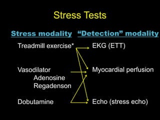
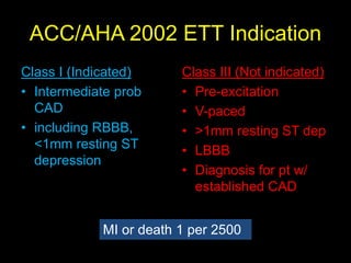
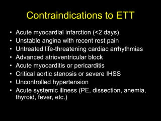
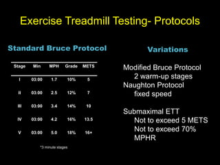
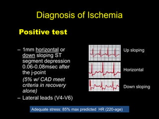
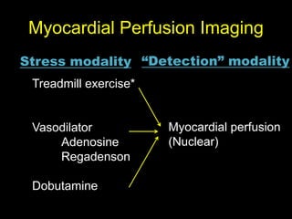
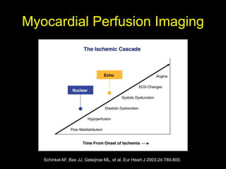
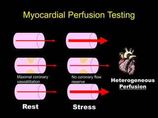
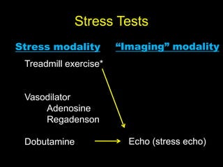
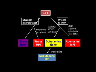
Stress Testing 
UTSW House Staff Didactic Series 
Anand Rohatgi, MD, MSCS, FACC, FAHA 
Assistant Professor 
Division of Cardiology
Stress Tests 
Stress modality “Detection” modality 
Treadmill exercise* 
Vasodilator 
Adenosine 
Regadenson 
Dobutamine 
EKG (ETT) 
Myocardial perfusion 
Echo (stress echo)
Probability 
Gibons at al, Progr Cardiol 1983;12:67 
Positive Predictive 
Value 
Probability of a subject 
with a positive test, 
actually having disease 
Depends upon 
Sensitivity 
Specificity 
Population prevalence 
or pretest likelihood
Pretest Probability 
Age Gender Typical 
Angina 
Atypical 
Angina 
Nonanginal 
CP 
Asymptoma 
tic 
30-39 Men Intermediate Intermediate Low Very Low 
40-49 High Intermediate Intermediate Low 
50-59 High Intermediate Intermediate Low 
60-69 High Intermediate Intermediate Low 
30-39 Women Intermediate Very Low Very Low Very Low 
40-49 Intermediate Low Very Low Very Low 
50-59 Intermediate Intermediate Low Very Low 
60-69 High Intermediate Intermediate Low 
Diamond et al, NEJM 1979;300:1350
ACC/AHA 2002 ETT Indication 
Class I (Indicated) 
• Intermediate prob 
CAD 
• including RBBB, 
<1mm resting ST 
depression 
Class III (Not indicated) 
• Pre-excitation 
• V-paced 
• >1mm resting ST dep 
• LBBB 
• Diagnosis for pt w/ 
established CAD 
MI or death 1 per 2500
Contraindications to ETT 
• Acute myocardial infarction (<2 days) 
• Unstable angina with recent rest pain 
• Untreated life-threatening cardiac arrhythmias 
• Advanced atrioventricular block 
• Acute myocarditis or pericarditis 
• Critical aortic stenosis or severe IHSS 
• Uncontrolled hypertension 
• Acute systemic illness (PE, dissection, anemia, 
thyroid, fever, etc.)
Exercise Treadmill Testing- Protocols 
Standard Bruce Protocol 
Stage Min MPH Grade METS 
I 03:00 1.7 10% 5 
II 03:00 2.5 12% 7 
III 03:00 3.4 14% 10 
IV 03:00 4.2 16% 13.5 
V 03:00 5.0 18% 16+ 
*3 minute stages 
Variations 
Modified Bruce Protocol 
2 warm-up stages 
Naughton Protocol 
fixed speed 
Submaximal ETT 
Not to exceed 5 METS 
Not to exceed 70% 
MPHR
Diagnosis of Ischemia 
Positive test 
– 1mm horizontal or 
down sloping ST 
segment depression 
0.06-0.08msec after 
the j-point 
(5% w/ CAD meet 
criteria in recovery 
alone) 
– Lateral leads (V4-V6) 
Up sloping 
Horizontal 
Down sloping 
Adequate stress: 85% max predicted HR (220-age)
Decreased Specificity 
• LVH with repolarization abnormalities 
– Decreased specificity with no change in sensitivity 
• Resting ST depression > 1mm 
• LBBB 
• RBBB (diagnostic accuracy preserved in V5, V6, II, AVF 
• Digoxin 
– ST depression in 25-40% of healthy subjects 
– 2 weeks required washout
Non-coronary Causes of ST 
segment depression 
• Severe aortic stenosis 
• Severe hypertension 
• Cardiomyopathy 
• Anemia 
• Hypokalemia 
• Severe hypoxia 
• Digitalis use 
• Sudden excessive 
exercise 
• Glucose load 
• Left ventricular 
hypertrophy 
• Hyperventilation 
• Mitral valve prolapse 
• Intraventricular 
conduction defect 
• Preexcitation syndrome 
• Severe volume overload 
• Supraventricular 
tachyarrhythmias
Thompson CA, et al. JACC 2000; 36:2140-5. Lauer MS, et al. Circulation 1996;93:1520-6
Prognostic Markers 
• Maximal exercise capacity 
• Chronotropic incompetence 
• HR recovery 
• Risk scores
Exercise Capacity 
MET= 02 uptake of 70kg 
man at rest for 1 min 
=3.5ml O2/kg/min 
Exercise capacity is 
one of the strongest 
prognostic markers 
Encompasses many 
different factors 
Each 1 MET increase = 
12% increased 
survival 
Stanford database of 6000 men 
>13 >11 
Ref 
<10 <8 
Myers et al, NEJM 2002;346:793
ETT in asymptomatic pts 
Class I 
• None. 
Class IIa 
• Evaluation of asymptomatic persons with diabetes mellitus who plan to start 
vigorous exercise (see page 39). (Level of Evidence: C) 
Class IIb 
• Evaluation of persons with multiple risk factors as a guide to risk-reduction therapy.* 
• Evaluation of asymptomatic men older than 45 years and women older than 55 
years: 
– Who plan to start vigorous exercise (especially if sedentary) or 
– Who are involved in occupations in which impairment might impact public safety or 
– Who are at high risk for CAD due to other diseases (e.g., peripheral vascular disease and 
chronic renal failure) 
Class III 
• Routine screening of asymptomatic men or women.
Myocardial Perfusion Imaging 
Stress modality “Detection” modality 
Treadmill exercise* 
Vasodilator 
Adenosine 
Regadenson 
Dobutamine 
Myocardial perfusion 
(Nuclear)
Myocardial Perfusion Imaging 
Schinkel AF, Bax JJ, Geleijnse ML, et al. Eur Heart J 2003;24:789-800.
Myocardial Perfusion Testing 
Maximal coronary 
vasodilitation 
Rest 
No coronary flow 
reserve 
Stress 
Heterogeneous 
Perfusion
Vasodilators 
• Dipyridamole 
– Increases adenosine levels 
– 50% with side effects, last 15-25 minutes 
• Adenosine 
– Coronary vasodilation via A2A receptor 
– 140mcg/kg/min x 6min 
– 80% with side effects: flushing 40%, AV block 
(7.6%), hypotension (5%), <10sec ½ life 
– CP non-specific 
– 1mmST depression 5-7%>CAD 
• Regadenoson 
– A2A agonist with lower affinity for receptors > 
side effects 
– Side effets of SOB, headache, flushing, last 15- 
30 min 
– Single 5ml injection 
Contra-indications 
• AV block (2nd or 3rd) 
•Bronchospasm 
•Methyl xanthines 
•ACS
Myocardial Perfusion Testing 
(Nuclear: SPECT) 
Protocol (Dual Isotope) 
• Resting images after Thallium-201 
injection 
• Stress, with Technetium-99 injected at 
peak exercise (Cardiolite/Myoview) 
• Post-stress images (with gated SPECT)
→ Revasc better 
Hachamovitch R, Hayes SW, Friedman JD, Cohen I, Berman DS. Circulation 2003;107:2900-6
Stress Tests 
Stress modality “Imaging” modality 
Treadmill exercise* 
Vasodilator 
Adenosine 
Regadenson 
Dobutamine Echo (stress echo)
Stress Echo 
Schinkel AF, Bax JJ, Geleijnse ML, et al. Eur Heart J 2003;24:789-800. 
Abnormal 
flow reserve 
Ischemia
Stress Echocardiography 
• Stress echo is used to 
assess ischemia 
• Wall motion 
abnormalities are the 
earliest response to 
ischemia 
Post-balloon inflation 
19 
30 
39 
0 50 
Chest 
Pain 
EKG 
change 
Wall 
Motion 
Seconds 
Hauser et al. JACC 
1985;5:193
Dobutamine Echo 
Mechanisms of Action 
– β1 agonist– inotropy and 
chronotropy (some 
vasodilatation) 
– induces ischemia at lower 
RPP than ETT, (RPP 
approximately 16-20K) 
– Begin at 10mcg/kg/min, 
increasing to 40 mcg/kgmin 
Side Effects 
– 3:1000 serious side effects 
• MI 
• Ventricular fibrillation 
– Atrial / Ventricular 
arrhythmia 
– Hypertension 
– Hypotension (cavity 
obliteration) 
– Headache / Tremor
Comparing SECHO and MPI 
Advantages Disadvantages 
MPI (Nuclear) 
Detects abnl flow reserve 
Peak-exercise images 
acquired 
Most studies complete 
Quantified LVEF and 
volumes 
Longer time than secho 
Radiation 
Lower spatial resolution 
Inferior wall diff to eval 
Balanced ischemia missed 
SECHO 
Safe 
No radiation 
Portable, faster 
Structural information 
Peak-exercise images 
difficult to acquire 
False-neg w/ rapid recovery 
Ischemic response needed 
15% cannot assess entire 
myocardium 
Afib, LBBB
ETT 
EKG not 
interpretable 
Stress 
Echo 
Poor echo 
arrhythmia 
Stress 
MPI 
Asthma 
COPD 
AV Block 
Dobutamine 
Echo 
LBBB 
V-paced 
arrhythmia 
Poor echo 
Adenosine 
MPI 
Unable 
to walk 
Poor echo 
Dobutamine 
MPI

Stresstesting housestaffdidactic_10092014[1]

  • 1.
    Stress Testing UTSWHouse Staff Didactic Series Anand Rohatgi, MD, MSCS, FACC, FAHA Assistant Professor Division of Cardiology
  • 2.
    Stress Tests Stressmodality “Detection” modality Treadmill exercise* Vasodilator Adenosine Regadenson Dobutamine EKG (ETT) Myocardial perfusion Echo (stress echo)
  • 3.
    Probability Gibons atal, Progr Cardiol 1983;12:67 Positive Predictive Value Probability of a subject with a positive test, actually having disease Depends upon Sensitivity Specificity Population prevalence or pretest likelihood
  • 4.
    Pretest Probability AgeGender Typical Angina Atypical Angina Nonanginal CP Asymptoma tic 30-39 Men Intermediate Intermediate Low Very Low 40-49 High Intermediate Intermediate Low 50-59 High Intermediate Intermediate Low 60-69 High Intermediate Intermediate Low 30-39 Women Intermediate Very Low Very Low Very Low 40-49 Intermediate Low Very Low Very Low 50-59 Intermediate Intermediate Low Very Low 60-69 High Intermediate Intermediate Low Diamond et al, NEJM 1979;300:1350
  • 5.
    ACC/AHA 2002 ETTIndication Class I (Indicated) • Intermediate prob CAD • including RBBB, <1mm resting ST depression Class III (Not indicated) • Pre-excitation • V-paced • >1mm resting ST dep • LBBB • Diagnosis for pt w/ established CAD MI or death 1 per 2500
  • 6.
    Contraindications to ETT • Acute myocardial infarction (<2 days) • Unstable angina with recent rest pain • Untreated life-threatening cardiac arrhythmias • Advanced atrioventricular block • Acute myocarditis or pericarditis • Critical aortic stenosis or severe IHSS • Uncontrolled hypertension • Acute systemic illness (PE, dissection, anemia, thyroid, fever, etc.)
  • 7.
    Exercise Treadmill Testing-Protocols Standard Bruce Protocol Stage Min MPH Grade METS I 03:00 1.7 10% 5 II 03:00 2.5 12% 7 III 03:00 3.4 14% 10 IV 03:00 4.2 16% 13.5 V 03:00 5.0 18% 16+ *3 minute stages Variations Modified Bruce Protocol 2 warm-up stages Naughton Protocol fixed speed Submaximal ETT Not to exceed 5 METS Not to exceed 70% MPHR
  • 8.
    Diagnosis of Ischemia Positive test – 1mm horizontal or down sloping ST segment depression 0.06-0.08msec after the j-point (5% w/ CAD meet criteria in recovery alone) – Lateral leads (V4-V6) Up sloping Horizontal Down sloping Adequate stress: 85% max predicted HR (220-age)
  • 9.
    Decreased Specificity •LVH with repolarization abnormalities – Decreased specificity with no change in sensitivity • Resting ST depression > 1mm • LBBB • RBBB (diagnostic accuracy preserved in V5, V6, II, AVF • Digoxin – ST depression in 25-40% of healthy subjects – 2 weeks required washout
  • 10.
    Non-coronary Causes ofST segment depression • Severe aortic stenosis • Severe hypertension • Cardiomyopathy • Anemia • Hypokalemia • Severe hypoxia • Digitalis use • Sudden excessive exercise • Glucose load • Left ventricular hypertrophy • Hyperventilation • Mitral valve prolapse • Intraventricular conduction defect • Preexcitation syndrome • Severe volume overload • Supraventricular tachyarrhythmias
  • 12.
    Thompson CA, etal. JACC 2000; 36:2140-5. Lauer MS, et al. Circulation 1996;93:1520-6
  • 13.
    Prognostic Markers •Maximal exercise capacity • Chronotropic incompetence • HR recovery • Risk scores
  • 14.
    Exercise Capacity MET=02 uptake of 70kg man at rest for 1 min =3.5ml O2/kg/min Exercise capacity is one of the strongest prognostic markers Encompasses many different factors Each 1 MET increase = 12% increased survival Stanford database of 6000 men >13 >11 Ref <10 <8 Myers et al, NEJM 2002;346:793
  • 15.
    ETT in asymptomaticpts Class I • None. Class IIa • Evaluation of asymptomatic persons with diabetes mellitus who plan to start vigorous exercise (see page 39). (Level of Evidence: C) Class IIb • Evaluation of persons with multiple risk factors as a guide to risk-reduction therapy.* • Evaluation of asymptomatic men older than 45 years and women older than 55 years: – Who plan to start vigorous exercise (especially if sedentary) or – Who are involved in occupations in which impairment might impact public safety or – Who are at high risk for CAD due to other diseases (e.g., peripheral vascular disease and chronic renal failure) Class III • Routine screening of asymptomatic men or women.
  • 16.
    Myocardial Perfusion Imaging Stress modality “Detection” modality Treadmill exercise* Vasodilator Adenosine Regadenson Dobutamine Myocardial perfusion (Nuclear)
  • 17.
    Myocardial Perfusion Imaging Schinkel AF, Bax JJ, Geleijnse ML, et al. Eur Heart J 2003;24:789-800.
  • 18.
    Myocardial Perfusion Testing Maximal coronary vasodilitation Rest No coronary flow reserve Stress Heterogeneous Perfusion
  • 19.
    Vasodilators • Dipyridamole – Increases adenosine levels – 50% with side effects, last 15-25 minutes • Adenosine – Coronary vasodilation via A2A receptor – 140mcg/kg/min x 6min – 80% with side effects: flushing 40%, AV block (7.6%), hypotension (5%), <10sec ½ life – CP non-specific – 1mmST depression 5-7%>CAD • Regadenoson – A2A agonist with lower affinity for receptors > side effects – Side effets of SOB, headache, flushing, last 15- 30 min – Single 5ml injection Contra-indications • AV block (2nd or 3rd) •Bronchospasm •Methyl xanthines •ACS
  • 20.
    Myocardial Perfusion Testing (Nuclear: SPECT) Protocol (Dual Isotope) • Resting images after Thallium-201 injection • Stress, with Technetium-99 injected at peak exercise (Cardiolite/Myoview) • Post-stress images (with gated SPECT)
  • 21.
    → Revasc better Hachamovitch R, Hayes SW, Friedman JD, Cohen I, Berman DS. Circulation 2003;107:2900-6
  • 22.
    Stress Tests Stressmodality “Imaging” modality Treadmill exercise* Vasodilator Adenosine Regadenson Dobutamine Echo (stress echo)
  • 23.
    Stress Echo SchinkelAF, Bax JJ, Geleijnse ML, et al. Eur Heart J 2003;24:789-800. Abnormal flow reserve Ischemia
  • 24.
    Stress Echocardiography •Stress echo is used to assess ischemia • Wall motion abnormalities are the earliest response to ischemia Post-balloon inflation 19 30 39 0 50 Chest Pain EKG change Wall Motion Seconds Hauser et al. JACC 1985;5:193
  • 25.
    Dobutamine Echo Mechanismsof Action – β1 agonist– inotropy and chronotropy (some vasodilatation) – induces ischemia at lower RPP than ETT, (RPP approximately 16-20K) – Begin at 10mcg/kg/min, increasing to 40 mcg/kgmin Side Effects – 3:1000 serious side effects • MI • Ventricular fibrillation – Atrial / Ventricular arrhythmia – Hypertension – Hypotension (cavity obliteration) – Headache / Tremor
  • 26.
    Comparing SECHO andMPI Advantages Disadvantages MPI (Nuclear) Detects abnl flow reserve Peak-exercise images acquired Most studies complete Quantified LVEF and volumes Longer time than secho Radiation Lower spatial resolution Inferior wall diff to eval Balanced ischemia missed SECHO Safe No radiation Portable, faster Structural information Peak-exercise images difficult to acquire False-neg w/ rapid recovery Ischemic response needed 15% cannot assess entire myocardium Afib, LBBB
  • 27.
    ETT EKG not interpretable Stress Echo Poor echo arrhythmia Stress MPI Asthma COPD AV Block Dobutamine Echo LBBB V-paced arrhythmia Poor echo Adenosine MPI Unable to walk Poor echo Dobutamine MPI