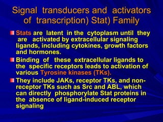
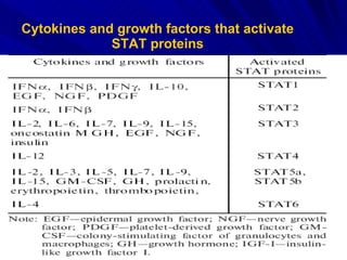
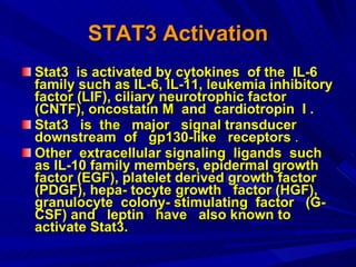
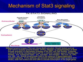
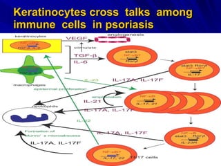
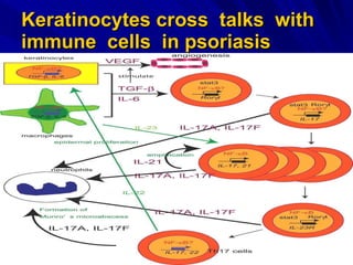
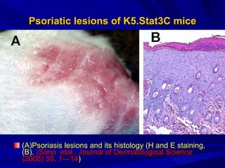
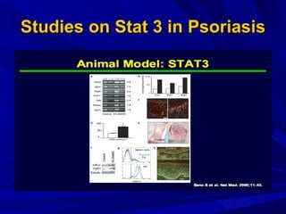
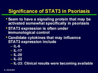
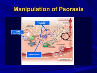
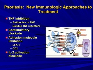
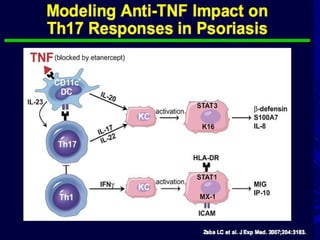
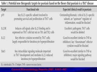
STAT 3  Protein &  Immuno Competent Cells in Psoriasis  Pathogenesis M.YOUSRY ABDEL-MAWLA,MD Zagazig  Faculty of  Medicine,Egypt.
Signal   transducers   and   activators   of   transcription )  Stat) Family Stats   are   latent   in   the   cytoplasm   until   they   are   activated   by   extracellular   signaling   ligands ,  including   cytokines ,  growth   factors   and   hormones. Binding  of  these  extracellular ligands to the  specific receptors leads to activation of various  Tyrosine kinases (TKs).  They include JAKs, receptor TKs, and non-receptor TKs such as Src and ABL, which can directly  phosphorylate Stat proteins in the  absence of ligand-induced receptor signaling
Cytokines and growth factors that activate STAT proteins
 
Structure of STAT3 protein.   Similar to other members of the STAT family, STAT3 is comprised of six domains: N-terminal domain, coiled-coil domain, DNA binding domain, linker domain, SH2 domain and Tran activation domain.
STAT3 Activation Stat3  is activated by cytokines  of the  IL-6 family such as IL-6, IL-11, leukemia inhibitory factor (LIF), ciliary neurotrophic factor (CNTF), oncostatin M  and  cardiotropin  I . Stat3  is  the  major  signal transducer  downstream  of  gp130-like  receptors  . Other  extracellular signaling  ligands  such  as IL-10 family members, epidermal growth  factor (EGF), platelet derived growth factor (PDGF), hepa- tocyte growth  factor (HGF),  granulocyte  colony- stimulating  factor  (G-CSF) and  leptin  have  also known to activate Stat3.
Mechanism of Stat3 signaling Different tyrosine kinases (TKs) can induce Stat3 activation. Growth factors such as EGF bind to receptor tyrosine kinases  (RTKs), followed  by phosphorylation of Stat3  through activation of intermediary kineases of the SRC and JAK families. Cytokines such as IL-6 family members bind to gp130, a common receptor subunit, thereby JAK families  and  subsequent Stat3  are  activated.  Non-receptor  TKs such  as  SRC and  ABL  can  directly phophorylate Stat3 in the absence of ligand-dependent receptor signaling.  In any pathway, two tyrosine phosphorylated Stat3 proteins form dimers, enter the nucleus and bind DNA to activate transcription of the target genes
Regulation of the STAT3 signal transduction pathway   Cytokine-binding induces receptor oligomerization that facilitates cross-phosphorylation and activation of receptor-associated JAK kinases.  Recruitment of STAT3 proteins to the activated receptor complex results in their activation, dimerization and translocation into the nucleus In the nucleus they to induce the expression of cytokine-responsive genes including, SOCS3 and to a lesser extent, SOCS1. SOCS proteins inhibit or terminate JAK/STAT signals by binding to tyrosine-phosphorylated JAKs and/or cytokine receptors and targeting them for degradation.  The STAT3signal can also be attenuated by PIAS3, a member of the  protein inhibitors of activated STATs (PIAS) family of proteins. PIAS3 binds selectively to activated STAT3 dimers and blocks their ability to activate gene transcription.
Regulation of the STAT3 signal transduction pathway
Regulation of intracellular Stat3 signalling .
JAK/Stat pathway activation and inhibition. Upon cytokine binding to its receptor, JAK/Stat pathway is activated (left) leading to sequential cell response.  The signaling process can be inhibited (right) by  (1)  JAK degradation through ubiquitin-proteasome system (Ub), ( 2)  dephosphorylate in cytoplasmic PTP1B or  (3)  nuclear phosphatase (NPTP), or ( 4)  by blocking the  Stat  dimer formation Deng et al.,  Current Cancer Drug Targets , 2007, 7 ,  91-107
Small Molecule Inhibitors of Stat3 Signaling Pathway Compelling evidence from mechanistic studies with antisense, RNA interference (RNAi), peptides, and small molecular inhibitors indicate that blocking Stat3 signaling can lead to successful suppression of tumor cell growth and apoptosis.  Thus, Stat3 is an attractive molecular target for the development of novel  therapeutics.  These  small  molecular  inhibitors, are divided into five classes of compounds.  They include  (1) Natural products and derivatives, such as curcumin, resveratrol and others.  (2) Tyrphostins.  (3) Patinum-containing complexes. (5) Azaspiranes.  Some compounds may have multiple targets including Stat3 protein.  Stat3 targeted small molecules will be beneficial for database development and template design for future drug development   (   Deng,  etal.,  Current Cancer Drug Targets , 2007, 7 ,  91-107 )
Effects of STAT3 Deficiency on Cellular Function Stuart  et  al.T he Journal of Immunology,  2009, 182: 21–28
Psoriasis Psoriasis  is a chronic  inflammatory  skin disease characterized by excessive  proliferation, abnormal  differentiation  of  epidermal  keratino- cytes, vascular  proliferation, and leukocyte infiltration  in  the  dermis  and  epidermis . It  has  been considered that  psoriasis  results  from  complex, aberrant  relationships  between  the  skin  and immune system  as  well  as  genetic predisposition and environmental factors
Psoriasis
Combined data on psoriasis susceptibility loci ND:not  determined   ( Pietrzak  etal Folia Histochem Cytobiol. 2008:46(1): 12 (11-21)
Psoriasis Clinical Presentation
Clinical  Manifestations of Psoriasis
Histopathology of Psoriasis Histology of healthy (A) and psoriatic (B) skin. Psoriatic skin shows epidermal acantohosis, elongation of rete ridges (indicated by arrows) with reciprocal elongation of intervening dermal papillae and inflammatory infiltrate (40X magnification ).
Immunopathology  of  Psoriasis Psoriasis is initiated and maintained through a multifaceted interplay between keratinocytes, blood vessels, gene expression, and the immune system. The development of psoriatic plaque begins with antigen recognition and uptake by antigen-presenting cells (APC)  In psoriasis the dendritic cells (DC) are the most common type of antigen-presenting cells.  Antigens are presented on the DC surface usually as MHC class II molecules, which can be recognized by T cell receptor of  CD4+ T  cells. In addition DCs can present antigens on MHC class I molecules, which lead to the activation of  CD8+ T  cells. DCs also enhance the production of adhesion and co-stimulatory molecules to facilitate its interaction with T cells ( Sabat et al. 2007  Exp Dermatol 16:779-798. ).
Immunopathology  of  Psoriasis Naive   T (CD4+ )cells  have to go through maturation in lymphatic tissues.  CD4+ T  cells have a strong affinity to MHC II-peptide complex and they stick together by forming an immunological synapsis.  Interaction between ICAM-1, produced by DCs, and LFA-1, from T cells, is one of the most important in order to facilitate the DC-T cell interaction.  Naive T cells  can mature either to  Th1, Th2, Th17  or regulatory T cells and subtype is guided by the selection of a soluble mediator present during the activation of naive T cells. After the activation,  T cells  express cutaneous lymphocyte-associated antigen ( CLA ), which directs  T cells  to inflamed skin.  P- and E-selectins expressed by endothelial cells are also important for T cell skin homing ( Sabat et al. 2007  Exp Dermatol 16:779-798. ).
Immunopathology  of  Psoriasis In inflamed skin,  T cells  enter the tissue and participate in the inflammation reaction.  Interaction between P- and E- selectins and CLA, as well as other selectin ligands, facilitate leukocytes rolling along the blood vessel wall as they decrease the rolling velocity.  Expressions of P- and E-selectins are upregulated in psoriatic skin, possibly stiffening the inflammation observed in psoriatic plaque.  T cells  recognize chemokines secreted by the endothelial cells, become active and a tight adhesion, facilitated by integrins produced by  T cells  and their ligands expressed by endothelial cells, is formed between the cells.  The most important molecule in skin homing is LFA-1 binding to ICAM-2. T cells  probably enter the endothelial wall through pores formed between endothelial cells in an integrin-dependent process called diapedesis   ( Sabat et al. 2007  Exp Dermatol 16:779-798. ).
Immunopathology  of  Psoriasis In the skin  T cells  are reactivated by different kinds of APCs, which also include keratinocytes. Interferon- (IFN-), produced by APCs, has an important role in  T cell  reactivation and proliferation.  T cells  in psoriatic skin have prolonged and increased IFN- response when compared to healthy skin. Interferon- enhances INF-expression in  T cells .  Also cytokines produced by APCs, especially  IL-23  and  IL-6 , have an important role in reactivation.  After reactivation  T cells   express a variety of cytokines.  T cell response leads to the activation of keratinocytes and the activation is carried out by  Th17  and different cytokines produced by macrophages, DCs and keratinocytes themselves.  Keratinocyte activation leads to their increased proliferation and alterations in the maturation process. Activated keratinocytes produce a vast variety of mediators, which further promote immigration of inflammatory cells and induce angiogenesis, thus enhancing phenomena relevant for the pathogenesis of psoriasis ( Sabat et al. 2007  Exp Dermatol 16:779-798 &.  Boehncke  et  al  JEADV 2010, 24 Suppl. 5, 2–24 ) .
One previous psoriasis model demonstrated that overexpression of the angiopoietin receptor  Tie2  in endothelial cells and keratinocytes led to the development of a psoriasiform phenotype.   Two new mouse models  have  been engineered whereby  Tie2  expression is confined to either endothelial cells or keratinocytes.  Both lines of mice have significant increases in dermal vasculature but only the KC- Tie2- overexpressing mice developed a cutaneous psoriasiform phenotype.  These mice spontaneously developed characteristic hallmarks of human psoriasis, including extensive acanthosis, increases in dermal CD4+ T cells, infiltrating epidermal CD8+ T cells, dermal dendritic cells and macrophages, and increased expression of cytokines and chemokines associated with psoriasis, including interferon- , tumor necrosis factor- , and interleukins 1 , 6, 12, 22, 23, and 17. Host-defense molecules, cathelicidin, β-defensin, and S100A8/A9, were also up-regulated in the hyperproliferative skin.  Therefore, confining  Tie2  overexpression solely to keratinocytes results in a mouse model that meets the clinical, histological, immunophenotypic, biochemical, and pharmacological criteria required for an animal model of human psoriasis  ( Wolfram  et  al, American Journal of Pathology.  2009 174:1443-1458 ) .
Following a trigger or stimulus, dendritic cells and T cells are activated, leading to the formation of an ‘immunological synapse’ that enhances their  interactions.  This ‘immunologic synapse’ results in the release of cytokines, chemokines and growth factors that trigger keratinocyte proliferation, altered differentiation and angiogenesis. Within the chronic psoriatic plaque, a vicious cycle of continuous T cell and dendritic cell activation is envisioned   ( Nickoloff & Nestle :  J Clin Invest 2004; 113: 1664–1675.).
Immunopathology of Psoriasis ( ( Nickoloff & Nestle : J Clin Invest 2004; 113: 1664–1675.).
Bidirectional flow of ‘information’ and cells in a mature psoriasis lesion There is close interdependence of the epidermis and dermal inflammatory infiltrate, as well as a balance between the innate and acquired immune systems.  Chemokines produced by keratinocytes in the epidermis act on both the innate and acquired immune systems, stimulating DCs, neutrophils and other innate mediators as well as T cells.  Keratinocytes also release cytokines and growth factors, leading to altered gene expression and regenerative hyperplasia.
Bidirectional flow of ‘information’ and cells in a mature psoriasis lesion Immune-system-derived cytokines, in turn, act on keratinocytes to either induce inflammatory genes or increase proliferation. Meanwhile, in the lymphoid-like tissue of the psoriatic dermis, molecules of the innate and acquired immune systems also interact.  The genetic underpinnings of psoriasis are known to be complex, with ten or more susceptibility loci, and these probably interact with various environmental factors that act on the skin and/or immune system .
 
Keratinocytes cross  talks  among immune  cells   in psoriasis
Psoriasis: Evidence of  T-Cell Mediation Early cells in psoriatic lesions Cyclosporine, anti-CD4 monoclonal antibodies  are used as treatment Blocking T cell:APC 2nd signal prevents psoriatic lesion Psoriasis altered in HIV infection Streptococcal superantigens can induce psoriasis
T-Cell in Psoriasis The pathogenesis of psoriasis is a multistep process, and an array of  cytokines  has an impressive role in these processes .  IL-2  and  IFN-γ  are two important cytokines, which secreted upon  Th1  activation.  These cytokines activate different signal transducers and augment transcription of a large group of immune related genes and may contribute to the overall pathogenic process ( Z Jadali et alIranian J Publ Health, Vol. 36, No.2, 2007, pp.87-91 )
T-Cell in Psoriasis The  Th1  character of psoriasis is supported by the development of skin lesions in quiescent patients treated with  IL-2  for cancer therapy.  Conversely, a therapeutic effect, accompanied by a shift toward Th2 cytokine expression patterns, is seen in psoriasis patients following administration of the  Th2  cytokine,  IL-10 .  T cells  cloned from psoriatic skin can variously produce  IFN -,  TGF -,  IL-2 ,  IL-3 ,  IL-6 ,  IL-8 , and  GM-CSF , although none of these are singly responsible for  T cell  stimulation of keratinocyte growth, which appears to involve  IFN - in concert with other cytokines . IL-7  ,  produced by both T cells & keratinocytes, is also present at elevated levels in lesional psoriatic compared with nonlesional and normal skin  (  Mollison  et  al  Journal of Investigative Dermatology  ,1999, 112: 729–738;) .
Serum cytokine levels of psoriatic patients and controls   ( Z Jadali et alIranian J Publ Health, Vol. 36, No.2, 2007, pp.87-91 )
Serum levels of s IL - 2R  in patients with  psoriasis vulgaris compared to  healthy controls   ( Halla M. Ragab  et al  J.  of American Science 2010;6(10):423-429 )
Serum levels of IL - 6  in patients with  psoriasis vulgaris compared to healthy controls   ( Halla M. Ragab  et al  J.  of American Science 2010;6(10):423-429 )
Serum  IL17 &  IL22  in psoriasis  Inas Almakhzangy & A. Gaballa Egyptian Dermatology Online Journal 5 (1): 4,  2009
T  Helper  1  versus 2  in psoriasis Psoriasis is an inflammatory skin disorder characterized by increased activation of CD4+ T lymphocytes, and systemic and local overexpression of pro-inflammatory cytokines such as interleukin 2 (IL-2), gamma interferon (IFN-), IL-6 and tumour necrosis factor alpha, indicating that immunopathogenesis of the disease is T helper 1 (Th1) mediated.  T helper cell precursors (Thp) can be skewed towards mutually exclusive Th1, Th2, Th17 and T regulatory cell (Treg) phenotypes on the basis of the cytokine environment .   Several studies suggest a pivotal role of bacterial superantigens in the initiation and/or exacerbation of this illness. In contrast to controls, psoriasis patients in the early course of disease were characterized by significantly increased expression of the pro-inflammatory cytokine IFN-, whilst a shift towards IL-10 secretion (Th2 response) was observed in those presenting with increased duration of disease.  These observations suggest a possible shift from a Th1 to a Th2 cytokine response with superantigen-associated progression for the duration of psoriasis, perhaps as an adaptive process by the immune system in an attempt to downregulate abnormal inflammatory Th1 immune responses   ( Jain  etal  2009  J Med Microbiol  58  :180-184).
PSORIASIS IS a TH17  DISEASE  :A  Hypothesis An initiating event such as trauma or skin surface microbes triggers  IL-23  production by keratinocytes and resident dendritic cells, which in turn stimulates proliferation of CCR4+ and CCR6+ Th17  cells found within skin.  These activated  Th17  cells secrete Th17 cytokines including  IL-22  and  IL-17A , which cause keratinocyte growth and activation, respectively.  Th17  cytokines also induce CCL20 production by keratinocytes, which fosters additional chemotaxis of CCR6++ Th17  cells and CCR6 dendritic cells from blood into skin. Cytokines released by these newly recruited cells maintain psoriatic  inflammation ( Fitch, etal., Current Rheumatology Reports 2007, 9:461–467 )
PSORIASIS IS a TH17  DISEASE  :A  Hypothesis CD4+ and CD8+ T-cell clones derived from lesional psoriatic skin in humans expressed  IL-17 , irrespective of their IFN-γ or IL-4 production.  IL-17  expression is detectable in biopsies from lesional psoriatic skin, but not in nonlesional control biopsies  Th17  cells are present in the inflammatory infiltrate in psoriatic plaques to  induce enhancement of neutrophil chemotaxis
PSORIASIS IS a TH17  DISEASE  :A  Hypothesis The expression of IL-23 is highly enhanced in lesional psoriatic skin .  This IL  23,  a  Th17 -inducing factor, is produced by keratinocytes, Langerhans cells  and macrophages.  The enhanced expression of both IL-23 and IL-17 within psoriatic lesions indicates that the IL-23/IL-17 cytokine axis is fully operational in the inflamed skin.
PSORIASIS IS a TH17  DISEASE  :A  Hypothesis IL-22, which mediates acanthosis, is preferentially expressed by  Th17  cells and, like  IL-17 , can be induced by IL-23. The production of  IL-17  and IL-22, however, appears to be differentially regulated.  Whereas IL-6 alone can induce IL-22, but not  IL-17 , the additional presence of TGFß dose dependently inhibits the expression of IL-22, while it promotes IL-17 production
STAT3  REGULATION  of CYTOKINE-MEDIATED GENERATION of TH  17 IL-6 functions to up-regulate IL-23R and that IL-23 synergized with IL-6 in promoting THi ( TH17 )generation.  STAT3, activated by both IL-6 and IL-23, plays a critical role in THi development.  A hyperactive form of STAT3 promoted THi development, whereas this differentiation process was greatly impaired in STAT3-deficient T cells.  Moreover, STAT3 regulated the expression of retinoic acid receptor-related orphan receptor IL-17 -T (RORt), a THi-specific transcriptional regulator.  STAT3 deficiency impaired ROR t expression and led to elevated expression of T-box expressed in T cells (T-bet) and Forkhead box P3 (Foxp3).  There is a pathway whereby cytokines regulate THi differentiation through a selective STAT transcription factor that functions to regulate lineage-specific gene expression ( Yang et  al.,2007  J.Biol Chem.,282,13:9358 &  Egwuagu :Cytokine 47 (2009) 149–156 ) .
Keratinocytes cross  talks  with immune  cells   in psoriasis
T  Helper  17  Activation  Mechanisms in Psoriasis (Fitch, etal., Current Rheumatology Reports 2007, 9:461–467 )
TH17 cells: effector cytokines and their function. Th17 cells  characterized by production of IL-17A, IL-17F, IL-22, and IL-21. Both IL-22 and IL-21 are not the exclusive cytokines produced byTh17 cells. IL- 17 A and IL- 17F  mediate tissue damages during organ  specific autoimmunity via variety of mechanism including the activation of matrix metalloproteinases and recruitment o f neutrophils. IL-22 induces skin inflammation. IL-21 mediates amplification of  T  helper 17   pathway.
Proinflammatory  Effects  of  IL17
Roles  of  IL-23 on  T  Helper 17  cells in Chronic  Inflammation  in  Psoriasis Adapted from Iwakura & Ishigame, J Clin Invest 116:1218-1222 (2006) Dendritic   cell Macrophage IL-23 IL-12 Th1 Th17 IFN  IL-1, IL-6 TNF Inflammatory  response Endothelial cells Stromal cells Epithelial cells Fibroblasts Macrophage IL-17, IL-6 IL-1 IL-6 IL-8 TNF IL-12R IL-23R IL-23R
Roles  of  IL-23 on  T  Helper 17  cells in Chronic  Inflammation  in  Psoriasis There is an important role for IL-23-induced  Th17  cells and the  Th17 -derived cytokines  IL-17  and IL-22 in the immunopathogenetic mechanisms underlying psoriasis.  A clinical improvement in most psoriasis patients was observed after therapy with blocking monoclonal antibodies against p40, the shared subunit of the heterodimeric cytokines IL-12 and IL-23.  The finding that this treatment causes a downregulation of p40 and IL-23 p19, but not of IL-12 p35 in psoriatic skin lesions suggests that interference with IL-23 rather than IL-12 activity is responsible for this clinical improvement .
Interleukin-23 (IL-23 ) helps to maintain the lesion by leading to the development of  Th17  cells.  These in turn produce  necrosis factor-alpha (TNF-a), I L-17  and  IL-22.  IL-22  is believed to drive many of the epidermal changes in  psoriasis .  Many autoimmune diseases, including  psoriasis , are characterized by high levels of  Th17 . Journal of Investigative Dermatology 2009;129:1339–1350 .
Toll like Receptor (TLR) in  Th17 -mediated Autoimmune Inflammation  ( Goriely S, Neurath M, Goldman M, Nature Rev Immunol 2007 )   IL-23 IL-12 IL-27 TLR3 TLR2 Th1 Th1 Th17 IFN-  Autoimmune Inflammation polyI : C PGN IFN- 
The IL23 – IL17 axis: a critical role in autoimmune inflammation IL-23 & IL-17 promote inflammatory bowel disease and autoimmune experimental encephalomyelitis IL-17 is overexpressed in multiple sclerosis, rheumatoid arthritis and  psoriasis Cua et al., Nature 421:744, 2003 Weaver et al., Immunity 24:677, 2006 Mensah-Brown et al., Eur J Immunol 36:216, 2006 S Hue et al. J Exp Med 11:2473, 2006
Genetic  Polymorphism Certain genetic changes in IL-12/23p40 and IL-23R will lead to enhanced IL-23 production and IL-23 receptor-mediated signaling and thus be correlative with psoriasis susceptibility in humans. Certain IL-12/23p40 and IL-23R polymorphisms correlate with certain disease presentations (e.g., guttate vs. pustular psoriasis) or with severity of disease   . ( Blauvelt  J Invest Dermatol. 2008 ; 128(5): 1064–1067 ).
Schematic of the related heterodimers IL-23 and IL-12 and their related heterodimeric cytokine receptors, indicating ( in red circles ) the subunits where polymorphisms in genes encoding these proteins have been linked to development of psoriasis   ( Blauvelt  J Invest Dermatol. 2008 128(5): 1064–1067 ).
Stat3 links activated keratinocytes and immunocytes required for development of psoriasis   Epidermal keratinocytes in psoriatic lesions are characterized by activated Stat3.  Transgenic mice with keratinocytes expressing a constitutively active Stat3 ( K5.Stat3C mice ) develop a skin phenotype either spontaneously, or in response to wounding, that closely resembles psoriasis. Keratinocytes from  K5.Stat3C mice  show upregulation of several molecules linked to the pathogenesis of psoriasis. In addition, the development of psoriatic lesions in  K5.Stat3C  mice requires cooperation between Stat3 activation in keratinocytes and activated T cells.  Finally, abrogation of Stat3 function by a decoy oligonucleotide inhibits the onset and reverses established psoriatic lesions in K5.Stat3C mice.  Thus, targeting Stat3  may be potentially therapeutic in the treatment of psoriasis.
A positive feedback mechanism for Th1/Th17 inflammation . Th1 cells, but,not Th2 cells, produce an endogenous ligand for Toll-like receptor (TLR) 4, namely extradomain A+ fibronectin containing extra type III domain A (FnEDA+). As TLR4 stimulation leads to production of proinflammatory cytokines that recruit (via altered endothelial adhesion molecule expression and chemokine production) more Th1/Th17 cells, a positive feedback mechanism for Th1/Th17 inflammation exists . FnEDA+ positive feedback loops are a potential driver  of Th1/Th17  in flammation  (John  P. McFadden et al Clinics in Dermatology (2011) 29, 257–265).
 
Skin trauma is one of the best-known stimuli for psoriasis plaque formation (the Koebner phenomenon).  After wound injury, TGF-β upregulates FnEDA+ and α5β1 integrin expres-macrophages) stimulation by FnEDA+ can lead to production of IL-1β, TNF-α, IL-6, IL-12, and IL-23. These cytokines will in turn lead to endothelial expression of intercellular adhesion molecule-1, E-selectin, and vascular cellular adhesion molecule-1, with augmentation of T-cell recruitment into the lesional skin. Such cytokine produc- tion should lead to promotion of Th1 and Th17 cell activation; and psoriasis is indeed characterized by such a cytokine and T-cell profile.
FnEDA+ will cause keratinocytes from psoriatic individuals (but not nonpsoriatic) to proliferate. Hyperprolifer-ating keratinocytes produce proliferative keratins such as keratins 6, 16, and 17. The observation of close homology between proliferative keratin and streptococcal M protein31 lead to the proposal that T cells that recognize epitopes in both proteins are involved in an autoimmune reaction in psoriasis.
Such an autoimmune reaction will lead to proinflammatory cytokine release, which in turn will lead to recruitment of Th1 and Th17 cells. Th1 cells will produce FnEDA+, completing the positive feedback loop. It remains to be established whether T cells within psoriasis lesions react to keratin, so the  a utoimmune loop remains as a hypothetic possibility,
Multiple genes/candidate genes variants have been associated with psoriasis. Many of these genes can be accommodated onto a model of psoriasis based on multiple  FnEDA+ loops . Multiple FnEDA+ loops  are operative in psoriasis and override regulatory attempts to  switch off cutaneous inflammation, which may explain why psoriatic inflammatory plaques have the potential to persist  indefinitely.
 
Experimental  Models  of  Psoriasis An  interesting transgenic mouse model for psoriasis is transgenic mouse with constitutively active Stat3 overexpression. Stat3, participates in cell proliferation, apoptosis, cell differentiation and other important biological functions  In addition, its expression is elevated in psoriasis and in lesional keratinocytes, particularly in the nuclei of keratinocytes . Stat3 is activated by many different cytokines, including members of IL-20 subfamily cytokines, namely IL-19, IL-20, IL-22 and IL-24, which are all upregulated in psoriatic skin . Of these, IL-22 is produced by Th17 lymphocytes, in contrast to the other related cytokines, and its production is induced by IL-23.  Overexpression of IL-23 is profound in lesional psoriatic skin and Stat3 activation via IL-22 seems to be important in the IL-23-induced epidermal acanthosis relevant in the pathogenesis of psoriasis ( Zhang et al 2007  J Invest Dermatol, 27:2544-2551 and  DANILENKO,2008  Vet Pathol 45:563–575 )
Experimental  Models  of  Psoriasis The scientists transplanted skin from their STAT3 mice to a mouse that produces no T cells, a key component of the immune system that is believed to be necessary for development of psoriasis.  The transplanted skin did not initially develop psoriasis lesions.  However, when the scientists injected activated T cells into the skin grafts on T cell-free mice, the mice then developed psoriasis following mild injury.  "This experiment showed it is necessary to have both activated STAT3 in keratinocytes and infiltrating, activated T cells to develop psoriasis. " Neither is sufficient alone."  ( DiGiovanni’s et al , the Journal Nature Medicine ,Jan.2005 )
Experimental  Models  of  Psoriasis Constitutive activation of Stat3 mice  ( K5.Stat3C mice )  increased the sensitivity of the epidermis to external stimuli, leading to a psoriatic phenotype, similar to the clinical Koebner phenomenon( Sano   etal., Journal of Dermatological Science (2008) 50, 1—14 ) .
Up-regulated molecules in keratinocytes of  K5.Stat3C  mice (Sano   etal., Journal of Dermatological Science (2008) 50, 1—14 )
Blocking the function of   STAT3   using antisense oligo-nucleotides inhibited the onset of, and reversed, established psoriatic lesions .  Further analysis revealed a dual requirement of both activated STAT3 in keratinocytes as well as in T cells, indicating that the pathogenesis of psoriasis is rooted in a co-operative process  involving STAT3-regulated genes in both skin cells and the immune  system . Phosphatyrosyl peptides block  STAT3-mediated DNA binding activity, gene regulation and cell transformation.  ( Varadwaj   et  al  2010  Egyptian  Dermatology Online Journal 6 (1 )  )
Development of psoriasis following wound healing . ( A)Psoriasis upon burn scar in a 74-year-old woman. (B) Epidermal keratinocytes of psoriatic lesions show activated Stat3. Nuclei of the lesional keratinocytes are positive for Stat3, suggesting activated status of Stat3 )  Sano   etal., Journal of Dermatological Science (2008) 50, 1—14 )
Psoriatic lesions of K5.Stat3C mice ( A)Psoriasis lesions and its histology (H and E staining, (B).   (Sano   etal., Journal of Dermatological Science (2008) 50, 1—14 )
A Tender  Piece of Information
Do Other Therapies Work Within This Framework? Anti–T-cell agents could affect  Th17  cells as  they would other T cells, but this needs to be  clarified  Anti-TNF  agents could decrease activity of  Th1 7 cells or work directly on keratinocyte responses
Studies on Stat 3 in Psoriasis
Stat 3 in Psoriasis


Manipulation of Psorasis
Psoriasis:  New Immunologic Approaches to Treatment TNF inhibition Antibodies to TNF Soluble TNF receptors Costimulatory blockade Adhesion molecule inhibition LFA-1 CD2  IL-2 activation blockade
Therapeutic Targets in Psoriasis  As a  T  Helper 17  Cell-Mediated Disease
Anti TNF approved for psoriasis treatment JEADV 2010, 24 (Suppl. 5), 2–24


IL-12/IL-23 Inhibitors in Psoriasis Kauffman et al., J Invest Dermatol 123: 1037, 2004 Krueger et al.,  N Engl J Med 356: 580, 2007  Torti et al., J Am Acad Dermatol, 10.1016/j.jaad.2007.07.016  
IL-12/IL-23 as Therapeutic Targets  in Psoriasis Role of IL-12/IL-23 in Mouse Models Experimental psoriasis is abolished by anti-p40 Ab therapy Transgenic overexpression of p40  results in skin inflammation IL-23 injection results in skin inflammation Expression of IL-12/IL-23 in Human Psoriasis p40 mRNA and protein are increased in psoriatic lesions p19 mRNA and protein are increased in psoriatic lesions IL-12 and IL-23 decrease after conventional psoriatic therapy A polymorphism in p40 gene is associated with susceptibility to psoriasis Clinical Studies with anti-p40 mAb Reviewed in  Torti et al., J Am Acad Dermatol, 2007 doi: 10.1016/j.jaad.2007.07.016 
Effect of a single 5 mg/kg dose of anti-p40 mAb in psoriasis Kauffman et al., J Invest Dermatol 123: 1037, 2004
Current Rheumatology Reports   2007 ,  9:461–467
Topical Macrolactam Ascomycin Analog(  ABT-281 )  in Psoriasis   Topical application of 3%  ABT-281  in acetone/olive oil  showed   its superior  efficacy, its rapid systemic clearance following uptake into the blood  stream . Its ability to inhibit cytokine biosynthesis of both Th1 and Th2 cell subsets, suggests that it will have a broad therapeutic value in inflammatory skin diseases, including psoriasis (   Mollison  et   al  Journal of Investigative Dermatology  ,1999, 112 , :729–738; ).
Future  Psoriasis  Therapy ( Wippel-Slupetzky&Stingl  Curr Probl Dermatol., 2009, 38, : 172–189 )
IL-17A blocker & early promise for psoriasis   Selective inhibition of the proinflammatory cytokine interleukin-17A using a novel fully human monoclonal antibody (AIN457) showed considerable early promise for treatment of psoriasis . Participants were randomized to a single intravenous infusion of AIN457 at 3 mg/kg or placebo and then followed for 12 weeks. The mean decrease in PASI score 4 weeks after the infusion was 58% in the AIN457-treated patients and 4% with placebo   ( B  Jancin Skin & Allergy News ,  2009  )
Message Home
Message Home STAT3  protein has emerged as an important determinant of whether the naïve T cell differentiates into regulatory ( Treg)  or an inflammatory ( Th17)  T cell lineage.  STAT3  also has potent anti-inflammatory effects and regulates critical cellular processes such as, cell growth, apoptosis and transcription of inflammatory genes.  Dysregulation of  STAT3  pathway has therefore been implicated in the development of chronic inflammatory diseases, as well as, a number of malignant and neurodegenerative diseases.  New insights from animal models of psoriasis as  an  exemplar of critical roles that  STAT3  pathways play in inflammatory diseases  including  psoriasis and on how inhibiting  STAT3  can be exploited to mitigate pathogenic autoimmunity( Egwuagu  Cytokine 47 (2009) 149–156 )
Message Home Stat3  is Key intracellular signaling molecule important  in  Th17  development and mediates  IL-22 –induced keratinocyte hyperproliferation. Blocking  of  stat3  pathway  is  good-to-excellent (similar to  TNF-a   inhibitors): major signaling pathway  inhibition  may  have  expected  good  clinical  results  in psoriasis  .
THANK  YOU

Stat3protein immunocompetent cells in psoriasisb pathogenesis.ppt

  • 1.
    STAT 3 Protein & Immuno Competent Cells in Psoriasis Pathogenesis M.YOUSRY ABDEL-MAWLA,MD Zagazig Faculty of Medicine,Egypt.
  • 2.
    Signal transducers and activators of transcription ) Stat) Family Stats are latent in the cytoplasm until they are activated by extracellular signaling ligands , including cytokines , growth factors and hormones. Binding of these extracellular ligands to the specific receptors leads to activation of various Tyrosine kinases (TKs). They include JAKs, receptor TKs, and non-receptor TKs such as Src and ABL, which can directly phosphorylate Stat proteins in the absence of ligand-induced receptor signaling
  • 3.
    Cytokines and growthfactors that activate STAT proteins
  • 4.
  • 5.
    Structure of STAT3protein. Similar to other members of the STAT family, STAT3 is comprised of six domains: N-terminal domain, coiled-coil domain, DNA binding domain, linker domain, SH2 domain and Tran activation domain.
  • 6.
    STAT3 Activation Stat3 is activated by cytokines of the IL-6 family such as IL-6, IL-11, leukemia inhibitory factor (LIF), ciliary neurotrophic factor (CNTF), oncostatin M and cardiotropin I . Stat3 is the major signal transducer downstream of gp130-like receptors . Other extracellular signaling ligands such as IL-10 family members, epidermal growth factor (EGF), platelet derived growth factor (PDGF), hepa- tocyte growth factor (HGF), granulocyte colony- stimulating factor (G-CSF) and leptin have also known to activate Stat3.
  • 7.
    Mechanism of Stat3signaling Different tyrosine kinases (TKs) can induce Stat3 activation. Growth factors such as EGF bind to receptor tyrosine kinases (RTKs), followed by phosphorylation of Stat3 through activation of intermediary kineases of the SRC and JAK families. Cytokines such as IL-6 family members bind to gp130, a common receptor subunit, thereby JAK families and subsequent Stat3 are activated. Non-receptor TKs such as SRC and ABL can directly phophorylate Stat3 in the absence of ligand-dependent receptor signaling. In any pathway, two tyrosine phosphorylated Stat3 proteins form dimers, enter the nucleus and bind DNA to activate transcription of the target genes
  • 8.
    Regulation of theSTAT3 signal transduction pathway Cytokine-binding induces receptor oligomerization that facilitates cross-phosphorylation and activation of receptor-associated JAK kinases. Recruitment of STAT3 proteins to the activated receptor complex results in their activation, dimerization and translocation into the nucleus In the nucleus they to induce the expression of cytokine-responsive genes including, SOCS3 and to a lesser extent, SOCS1. SOCS proteins inhibit or terminate JAK/STAT signals by binding to tyrosine-phosphorylated JAKs and/or cytokine receptors and targeting them for degradation. The STAT3signal can also be attenuated by PIAS3, a member of the protein inhibitors of activated STATs (PIAS) family of proteins. PIAS3 binds selectively to activated STAT3 dimers and blocks their ability to activate gene transcription.
  • 9.
    Regulation of theSTAT3 signal transduction pathway
  • 10.
    Regulation of intracellularStat3 signalling .
  • 11.
    JAK/Stat pathway activationand inhibition. Upon cytokine binding to its receptor, JAK/Stat pathway is activated (left) leading to sequential cell response. The signaling process can be inhibited (right) by (1) JAK degradation through ubiquitin-proteasome system (Ub), ( 2) dephosphorylate in cytoplasmic PTP1B or (3) nuclear phosphatase (NPTP), or ( 4) by blocking the Stat dimer formation Deng et al., Current Cancer Drug Targets , 2007, 7 , 91-107
  • 12.
    Small Molecule Inhibitorsof Stat3 Signaling Pathway Compelling evidence from mechanistic studies with antisense, RNA interference (RNAi), peptides, and small molecular inhibitors indicate that blocking Stat3 signaling can lead to successful suppression of tumor cell growth and apoptosis. Thus, Stat3 is an attractive molecular target for the development of novel therapeutics. These small molecular inhibitors, are divided into five classes of compounds. They include (1) Natural products and derivatives, such as curcumin, resveratrol and others. (2) Tyrphostins. (3) Patinum-containing complexes. (5) Azaspiranes. Some compounds may have multiple targets including Stat3 protein. Stat3 targeted small molecules will be beneficial for database development and template design for future drug development ( Deng, etal., Current Cancer Drug Targets , 2007, 7 , 91-107 )
  • 13.
    Effects of STAT3Deficiency on Cellular Function Stuart et al.T he Journal of Immunology, 2009, 182: 21–28
  • 14.
    Psoriasis Psoriasis is a chronic inflammatory skin disease characterized by excessive proliferation, abnormal differentiation of epidermal keratino- cytes, vascular proliferation, and leukocyte infiltration in the dermis and epidermis . It has been considered that psoriasis results from complex, aberrant relationships between the skin and immune system as well as genetic predisposition and environmental factors
  • 15.
  • 16.
    Combined data onpsoriasis susceptibility loci ND:not determined ( Pietrzak etal Folia Histochem Cytobiol. 2008:46(1): 12 (11-21)
  • 17.
  • 18.
  • 19.
    Histopathology of PsoriasisHistology of healthy (A) and psoriatic (B) skin. Psoriatic skin shows epidermal acantohosis, elongation of rete ridges (indicated by arrows) with reciprocal elongation of intervening dermal papillae and inflammatory infiltrate (40X magnification ).
  • 20.
    Immunopathology of Psoriasis Psoriasis is initiated and maintained through a multifaceted interplay between keratinocytes, blood vessels, gene expression, and the immune system. The development of psoriatic plaque begins with antigen recognition and uptake by antigen-presenting cells (APC) In psoriasis the dendritic cells (DC) are the most common type of antigen-presenting cells. Antigens are presented on the DC surface usually as MHC class II molecules, which can be recognized by T cell receptor of CD4+ T cells. In addition DCs can present antigens on MHC class I molecules, which lead to the activation of CD8+ T cells. DCs also enhance the production of adhesion and co-stimulatory molecules to facilitate its interaction with T cells ( Sabat et al. 2007 Exp Dermatol 16:779-798. ).
  • 21.
    Immunopathology of Psoriasis Naive T (CD4+ )cells have to go through maturation in lymphatic tissues. CD4+ T cells have a strong affinity to MHC II-peptide complex and they stick together by forming an immunological synapsis. Interaction between ICAM-1, produced by DCs, and LFA-1, from T cells, is one of the most important in order to facilitate the DC-T cell interaction. Naive T cells can mature either to Th1, Th2, Th17 or regulatory T cells and subtype is guided by the selection of a soluble mediator present during the activation of naive T cells. After the activation, T cells express cutaneous lymphocyte-associated antigen ( CLA ), which directs T cells to inflamed skin. P- and E-selectins expressed by endothelial cells are also important for T cell skin homing ( Sabat et al. 2007 Exp Dermatol 16:779-798. ).
  • 22.
    Immunopathology of Psoriasis In inflamed skin, T cells enter the tissue and participate in the inflammation reaction. Interaction between P- and E- selectins and CLA, as well as other selectin ligands, facilitate leukocytes rolling along the blood vessel wall as they decrease the rolling velocity. Expressions of P- and E-selectins are upregulated in psoriatic skin, possibly stiffening the inflammation observed in psoriatic plaque. T cells recognize chemokines secreted by the endothelial cells, become active and a tight adhesion, facilitated by integrins produced by T cells and their ligands expressed by endothelial cells, is formed between the cells. The most important molecule in skin homing is LFA-1 binding to ICAM-2. T cells probably enter the endothelial wall through pores formed between endothelial cells in an integrin-dependent process called diapedesis ( Sabat et al. 2007 Exp Dermatol 16:779-798. ).
  • 23.
    Immunopathology of Psoriasis In the skin T cells are reactivated by different kinds of APCs, which also include keratinocytes. Interferon- (IFN-), produced by APCs, has an important role in T cell reactivation and proliferation. T cells in psoriatic skin have prolonged and increased IFN- response when compared to healthy skin. Interferon- enhances INF-expression in T cells . Also cytokines produced by APCs, especially IL-23 and IL-6 , have an important role in reactivation. After reactivation T cells express a variety of cytokines. T cell response leads to the activation of keratinocytes and the activation is carried out by Th17 and different cytokines produced by macrophages, DCs and keratinocytes themselves. Keratinocyte activation leads to their increased proliferation and alterations in the maturation process. Activated keratinocytes produce a vast variety of mediators, which further promote immigration of inflammatory cells and induce angiogenesis, thus enhancing phenomena relevant for the pathogenesis of psoriasis ( Sabat et al. 2007 Exp Dermatol 16:779-798 &. Boehncke et al JEADV 2010, 24 Suppl. 5, 2–24 ) .
  • 24.
    One previous psoriasismodel demonstrated that overexpression of the angiopoietin receptor Tie2 in endothelial cells and keratinocytes led to the development of a psoriasiform phenotype.   Two new mouse models have been engineered whereby Tie2 expression is confined to either endothelial cells or keratinocytes. Both lines of mice have significant increases in dermal vasculature but only the KC- Tie2- overexpressing mice developed a cutaneous psoriasiform phenotype. These mice spontaneously developed characteristic hallmarks of human psoriasis, including extensive acanthosis, increases in dermal CD4+ T cells, infiltrating epidermal CD8+ T cells, dermal dendritic cells and macrophages, and increased expression of cytokines and chemokines associated with psoriasis, including interferon- , tumor necrosis factor- , and interleukins 1 , 6, 12, 22, 23, and 17. Host-defense molecules, cathelicidin, β-defensin, and S100A8/A9, were also up-regulated in the hyperproliferative skin. Therefore, confining Tie2 overexpression solely to keratinocytes results in a mouse model that meets the clinical, histological, immunophenotypic, biochemical, and pharmacological criteria required for an animal model of human psoriasis ( Wolfram et al, American Journal of Pathology.  2009 174:1443-1458 ) .
  • 25.
    Following a triggeror stimulus, dendritic cells and T cells are activated, leading to the formation of an ‘immunological synapse’ that enhances their interactions. This ‘immunologic synapse’ results in the release of cytokines, chemokines and growth factors that trigger keratinocyte proliferation, altered differentiation and angiogenesis. Within the chronic psoriatic plaque, a vicious cycle of continuous T cell and dendritic cell activation is envisioned ( Nickoloff & Nestle : J Clin Invest 2004; 113: 1664–1675.).
  • 26.
    Immunopathology of Psoriasis( ( Nickoloff & Nestle : J Clin Invest 2004; 113: 1664–1675.).
  • 27.
    Bidirectional flow of‘information’ and cells in a mature psoriasis lesion There is close interdependence of the epidermis and dermal inflammatory infiltrate, as well as a balance between the innate and acquired immune systems. Chemokines produced by keratinocytes in the epidermis act on both the innate and acquired immune systems, stimulating DCs, neutrophils and other innate mediators as well as T cells. Keratinocytes also release cytokines and growth factors, leading to altered gene expression and regenerative hyperplasia.
  • 28.
    Bidirectional flow of‘information’ and cells in a mature psoriasis lesion Immune-system-derived cytokines, in turn, act on keratinocytes to either induce inflammatory genes or increase proliferation. Meanwhile, in the lymphoid-like tissue of the psoriatic dermis, molecules of the innate and acquired immune systems also interact. The genetic underpinnings of psoriasis are known to be complex, with ten or more susceptibility loci, and these probably interact with various environmental factors that act on the skin and/or immune system .
  • 29.
  • 30.
    Keratinocytes cross talks among immune cells in psoriasis
  • 31.
    Psoriasis: Evidence of T-Cell Mediation Early cells in psoriatic lesions Cyclosporine, anti-CD4 monoclonal antibodies are used as treatment Blocking T cell:APC 2nd signal prevents psoriatic lesion Psoriasis altered in HIV infection Streptococcal superantigens can induce psoriasis
  • 32.
    T-Cell in PsoriasisThe pathogenesis of psoriasis is a multistep process, and an array of cytokines has an impressive role in these processes . IL-2 and IFN-γ are two important cytokines, which secreted upon Th1 activation. These cytokines activate different signal transducers and augment transcription of a large group of immune related genes and may contribute to the overall pathogenic process ( Z Jadali et alIranian J Publ Health, Vol. 36, No.2, 2007, pp.87-91 )
  • 33.
    T-Cell in PsoriasisThe Th1 character of psoriasis is supported by the development of skin lesions in quiescent patients treated with IL-2 for cancer therapy. Conversely, a therapeutic effect, accompanied by a shift toward Th2 cytokine expression patterns, is seen in psoriasis patients following administration of the Th2 cytokine, IL-10 . T cells cloned from psoriatic skin can variously produce IFN -, TGF -, IL-2 , IL-3 , IL-6 , IL-8 , and GM-CSF , although none of these are singly responsible for T cell stimulation of keratinocyte growth, which appears to involve IFN - in concert with other cytokines . IL-7 , produced by both T cells & keratinocytes, is also present at elevated levels in lesional psoriatic compared with nonlesional and normal skin ( Mollison et al Journal of Investigative Dermatology  ,1999, 112: 729–738;) .
  • 34.
    Serum cytokine levelsof psoriatic patients and controls ( Z Jadali et alIranian J Publ Health, Vol. 36, No.2, 2007, pp.87-91 )
  • 35.
    Serum levels ofs IL - 2R in patients with psoriasis vulgaris compared to healthy controls ( Halla M. Ragab et al J. of American Science 2010;6(10):423-429 )
  • 36.
    Serum levels ofIL - 6 in patients with psoriasis vulgaris compared to healthy controls ( Halla M. Ragab et al J. of American Science 2010;6(10):423-429 )
  • 37.
    Serum IL17& IL22 in psoriasis Inas Almakhzangy & A. Gaballa Egyptian Dermatology Online Journal 5 (1): 4, 2009
  • 38.
    T Helper 1 versus 2 in psoriasis Psoriasis is an inflammatory skin disorder characterized by increased activation of CD4+ T lymphocytes, and systemic and local overexpression of pro-inflammatory cytokines such as interleukin 2 (IL-2), gamma interferon (IFN-), IL-6 and tumour necrosis factor alpha, indicating that immunopathogenesis of the disease is T helper 1 (Th1) mediated. T helper cell precursors (Thp) can be skewed towards mutually exclusive Th1, Th2, Th17 and T regulatory cell (Treg) phenotypes on the basis of the cytokine environment .   Several studies suggest a pivotal role of bacterial superantigens in the initiation and/or exacerbation of this illness. In contrast to controls, psoriasis patients in the early course of disease were characterized by significantly increased expression of the pro-inflammatory cytokine IFN-, whilst a shift towards IL-10 secretion (Th2 response) was observed in those presenting with increased duration of disease. These observations suggest a possible shift from a Th1 to a Th2 cytokine response with superantigen-associated progression for the duration of psoriasis, perhaps as an adaptive process by the immune system in an attempt to downregulate abnormal inflammatory Th1 immune responses ( Jain etal 2009 J Med Microbiol  58  :180-184).
  • 39.
    PSORIASIS IS aTH17 DISEASE :A Hypothesis An initiating event such as trauma or skin surface microbes triggers IL-23 production by keratinocytes and resident dendritic cells, which in turn stimulates proliferation of CCR4+ and CCR6+ Th17 cells found within skin. These activated Th17 cells secrete Th17 cytokines including IL-22 and IL-17A , which cause keratinocyte growth and activation, respectively. Th17 cytokines also induce CCL20 production by keratinocytes, which fosters additional chemotaxis of CCR6++ Th17 cells and CCR6 dendritic cells from blood into skin. Cytokines released by these newly recruited cells maintain psoriatic inflammation ( Fitch, etal., Current Rheumatology Reports 2007, 9:461–467 )
  • 40.
    PSORIASIS IS aTH17 DISEASE :A Hypothesis CD4+ and CD8+ T-cell clones derived from lesional psoriatic skin in humans expressed IL-17 , irrespective of their IFN-γ or IL-4 production. IL-17 expression is detectable in biopsies from lesional psoriatic skin, but not in nonlesional control biopsies Th17 cells are present in the inflammatory infiltrate in psoriatic plaques to induce enhancement of neutrophil chemotaxis
  • 41.
    PSORIASIS IS aTH17 DISEASE :A Hypothesis The expression of IL-23 is highly enhanced in lesional psoriatic skin . This IL 23, a Th17 -inducing factor, is produced by keratinocytes, Langerhans cells and macrophages.  The enhanced expression of both IL-23 and IL-17 within psoriatic lesions indicates that the IL-23/IL-17 cytokine axis is fully operational in the inflamed skin.
  • 42.
    PSORIASIS IS aTH17 DISEASE :A Hypothesis IL-22, which mediates acanthosis, is preferentially expressed by Th17 cells and, like IL-17 , can be induced by IL-23. The production of IL-17 and IL-22, however, appears to be differentially regulated. Whereas IL-6 alone can induce IL-22, but not IL-17 , the additional presence of TGFß dose dependently inhibits the expression of IL-22, while it promotes IL-17 production
  • 43.
    STAT3 REGULATION of CYTOKINE-MEDIATED GENERATION of TH 17 IL-6 functions to up-regulate IL-23R and that IL-23 synergized with IL-6 in promoting THi ( TH17 )generation. STAT3, activated by both IL-6 and IL-23, plays a critical role in THi development. A hyperactive form of STAT3 promoted THi development, whereas this differentiation process was greatly impaired in STAT3-deficient T cells. Moreover, STAT3 regulated the expression of retinoic acid receptor-related orphan receptor IL-17 -T (RORt), a THi-specific transcriptional regulator. STAT3 deficiency impaired ROR t expression and led to elevated expression of T-box expressed in T cells (T-bet) and Forkhead box P3 (Foxp3). There is a pathway whereby cytokines regulate THi differentiation through a selective STAT transcription factor that functions to regulate lineage-specific gene expression ( Yang et al.,2007 J.Biol Chem.,282,13:9358 & Egwuagu :Cytokine 47 (2009) 149–156 ) .
  • 44.
    Keratinocytes cross talks with immune cells in psoriasis
  • 45.
    T Helper 17 Activation Mechanisms in Psoriasis (Fitch, etal., Current Rheumatology Reports 2007, 9:461–467 )
  • 46.
    TH17 cells: effectorcytokines and their function. Th17 cells characterized by production of IL-17A, IL-17F, IL-22, and IL-21. Both IL-22 and IL-21 are not the exclusive cytokines produced byTh17 cells. IL- 17 A and IL- 17F mediate tissue damages during organ specific autoimmunity via variety of mechanism including the activation of matrix metalloproteinases and recruitment o f neutrophils. IL-22 induces skin inflammation. IL-21 mediates amplification of T helper 17 pathway.
  • 47.
  • 48.
    Roles of IL-23 on T Helper 17 cells in Chronic Inflammation in Psoriasis Adapted from Iwakura & Ishigame, J Clin Invest 116:1218-1222 (2006) Dendritic cell Macrophage IL-23 IL-12 Th1 Th17 IFN  IL-1, IL-6 TNF Inflammatory response Endothelial cells Stromal cells Epithelial cells Fibroblasts Macrophage IL-17, IL-6 IL-1 IL-6 IL-8 TNF IL-12R IL-23R IL-23R
  • 49.
    Roles of IL-23 on T Helper 17 cells in Chronic Inflammation in Psoriasis There is an important role for IL-23-induced Th17 cells and the Th17 -derived cytokines IL-17 and IL-22 in the immunopathogenetic mechanisms underlying psoriasis. A clinical improvement in most psoriasis patients was observed after therapy with blocking monoclonal antibodies against p40, the shared subunit of the heterodimeric cytokines IL-12 and IL-23. The finding that this treatment causes a downregulation of p40 and IL-23 p19, but not of IL-12 p35 in psoriatic skin lesions suggests that interference with IL-23 rather than IL-12 activity is responsible for this clinical improvement .
  • 50.
    Interleukin-23 (IL-23 )helps to maintain the lesion by leading to the development of Th17 cells. These in turn produce necrosis factor-alpha (TNF-a), I L-17 and IL-22. IL-22 is believed to drive many of the epidermal changes in psoriasis . Many autoimmune diseases, including psoriasis , are characterized by high levels of Th17 . Journal of Investigative Dermatology 2009;129:1339–1350 .
  • 51.
    Toll like Receptor(TLR) in Th17 -mediated Autoimmune Inflammation ( Goriely S, Neurath M, Goldman M, Nature Rev Immunol 2007 ) IL-23 IL-12 IL-27 TLR3 TLR2 Th1 Th1 Th17 IFN-  Autoimmune Inflammation polyI : C PGN IFN- 
  • 52.
    The IL23 –IL17 axis: a critical role in autoimmune inflammation IL-23 & IL-17 promote inflammatory bowel disease and autoimmune experimental encephalomyelitis IL-17 is overexpressed in multiple sclerosis, rheumatoid arthritis and psoriasis Cua et al., Nature 421:744, 2003 Weaver et al., Immunity 24:677, 2006 Mensah-Brown et al., Eur J Immunol 36:216, 2006 S Hue et al. J Exp Med 11:2473, 2006
  • 53.
    Genetic PolymorphismCertain genetic changes in IL-12/23p40 and IL-23R will lead to enhanced IL-23 production and IL-23 receptor-mediated signaling and thus be correlative with psoriasis susceptibility in humans. Certain IL-12/23p40 and IL-23R polymorphisms correlate with certain disease presentations (e.g., guttate vs. pustular psoriasis) or with severity of disease . ( Blauvelt J Invest Dermatol. 2008 ; 128(5): 1064–1067 ).
  • 54.
    Schematic of therelated heterodimers IL-23 and IL-12 and their related heterodimeric cytokine receptors, indicating ( in red circles ) the subunits where polymorphisms in genes encoding these proteins have been linked to development of psoriasis ( Blauvelt J Invest Dermatol. 2008 128(5): 1064–1067 ).
  • 55.
    Stat3 links activatedkeratinocytes and immunocytes required for development of psoriasis Epidermal keratinocytes in psoriatic lesions are characterized by activated Stat3. Transgenic mice with keratinocytes expressing a constitutively active Stat3 ( K5.Stat3C mice ) develop a skin phenotype either spontaneously, or in response to wounding, that closely resembles psoriasis. Keratinocytes from K5.Stat3C mice show upregulation of several molecules linked to the pathogenesis of psoriasis. In addition, the development of psoriatic lesions in K5.Stat3C mice requires cooperation between Stat3 activation in keratinocytes and activated T cells. Finally, abrogation of Stat3 function by a decoy oligonucleotide inhibits the onset and reverses established psoriatic lesions in K5.Stat3C mice. Thus, targeting Stat3 may be potentially therapeutic in the treatment of psoriasis.
  • 56.
    A positive feedbackmechanism for Th1/Th17 inflammation . Th1 cells, but,not Th2 cells, produce an endogenous ligand for Toll-like receptor (TLR) 4, namely extradomain A+ fibronectin containing extra type III domain A (FnEDA+). As TLR4 stimulation leads to production of proinflammatory cytokines that recruit (via altered endothelial adhesion molecule expression and chemokine production) more Th1/Th17 cells, a positive feedback mechanism for Th1/Th17 inflammation exists . FnEDA+ positive feedback loops are a potential driver of Th1/Th17 in flammation (John P. McFadden et al Clinics in Dermatology (2011) 29, 257–265).
  • 57.
  • 58.
    Skin trauma isone of the best-known stimuli for psoriasis plaque formation (the Koebner phenomenon). After wound injury, TGF-β upregulates FnEDA+ and α5β1 integrin expres-macrophages) stimulation by FnEDA+ can lead to production of IL-1β, TNF-α, IL-6, IL-12, and IL-23. These cytokines will in turn lead to endothelial expression of intercellular adhesion molecule-1, E-selectin, and vascular cellular adhesion molecule-1, with augmentation of T-cell recruitment into the lesional skin. Such cytokine produc- tion should lead to promotion of Th1 and Th17 cell activation; and psoriasis is indeed characterized by such a cytokine and T-cell profile.
  • 59.
    FnEDA+ will causekeratinocytes from psoriatic individuals (but not nonpsoriatic) to proliferate. Hyperprolifer-ating keratinocytes produce proliferative keratins such as keratins 6, 16, and 17. The observation of close homology between proliferative keratin and streptococcal M protein31 lead to the proposal that T cells that recognize epitopes in both proteins are involved in an autoimmune reaction in psoriasis.
  • 60.
    Such an autoimmunereaction will lead to proinflammatory cytokine release, which in turn will lead to recruitment of Th1 and Th17 cells. Th1 cells will produce FnEDA+, completing the positive feedback loop. It remains to be established whether T cells within psoriasis lesions react to keratin, so the a utoimmune loop remains as a hypothetic possibility,
  • 61.
    Multiple genes/candidate genesvariants have been associated with psoriasis. Many of these genes can be accommodated onto a model of psoriasis based on multiple FnEDA+ loops . Multiple FnEDA+ loops are operative in psoriasis and override regulatory attempts to switch off cutaneous inflammation, which may explain why psoriatic inflammatory plaques have the potential to persist indefinitely.
  • 62.
  • 63.
    Experimental Models of Psoriasis An interesting transgenic mouse model for psoriasis is transgenic mouse with constitutively active Stat3 overexpression. Stat3, participates in cell proliferation, apoptosis, cell differentiation and other important biological functions In addition, its expression is elevated in psoriasis and in lesional keratinocytes, particularly in the nuclei of keratinocytes . Stat3 is activated by many different cytokines, including members of IL-20 subfamily cytokines, namely IL-19, IL-20, IL-22 and IL-24, which are all upregulated in psoriatic skin . Of these, IL-22 is produced by Th17 lymphocytes, in contrast to the other related cytokines, and its production is induced by IL-23. Overexpression of IL-23 is profound in lesional psoriatic skin and Stat3 activation via IL-22 seems to be important in the IL-23-induced epidermal acanthosis relevant in the pathogenesis of psoriasis ( Zhang et al 2007 J Invest Dermatol, 27:2544-2551 and DANILENKO,2008 Vet Pathol 45:563–575 )
  • 64.
    Experimental Models of Psoriasis The scientists transplanted skin from their STAT3 mice to a mouse that produces no T cells, a key component of the immune system that is believed to be necessary for development of psoriasis. The transplanted skin did not initially develop psoriasis lesions. However, when the scientists injected activated T cells into the skin grafts on T cell-free mice, the mice then developed psoriasis following mild injury. "This experiment showed it is necessary to have both activated STAT3 in keratinocytes and infiltrating, activated T cells to develop psoriasis. " Neither is sufficient alone."  ( DiGiovanni’s et al , the Journal Nature Medicine ,Jan.2005 )
  • 65.
    Experimental Models of Psoriasis Constitutive activation of Stat3 mice ( K5.Stat3C mice ) increased the sensitivity of the epidermis to external stimuli, leading to a psoriatic phenotype, similar to the clinical Koebner phenomenon( Sano etal., Journal of Dermatological Science (2008) 50, 1—14 ) .
  • 66.
    Up-regulated molecules inkeratinocytes of K5.Stat3C mice (Sano etal., Journal of Dermatological Science (2008) 50, 1—14 )
  • 67.
    Blocking the functionof STAT3 using antisense oligo-nucleotides inhibited the onset of, and reversed, established psoriatic lesions . Further analysis revealed a dual requirement of both activated STAT3 in keratinocytes as well as in T cells, indicating that the pathogenesis of psoriasis is rooted in a co-operative process involving STAT3-regulated genes in both skin cells and the immune system . Phosphatyrosyl peptides block STAT3-mediated DNA binding activity, gene regulation and cell transformation. ( Varadwaj et al 2010 Egyptian Dermatology Online Journal 6 (1 ) )
  • 68.
    Development of psoriasisfollowing wound healing . ( A)Psoriasis upon burn scar in a 74-year-old woman. (B) Epidermal keratinocytes of psoriatic lesions show activated Stat3. Nuclei of the lesional keratinocytes are positive for Stat3, suggesting activated status of Stat3 ) Sano etal., Journal of Dermatological Science (2008) 50, 1—14 )
  • 69.
    Psoriatic lesions ofK5.Stat3C mice ( A)Psoriasis lesions and its histology (H and E staining, (B). (Sano etal., Journal of Dermatological Science (2008) 50, 1—14 )
  • 70.
    A Tender Piece of Information
  • 71.
    Do Other TherapiesWork Within This Framework? Anti–T-cell agents could affect Th17 cells as they would other T cells, but this needs to be clarified Anti-TNF agents could decrease activity of Th1 7 cells or work directly on keratinocyte responses
  • 72.
    Studies on Stat3 in Psoriasis
  • 73.
    Stat 3 inPsoriasis
  • 74.
  • 75.
  • 76.
  • 77.
  • 78.
    Psoriasis: NewImmunologic Approaches to Treatment TNF inhibition Antibodies to TNF Soluble TNF receptors Costimulatory blockade Adhesion molecule inhibition LFA-1 CD2 IL-2 activation blockade
  • 79.
    Therapeutic Targets inPsoriasis As a T Helper 17 Cell-Mediated Disease
  • 80.
    Anti TNF approvedfor psoriasis treatment JEADV 2010, 24 (Suppl. 5), 2–24
  • 81.
  • 82.
  • 83.
  • 84.
    IL-12/IL-23 Inhibitors inPsoriasis Kauffman et al., J Invest Dermatol 123: 1037, 2004 Krueger et al., N Engl J Med 356: 580, 2007 Torti et al., J Am Acad Dermatol, 10.1016/j.jaad.2007.07.016  
  • 85.
    IL-12/IL-23 as TherapeuticTargets in Psoriasis Role of IL-12/IL-23 in Mouse Models Experimental psoriasis is abolished by anti-p40 Ab therapy Transgenic overexpression of p40 results in skin inflammation IL-23 injection results in skin inflammation Expression of IL-12/IL-23 in Human Psoriasis p40 mRNA and protein are increased in psoriatic lesions p19 mRNA and protein are increased in psoriatic lesions IL-12 and IL-23 decrease after conventional psoriatic therapy A polymorphism in p40 gene is associated with susceptibility to psoriasis Clinical Studies with anti-p40 mAb Reviewed in Torti et al., J Am Acad Dermatol, 2007 doi: 10.1016/j.jaad.2007.07.016 
  • 86.
    Effect of asingle 5 mg/kg dose of anti-p40 mAb in psoriasis Kauffman et al., J Invest Dermatol 123: 1037, 2004
  • 87.
    Current Rheumatology Reports 2007 , 9:461–467
  • 88.
    Topical Macrolactam AscomycinAnalog( ABT-281 ) in Psoriasis Topical application of 3% ABT-281 in acetone/olive oil showed its superior efficacy, its rapid systemic clearance following uptake into the blood stream . Its ability to inhibit cytokine biosynthesis of both Th1 and Th2 cell subsets, suggests that it will have a broad therapeutic value in inflammatory skin diseases, including psoriasis ( Mollison et al Journal of Investigative Dermatology  ,1999, 112 , :729–738; ).
  • 89.
    Future Psoriasis Therapy ( Wippel-Slupetzky&Stingl Curr Probl Dermatol., 2009, 38, : 172–189 )
  • 90.
    Future Psoriasis Therapy ( Wippel-Slupetzky&Stingl Curr Probl Dermatol., 2009, 38, : 172–189 )
  • 91.
    IL-17A blocker &early promise for psoriasis Selective inhibition of the proinflammatory cytokine interleukin-17A using a novel fully human monoclonal antibody (AIN457) showed considerable early promise for treatment of psoriasis . Participants were randomized to a single intravenous infusion of AIN457 at 3 mg/kg or placebo and then followed for 12 weeks. The mean decrease in PASI score 4 weeks after the infusion was 58% in the AIN457-treated patients and 4% with placebo ( B Jancin Skin & Allergy News ,  2009  )
  • 92.
  • 93.
    Message Home STAT3 protein has emerged as an important determinant of whether the naïve T cell differentiates into regulatory ( Treg) or an inflammatory ( Th17) T cell lineage. STAT3 also has potent anti-inflammatory effects and regulates critical cellular processes such as, cell growth, apoptosis and transcription of inflammatory genes. Dysregulation of STAT3 pathway has therefore been implicated in the development of chronic inflammatory diseases, as well as, a number of malignant and neurodegenerative diseases. New insights from animal models of psoriasis as an exemplar of critical roles that STAT3 pathways play in inflammatory diseases including psoriasis and on how inhibiting STAT3 can be exploited to mitigate pathogenic autoimmunity( Egwuagu Cytokine 47 (2009) 149–156 )
  • 94.
    Message Home Stat3 is Key intracellular signaling molecule important in Th17 development and mediates IL-22 –induced keratinocyte hyperproliferation. Blocking of stat3 pathway is good-to-excellent (similar to TNF-a inhibitors): major signaling pathway inhibition may have expected good clinical results in psoriasis .
  • 95.

Editor's Notes