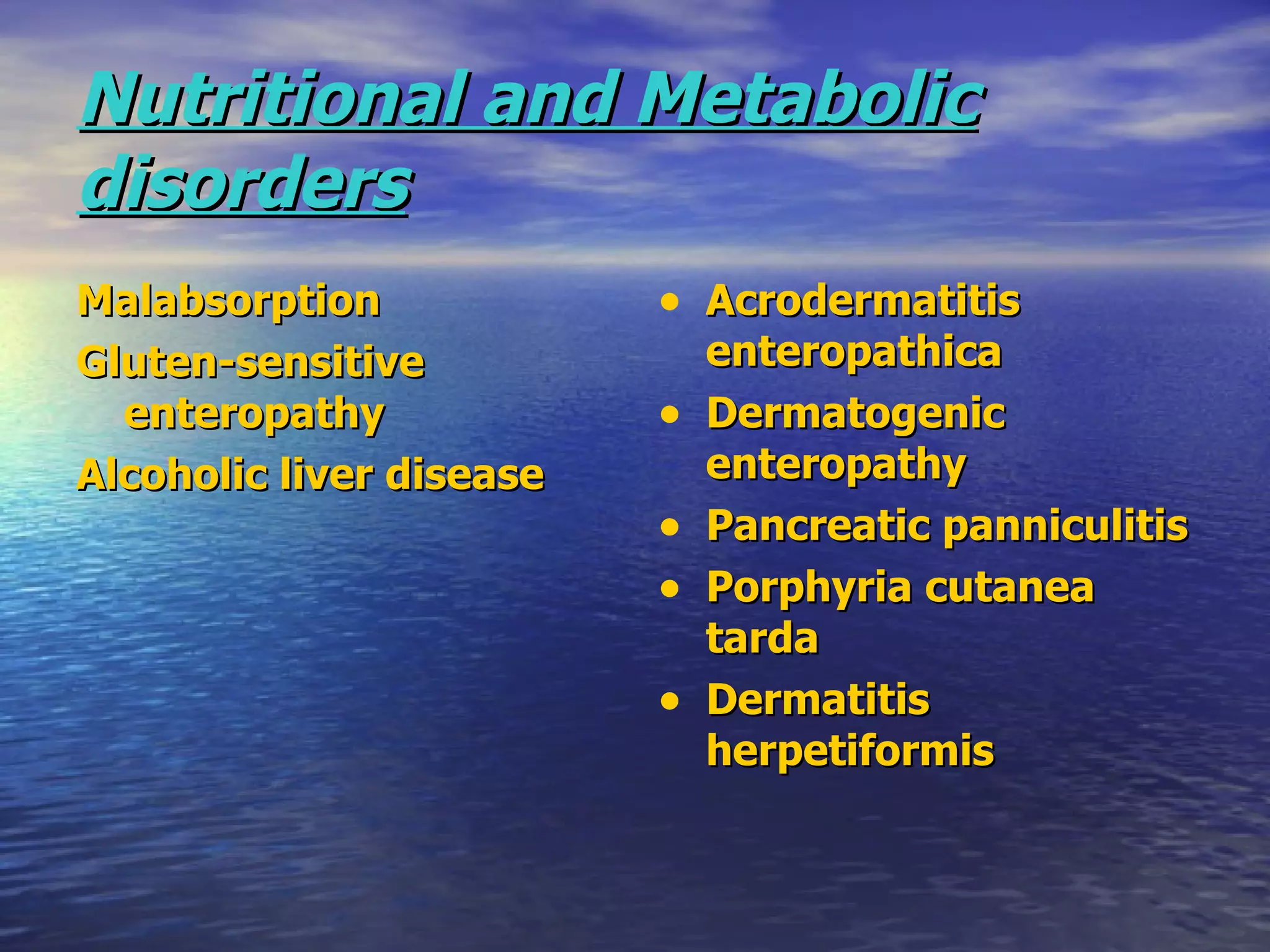
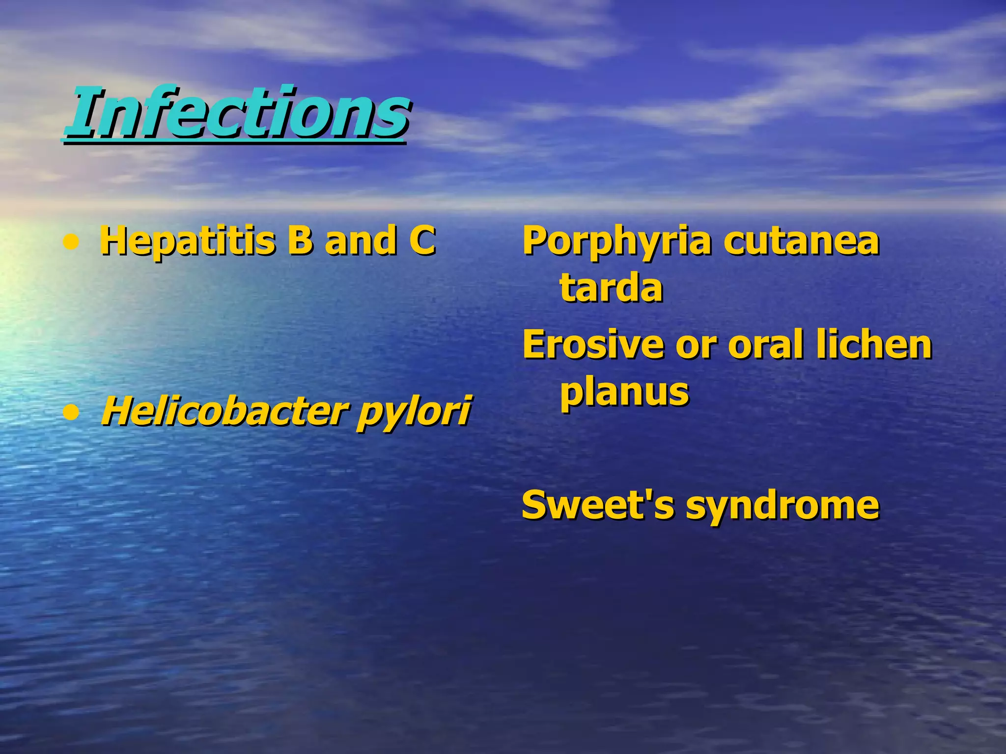
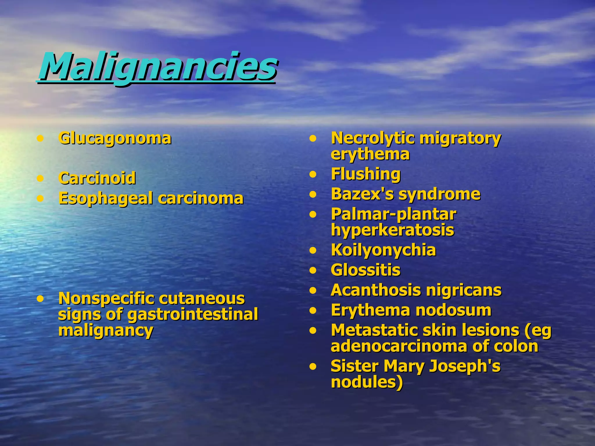
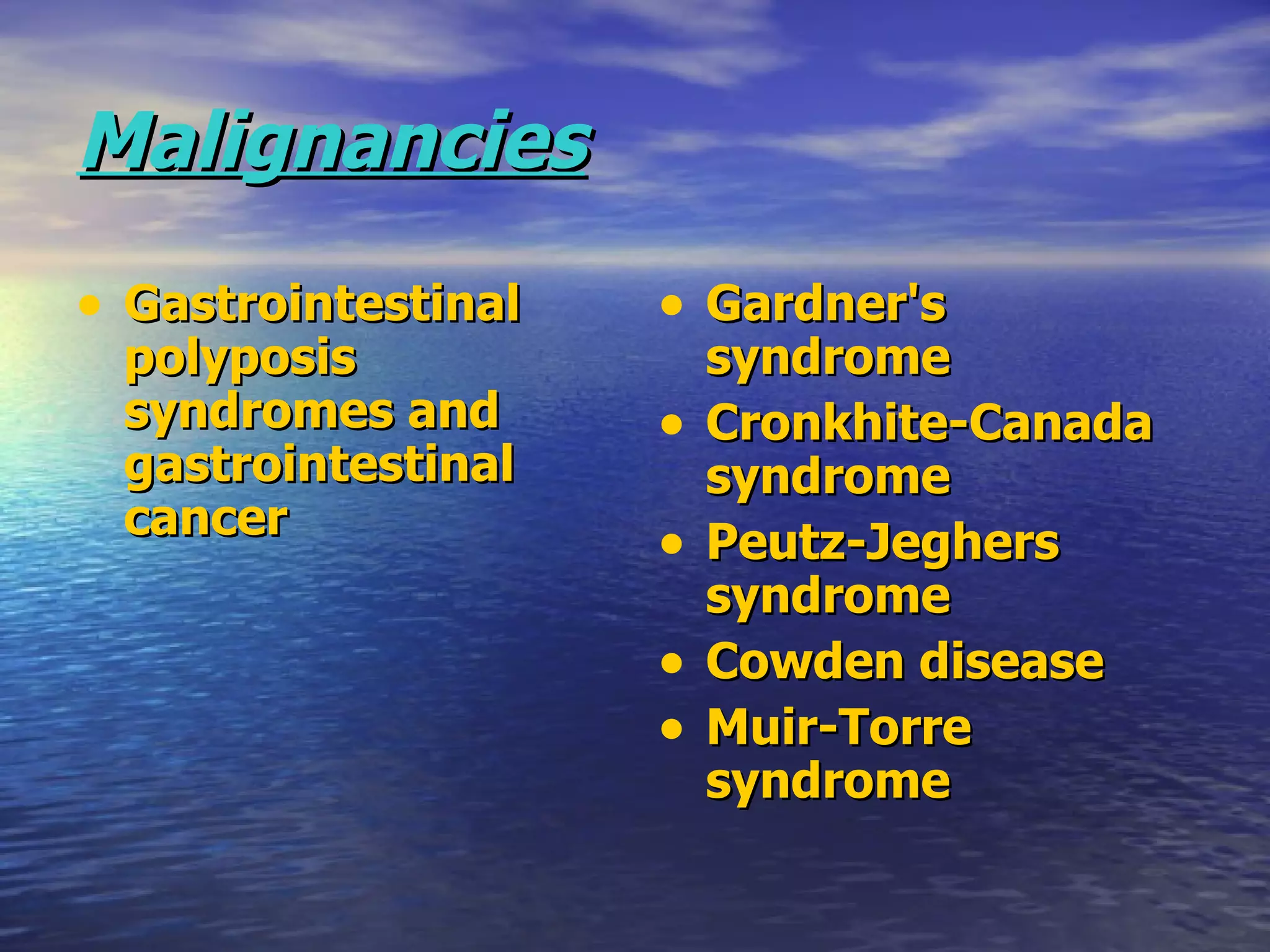
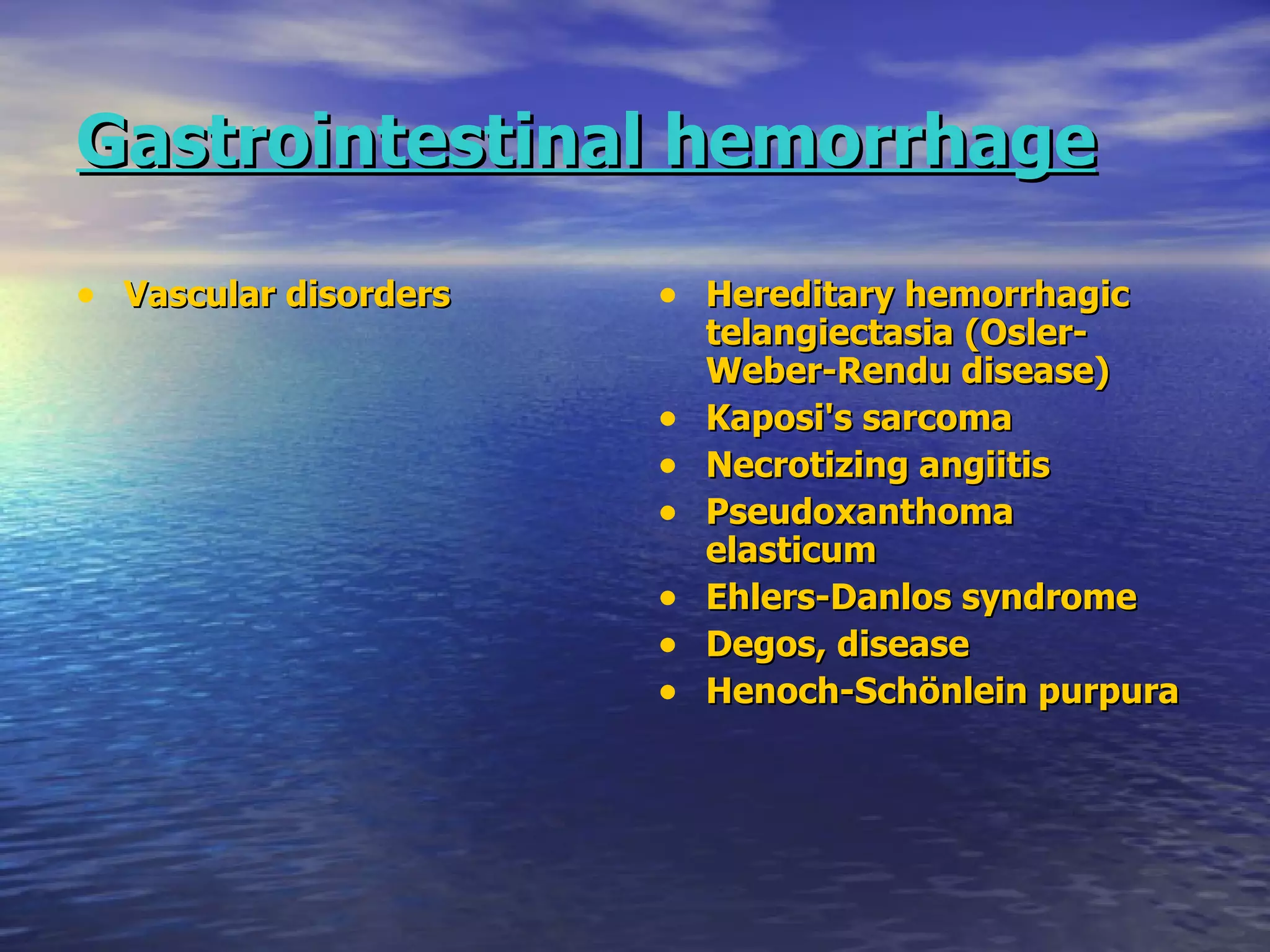
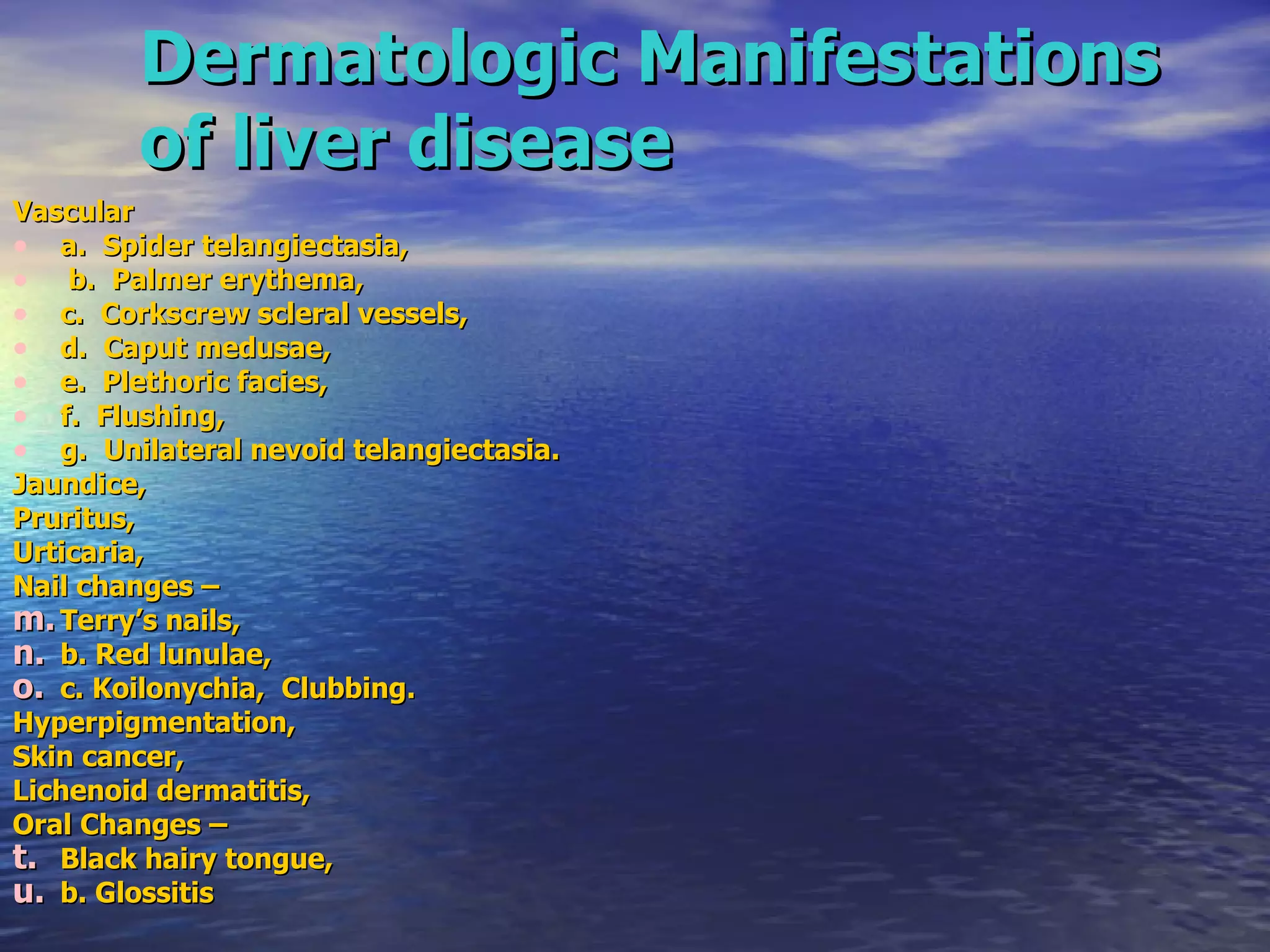
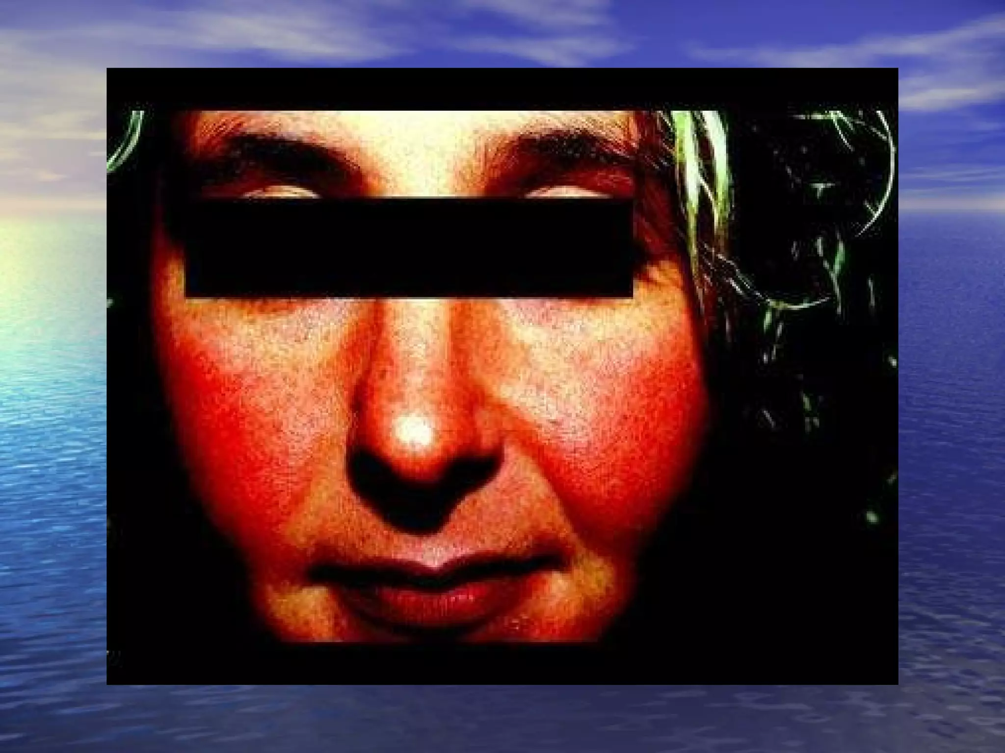
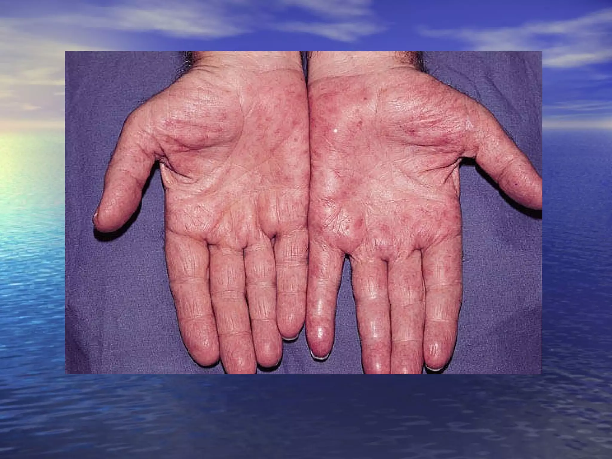
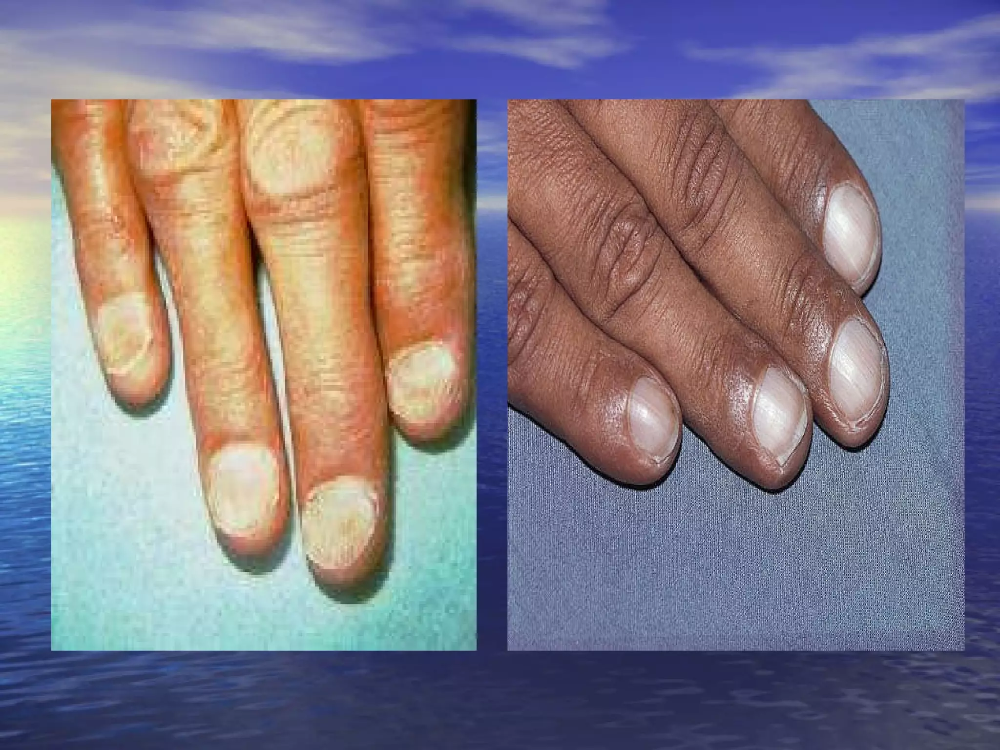
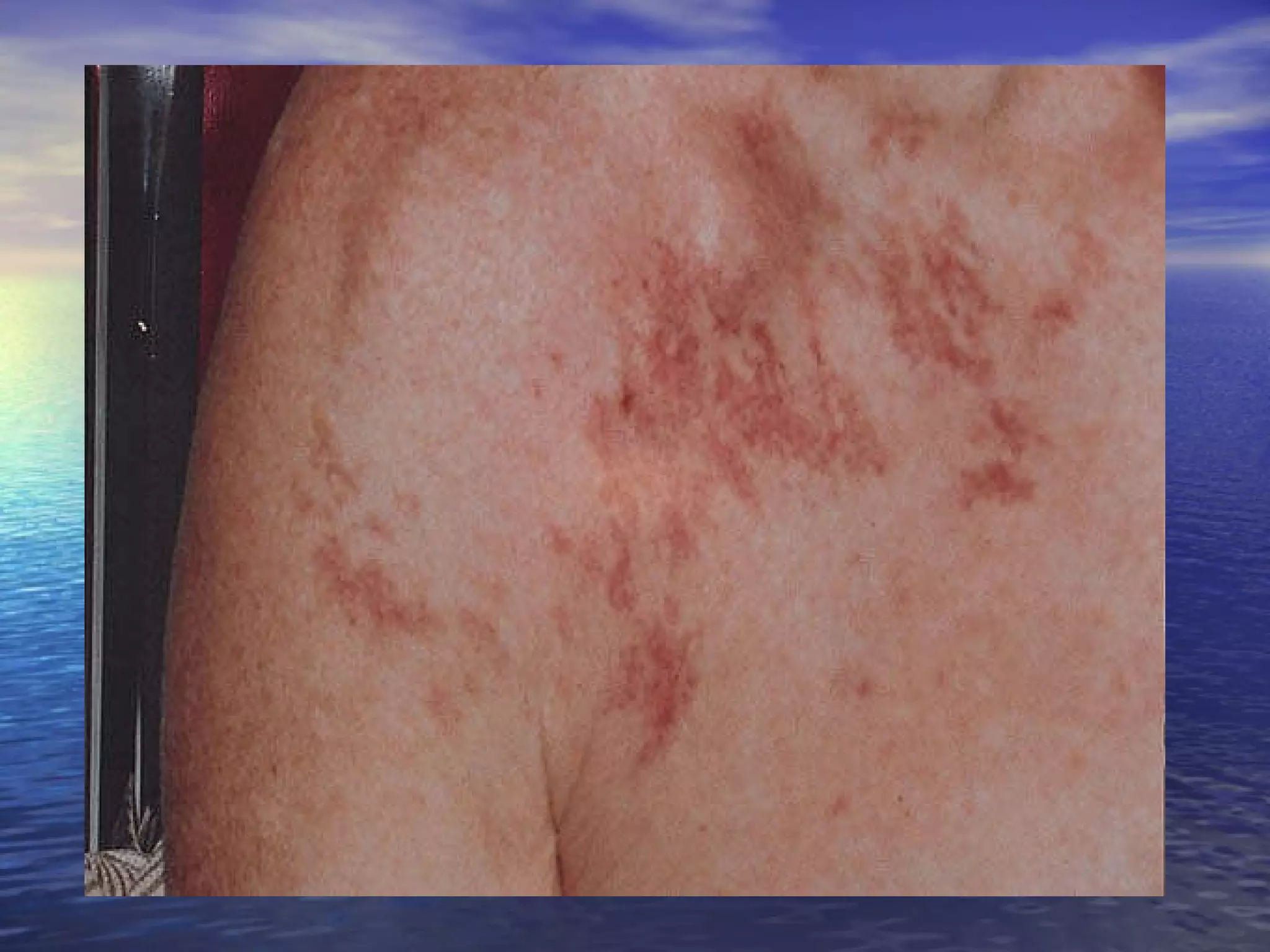
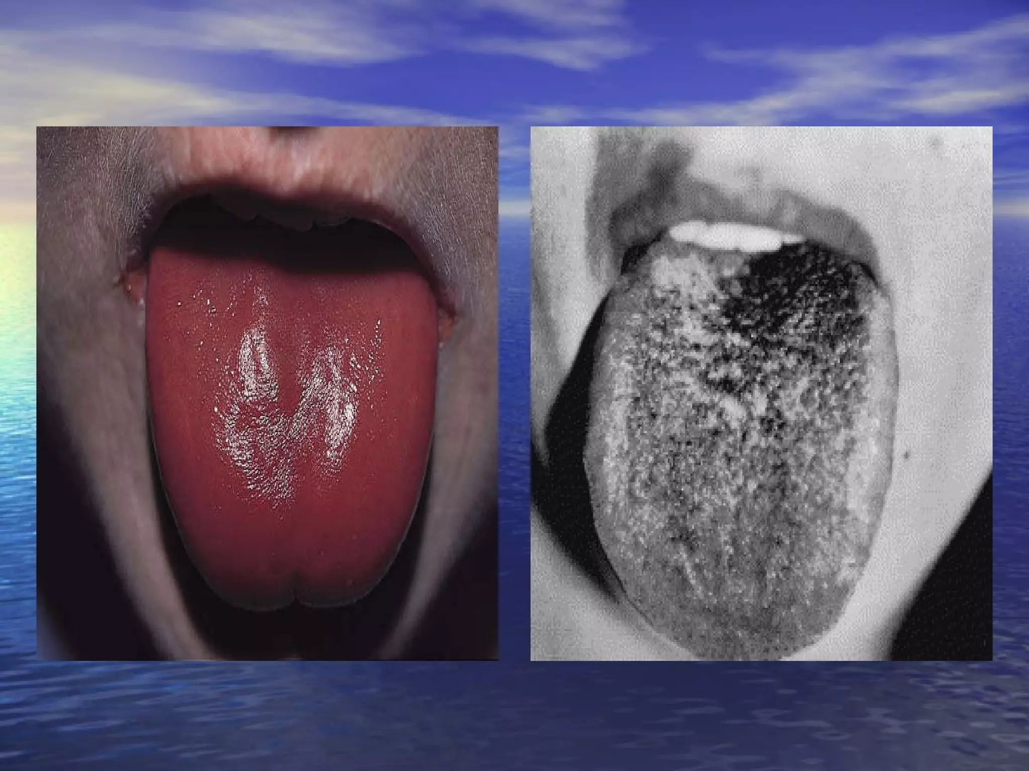
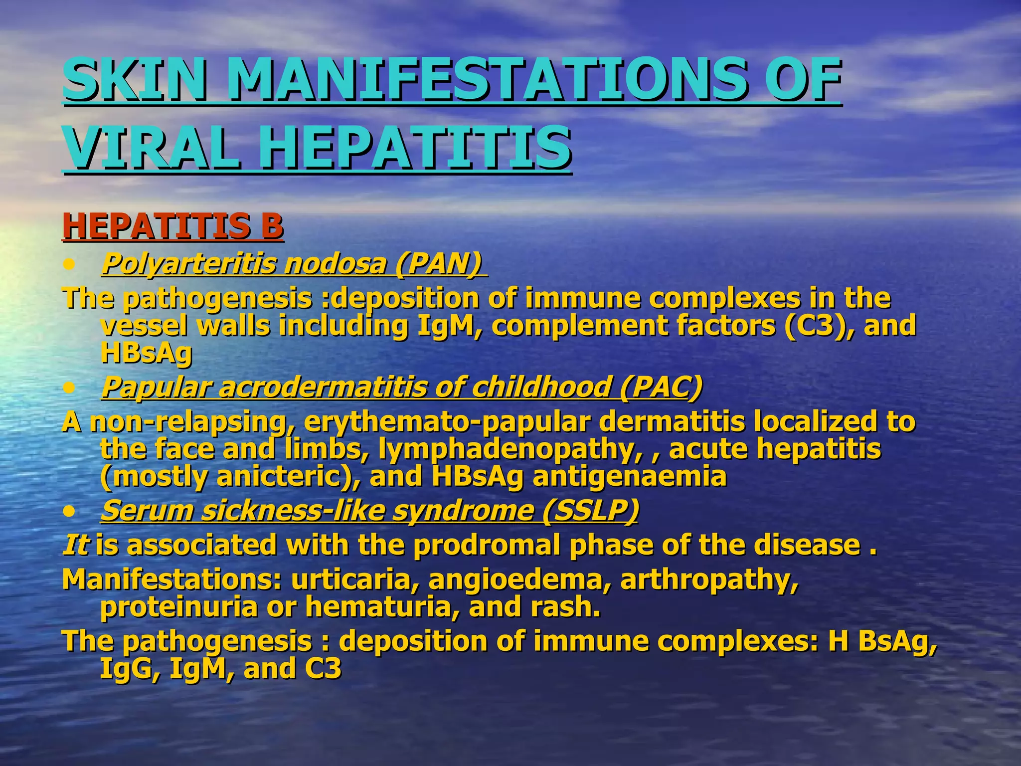
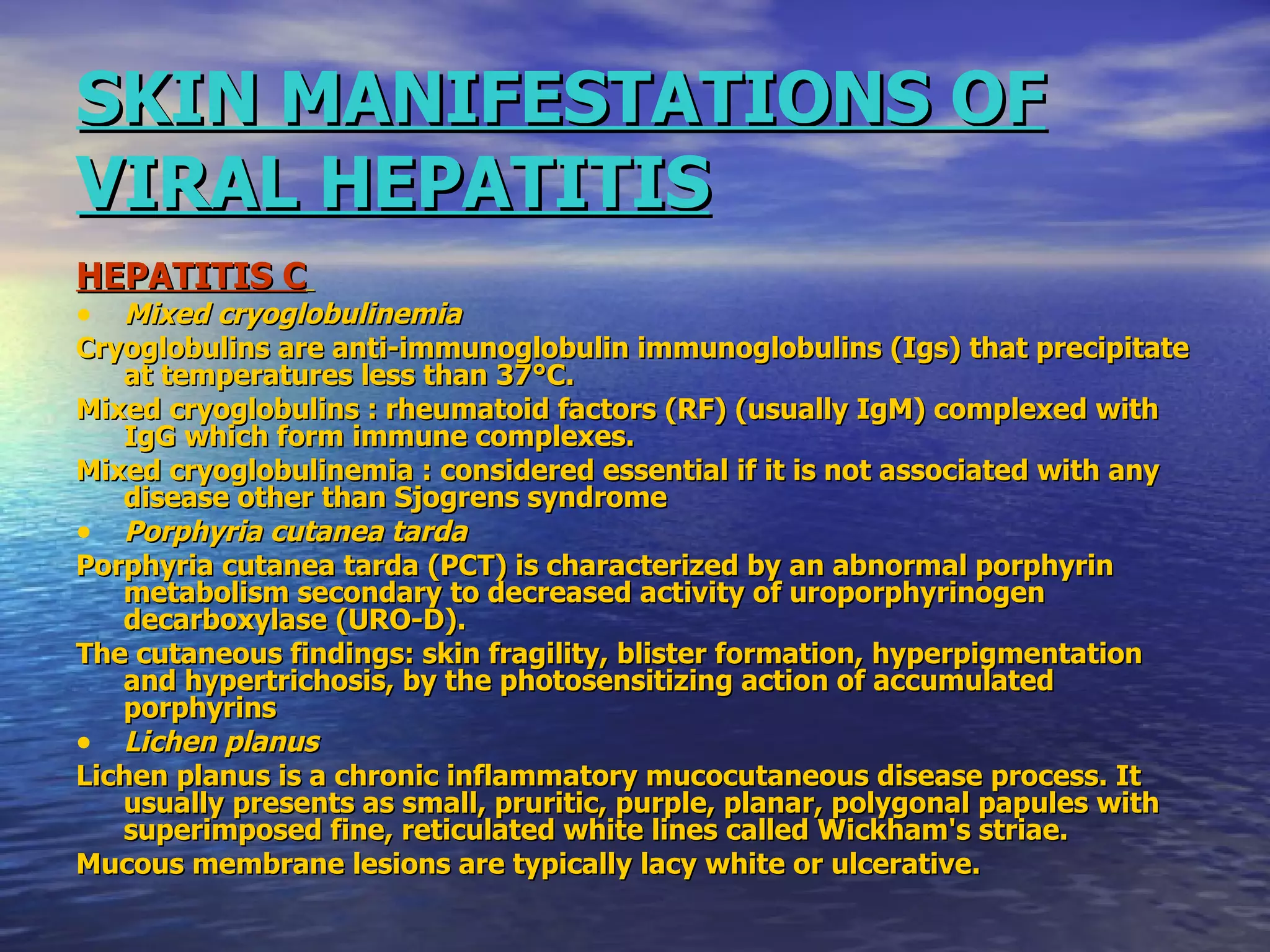
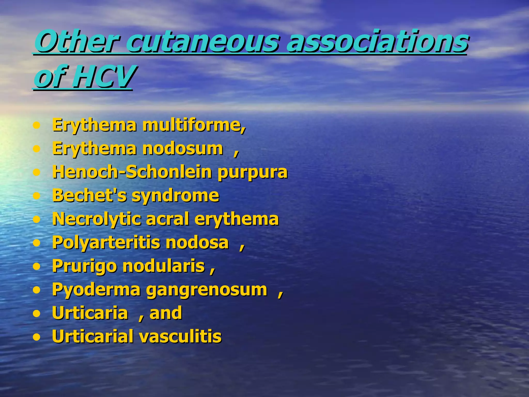
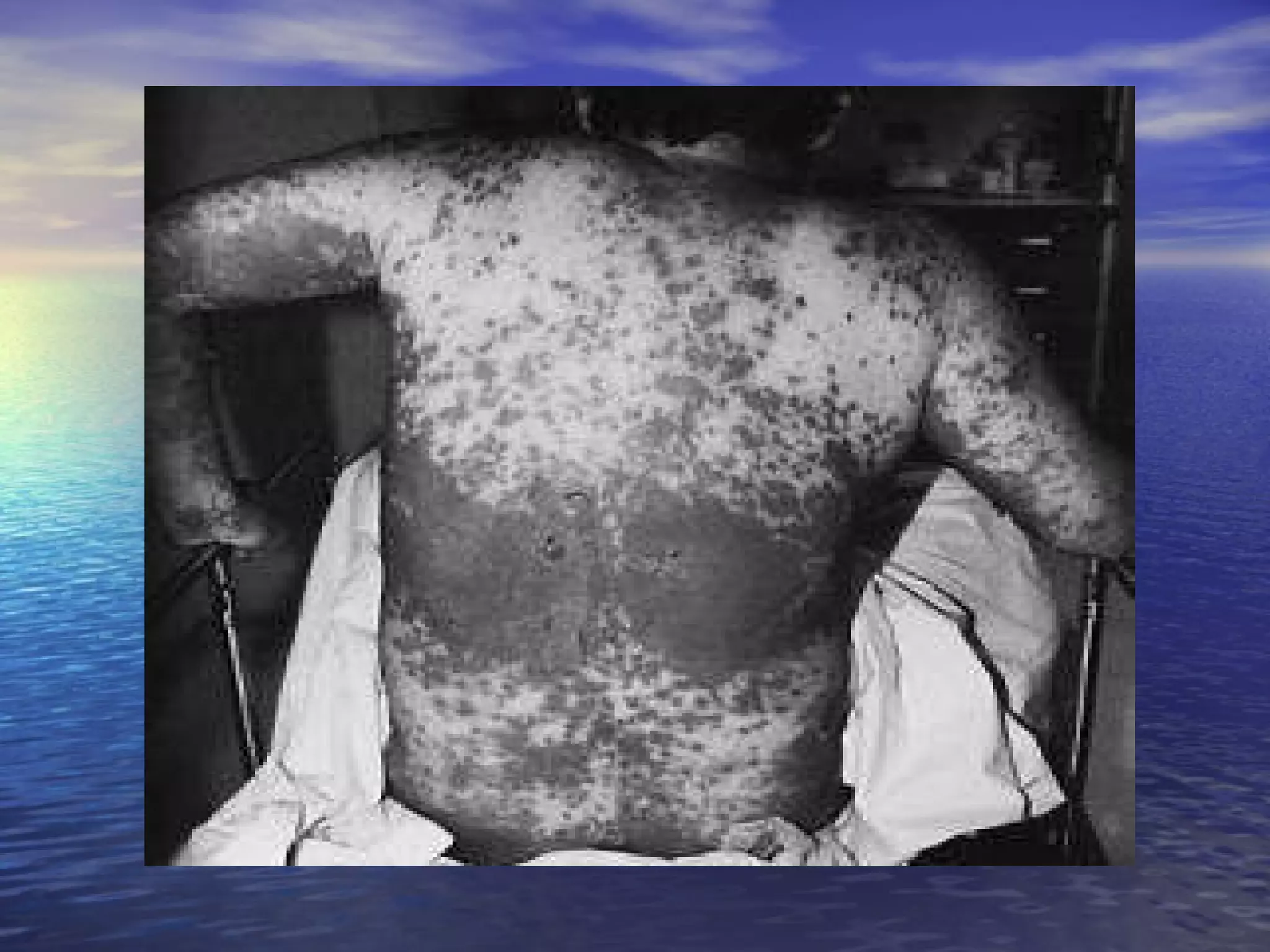
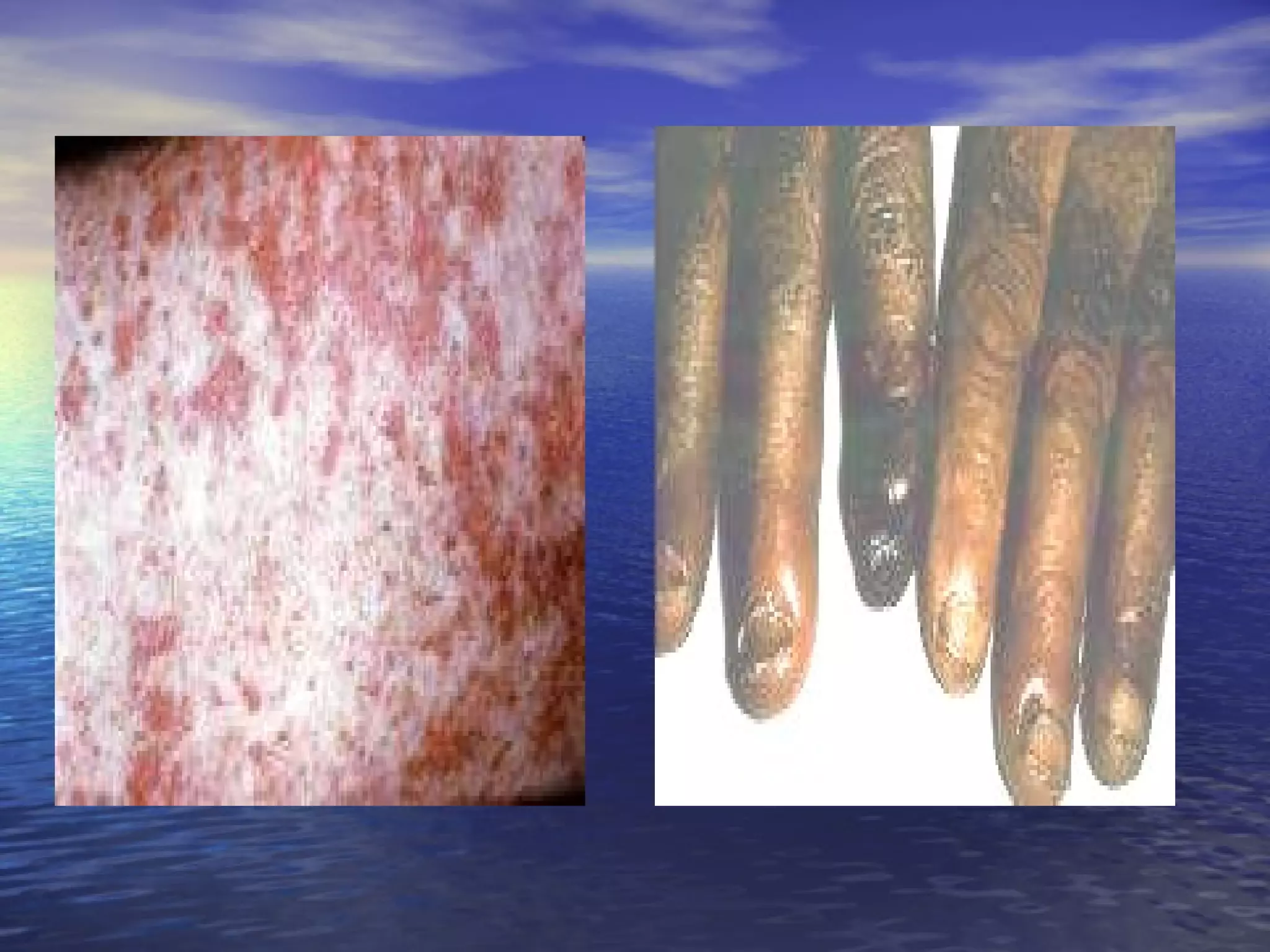
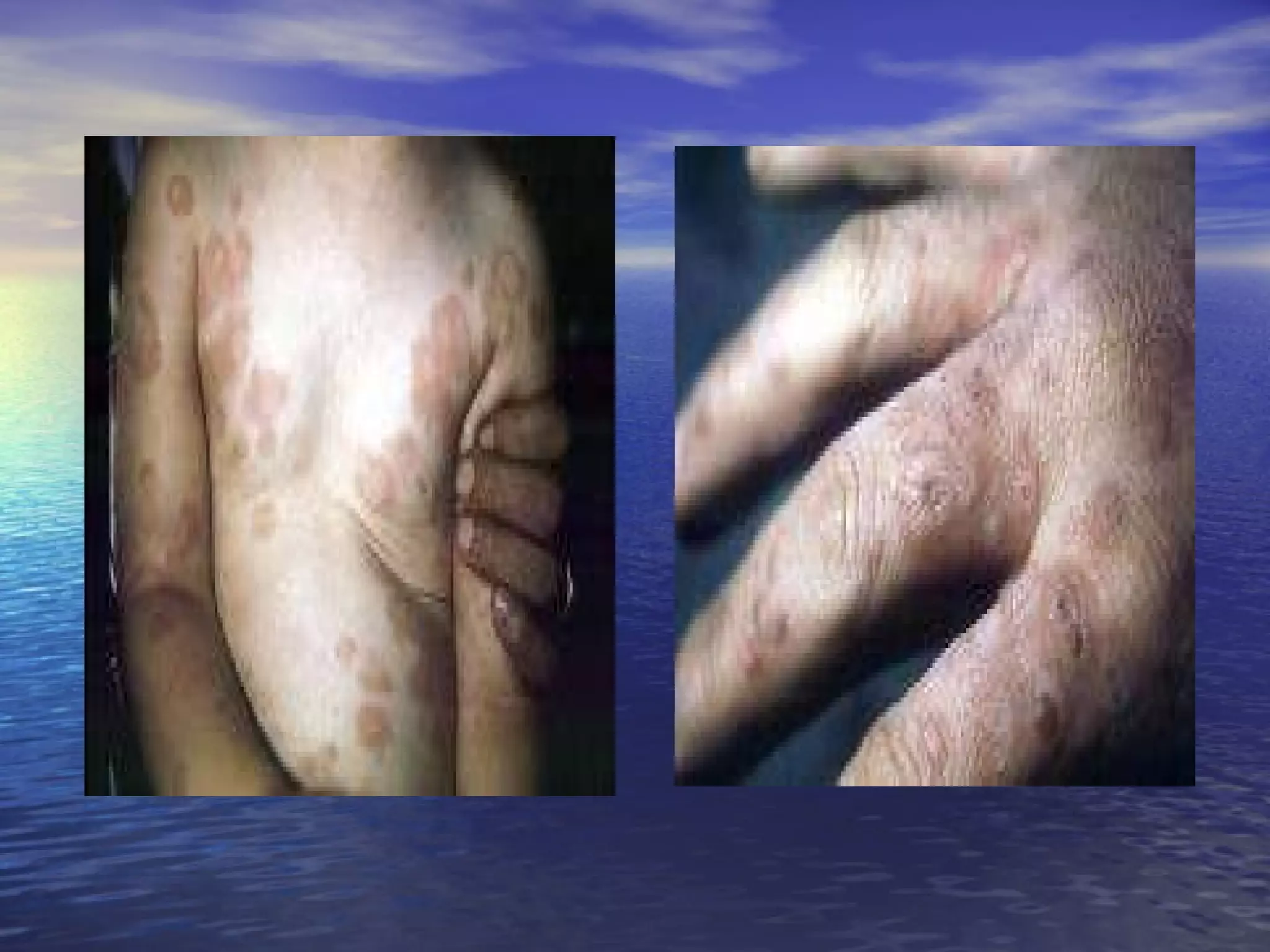
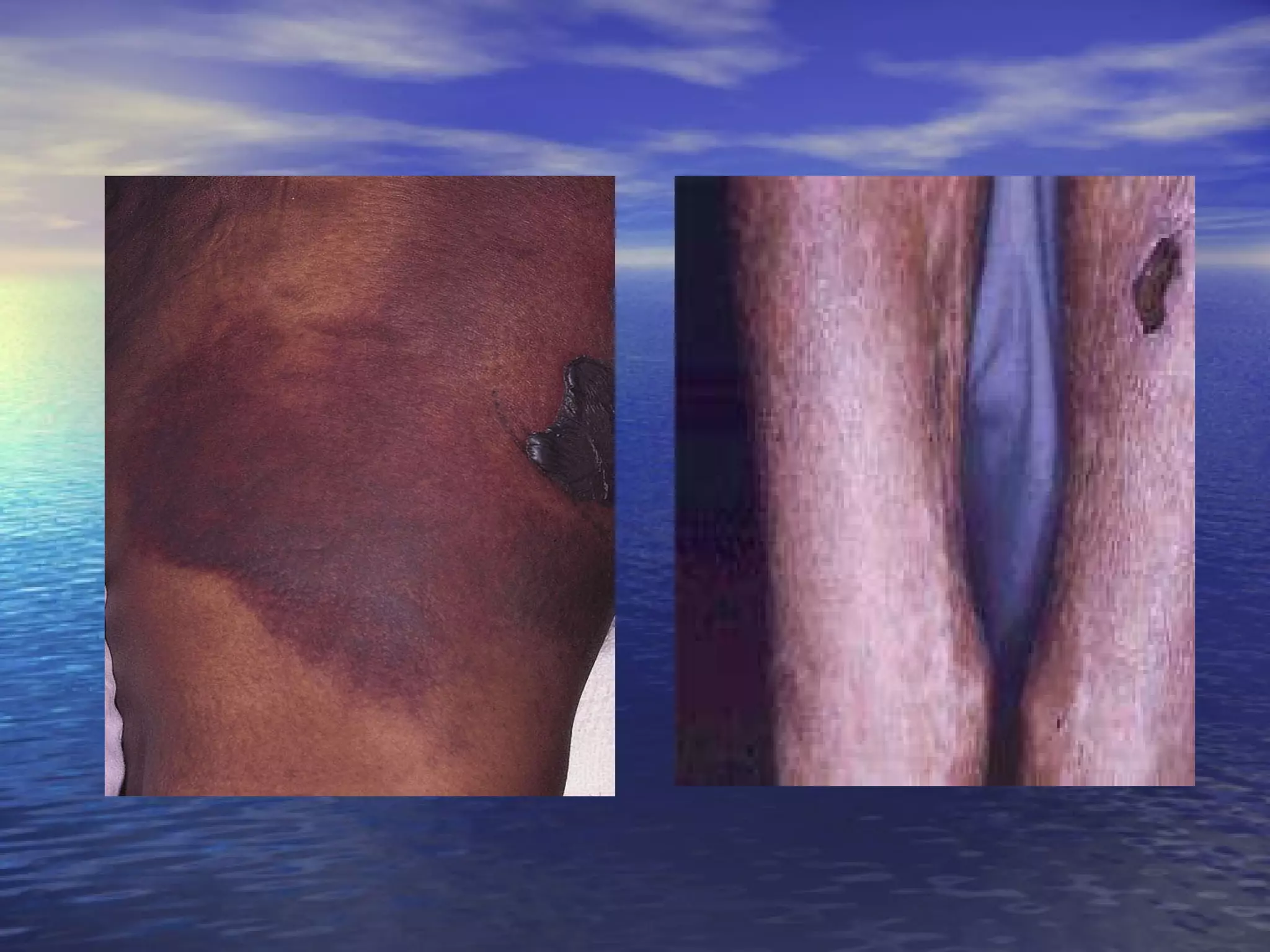
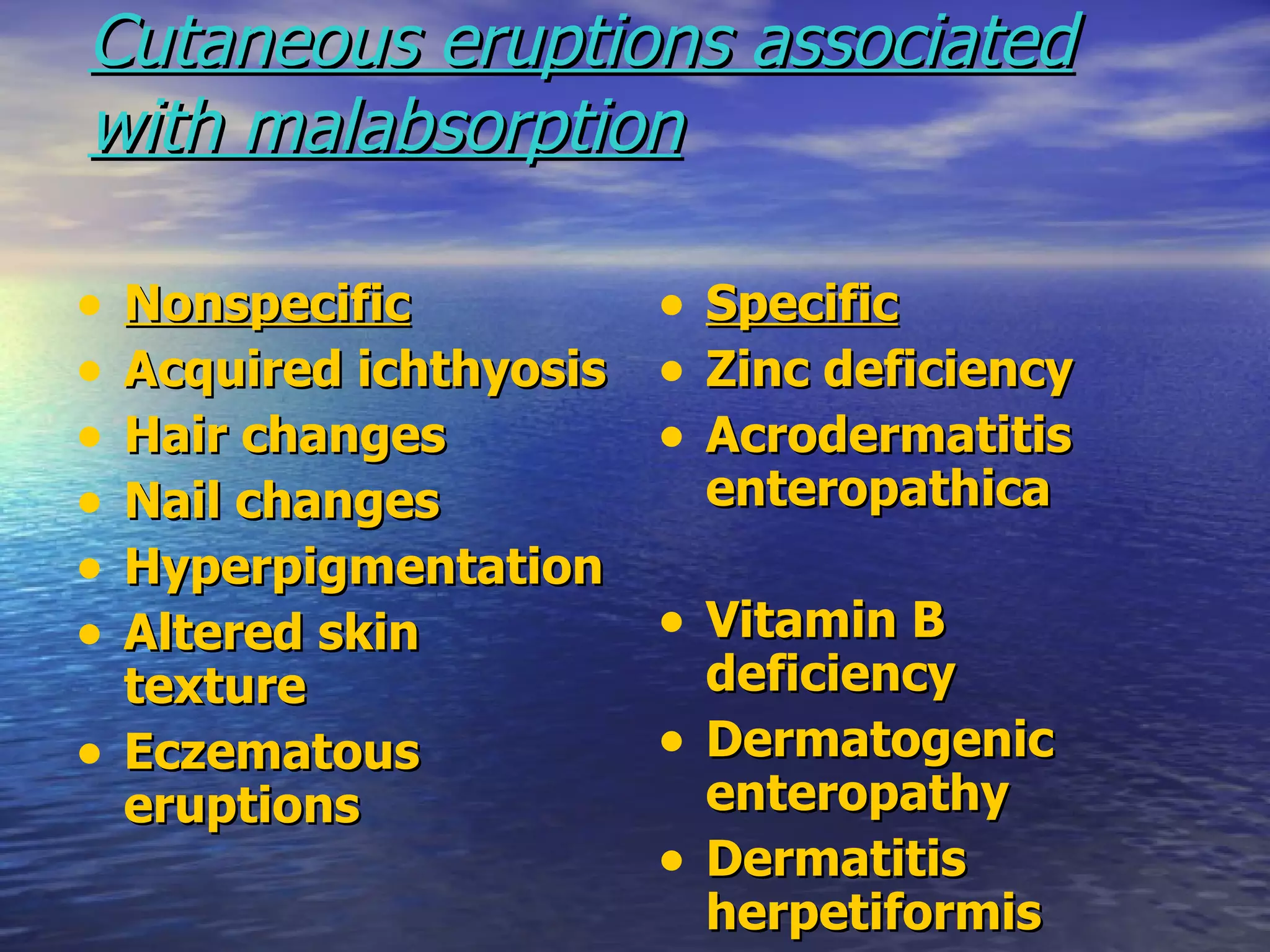
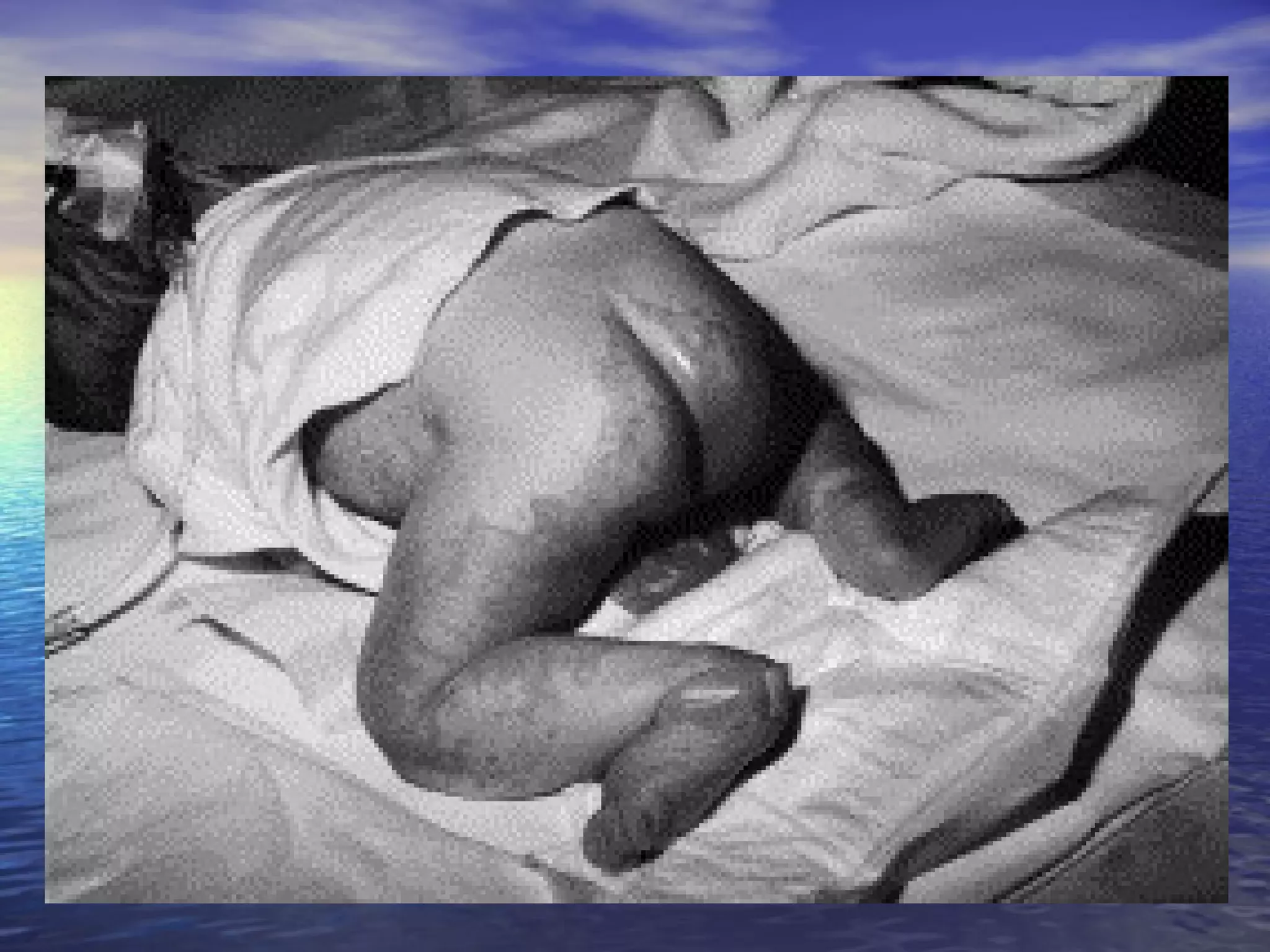
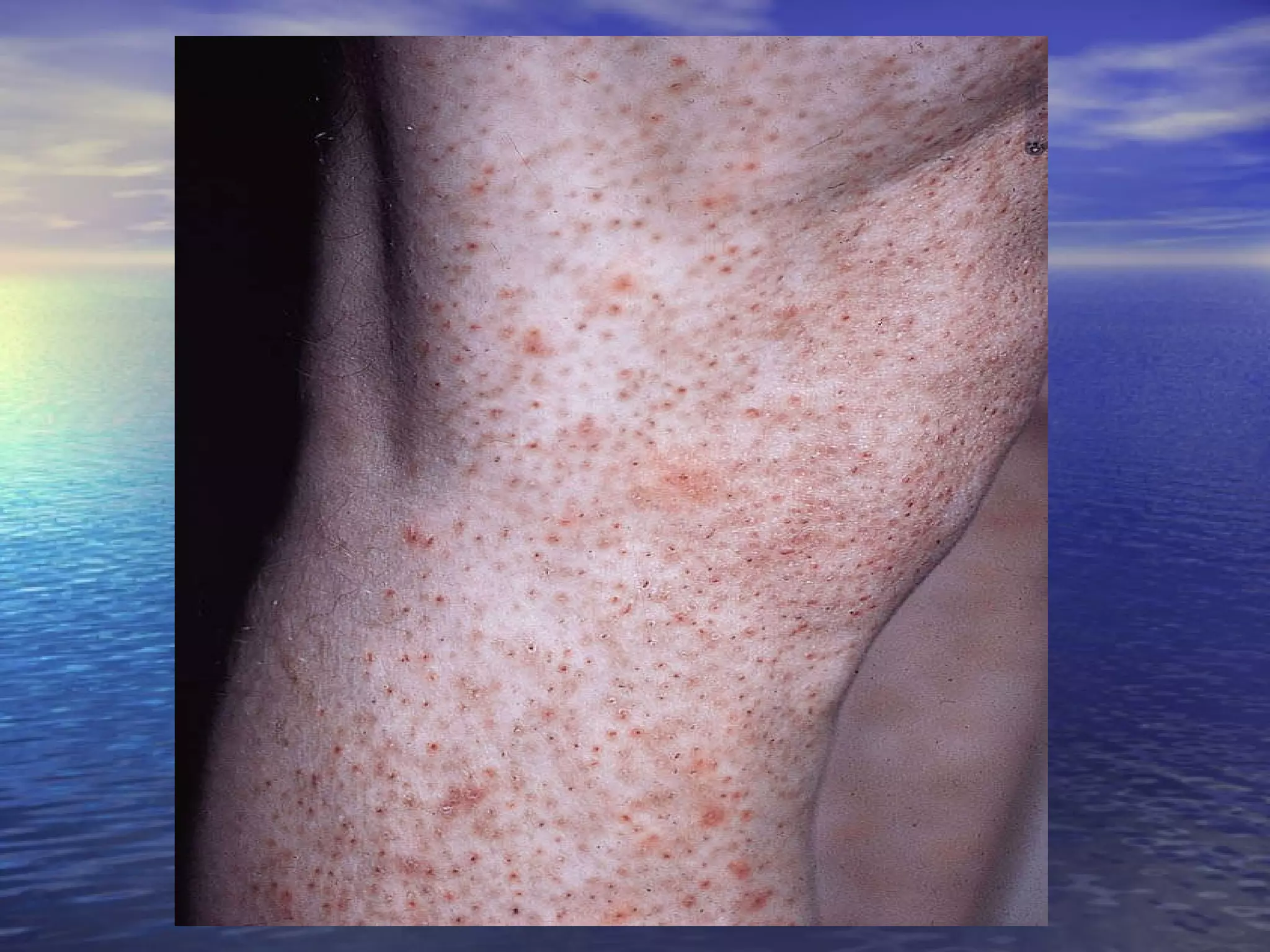
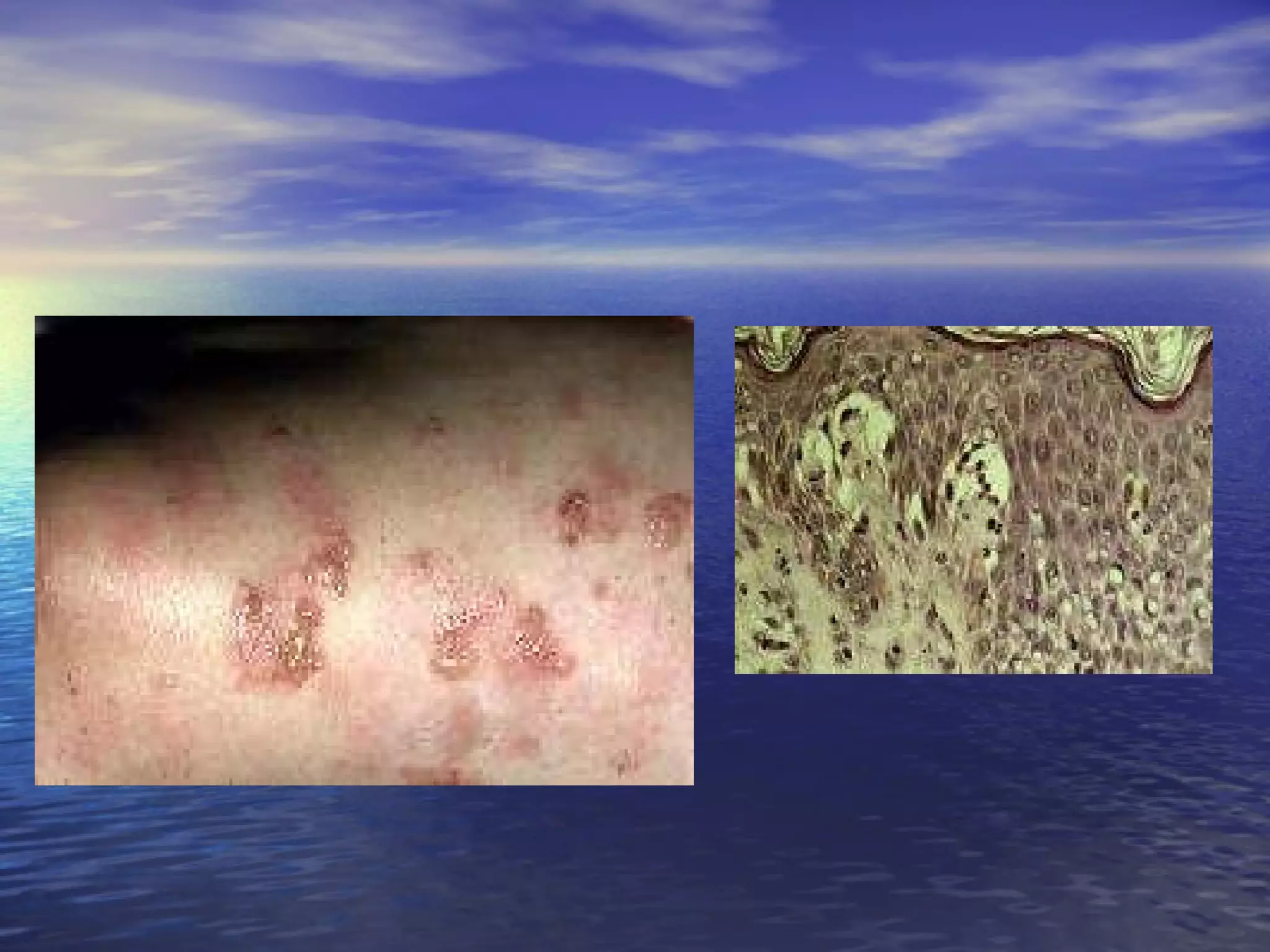
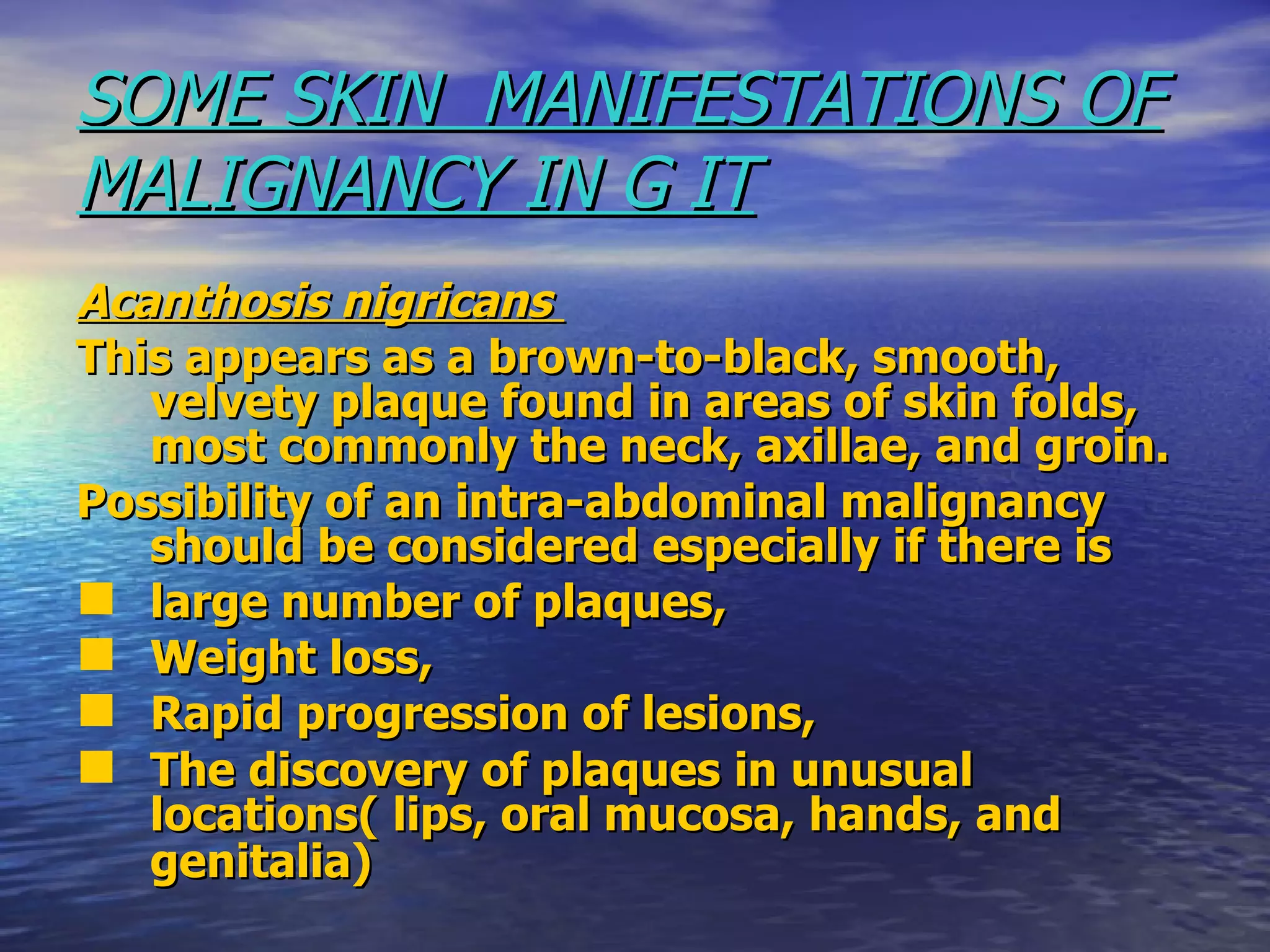
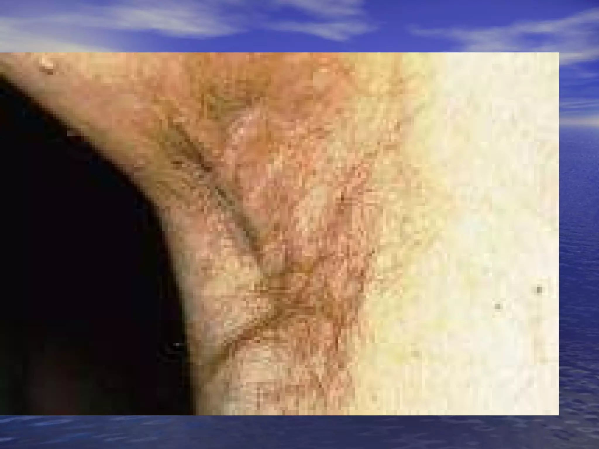
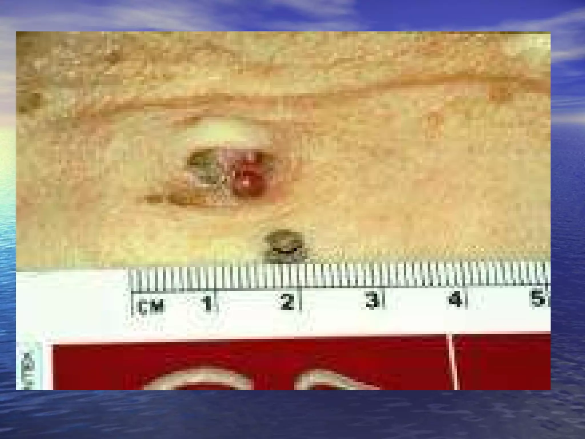
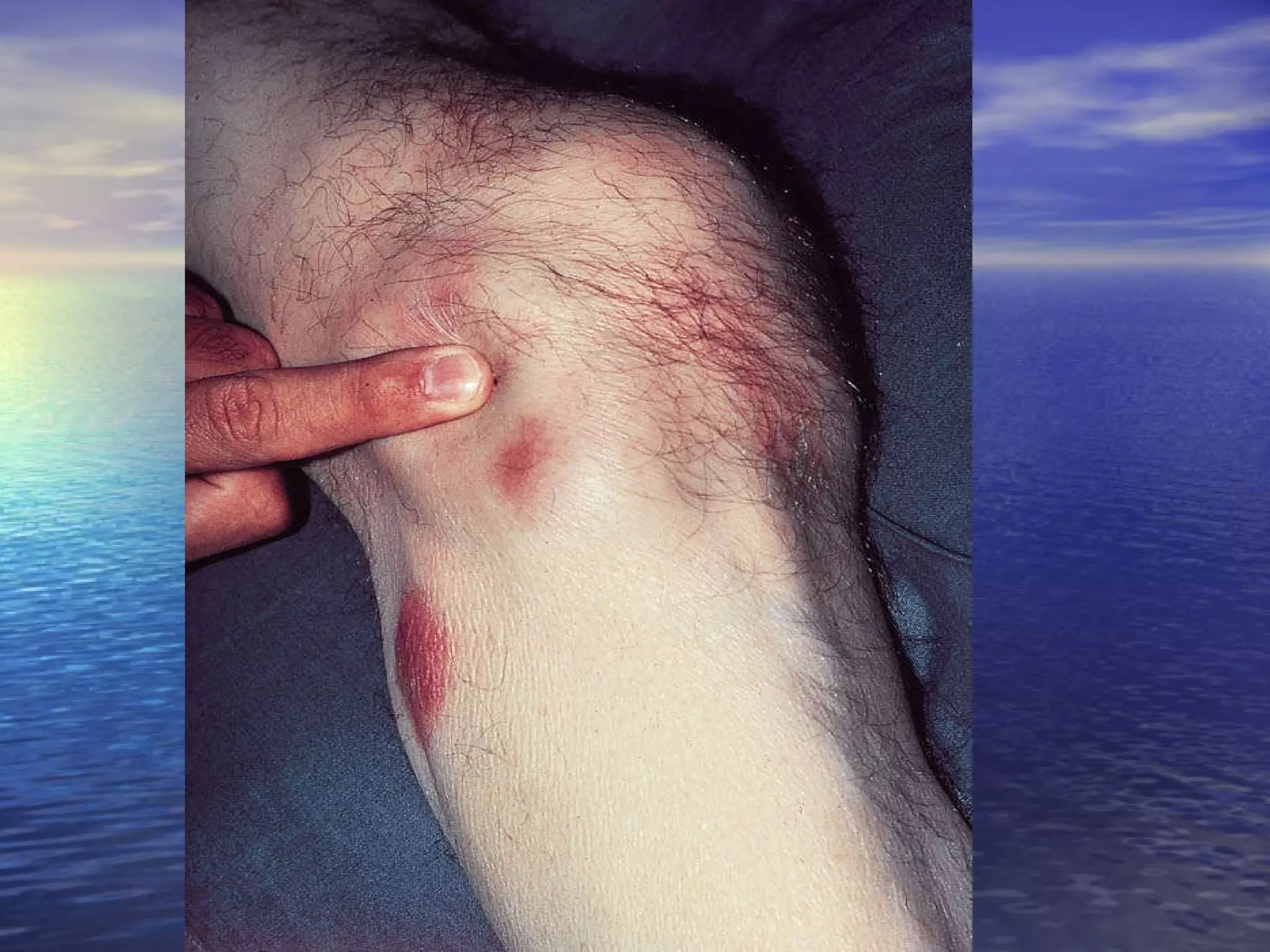
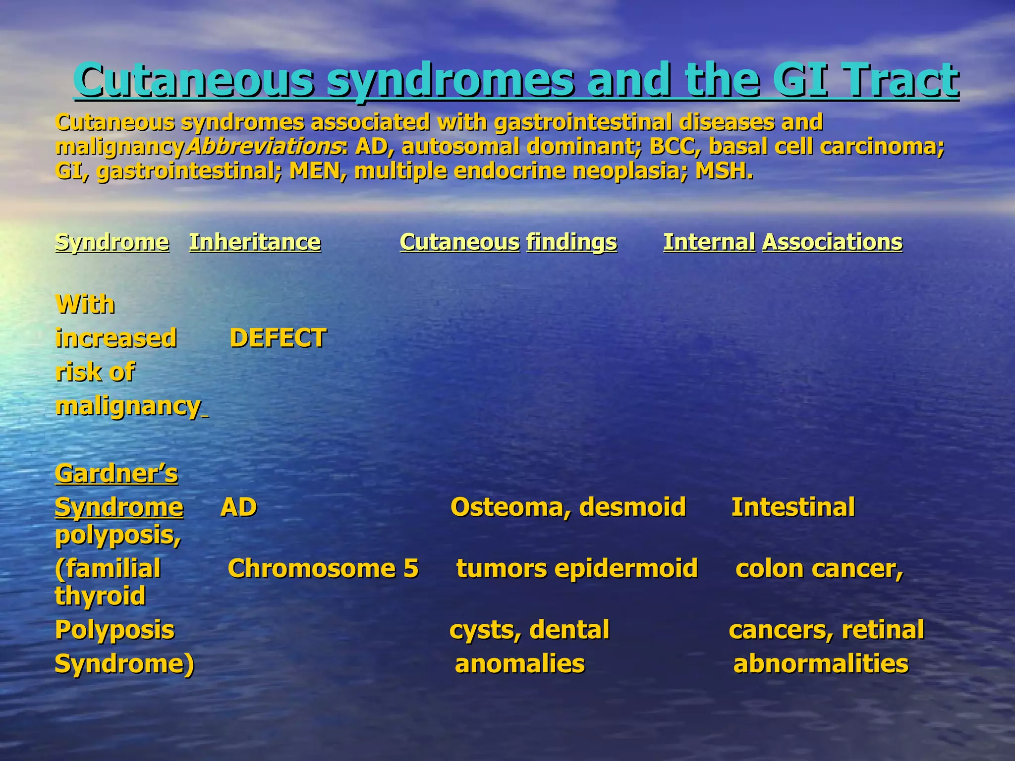
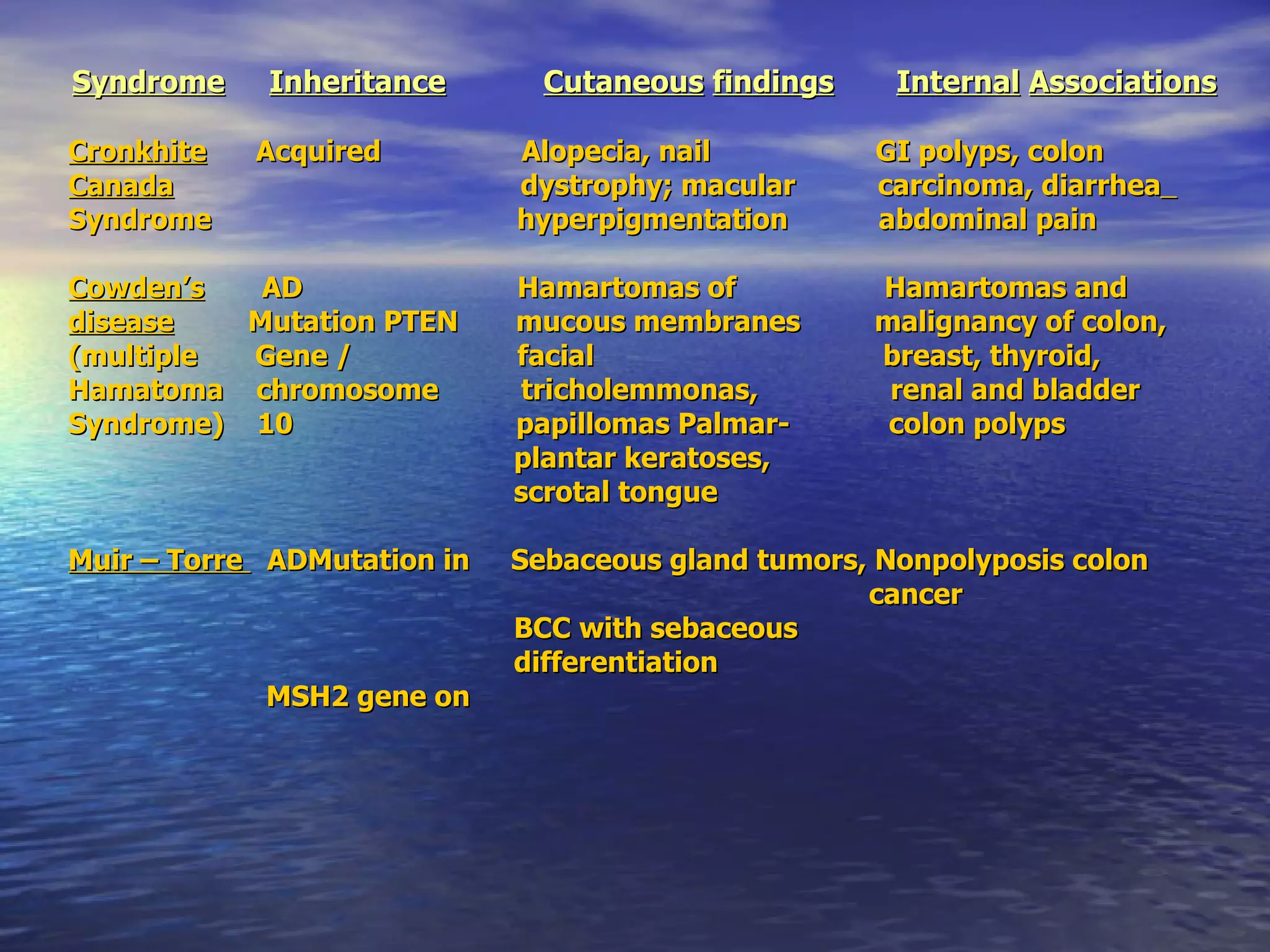
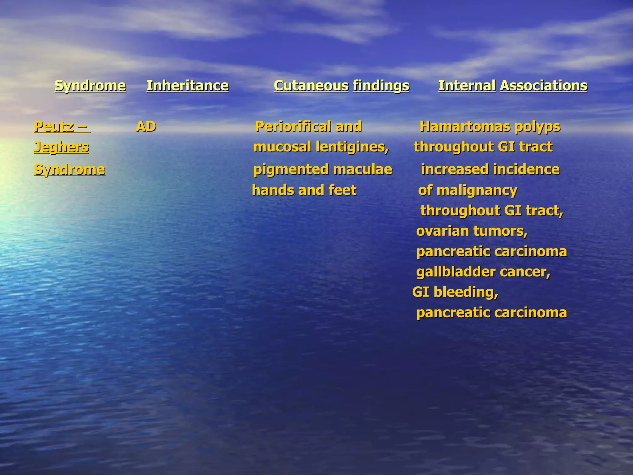
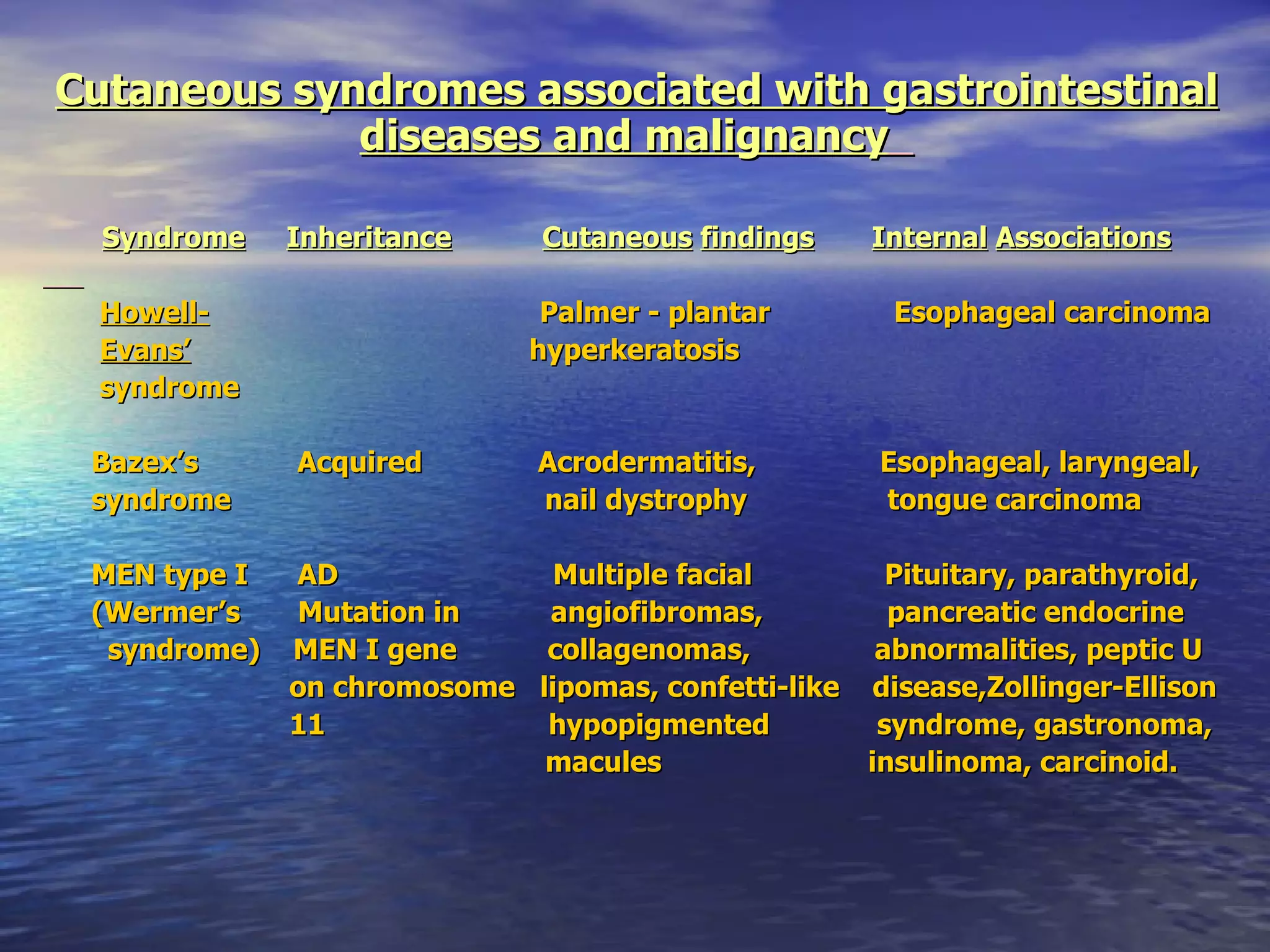
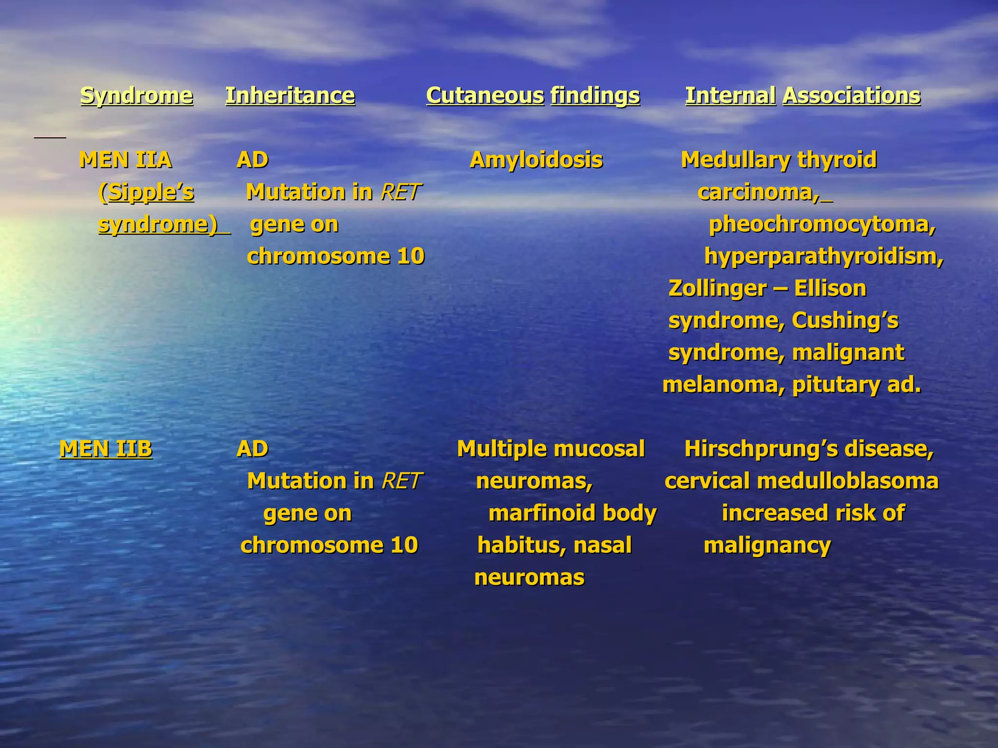
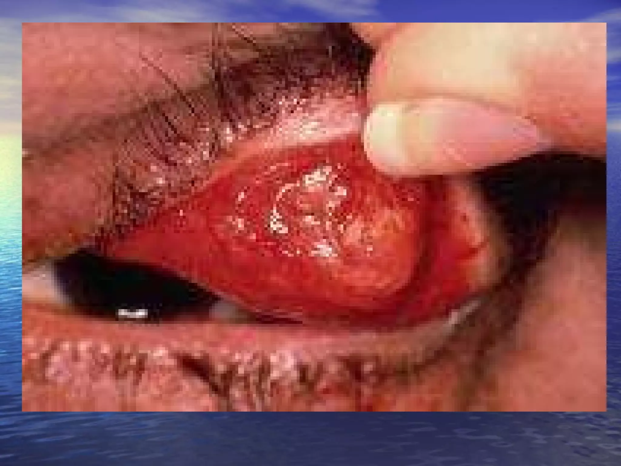
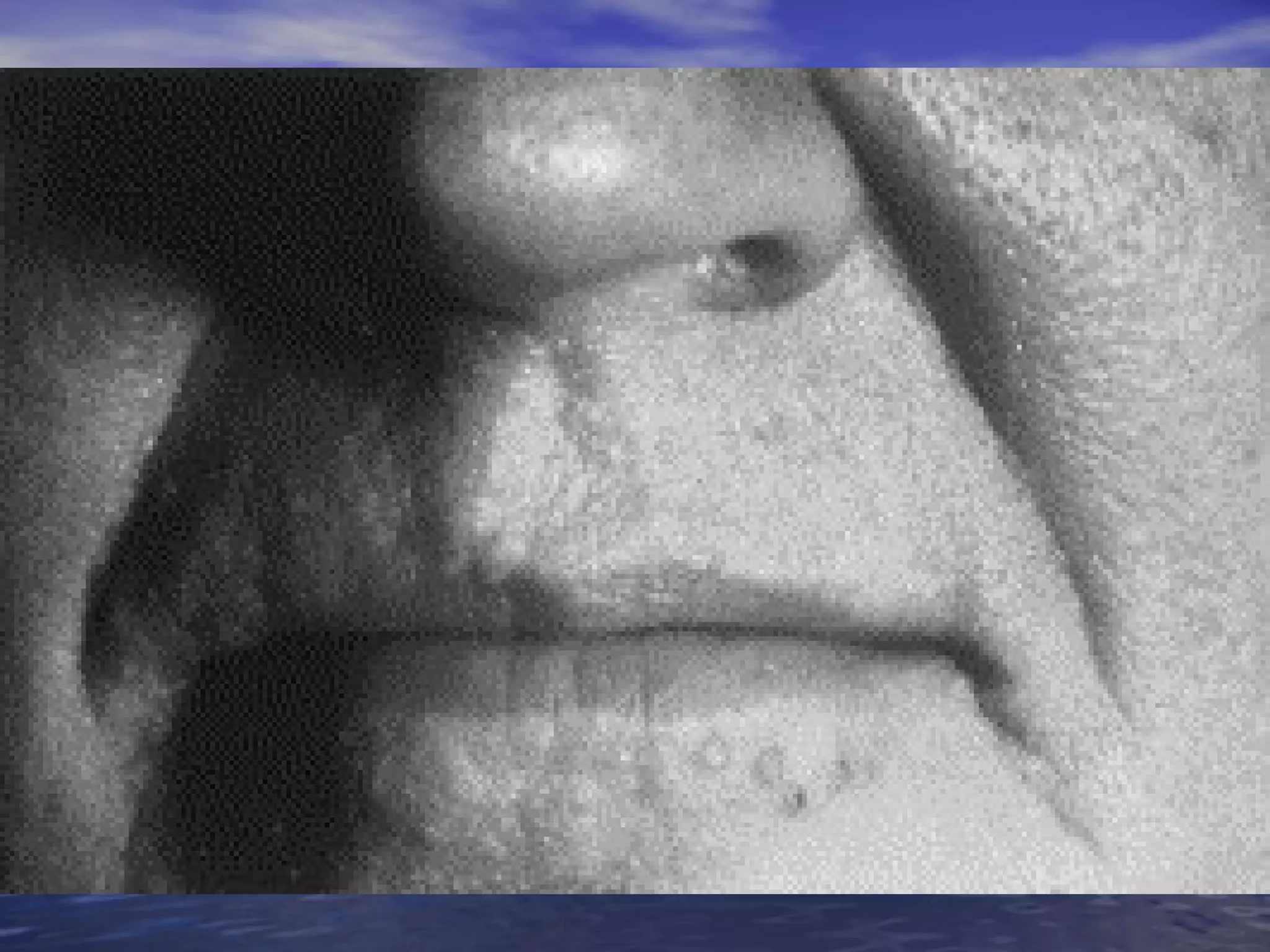
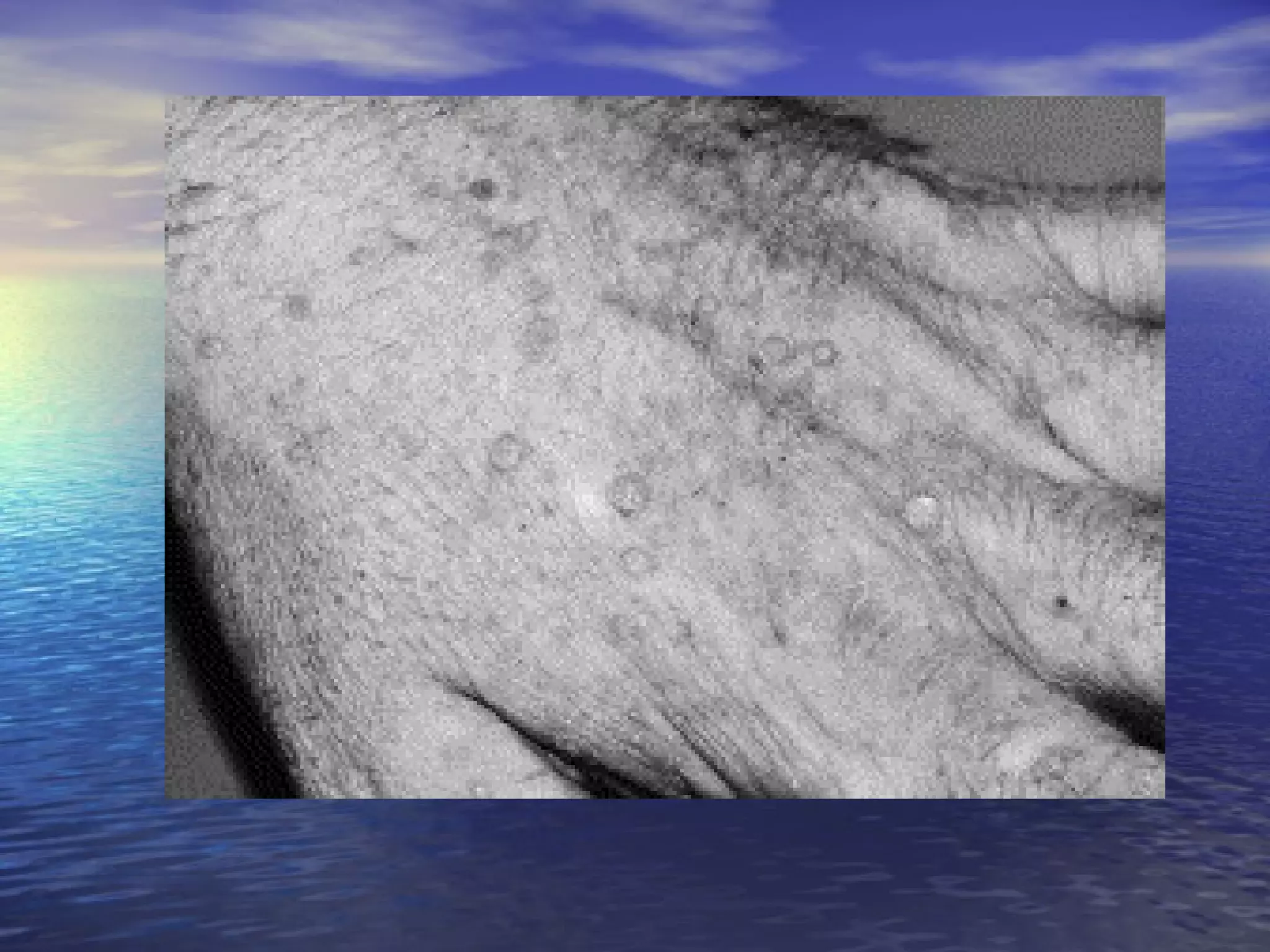
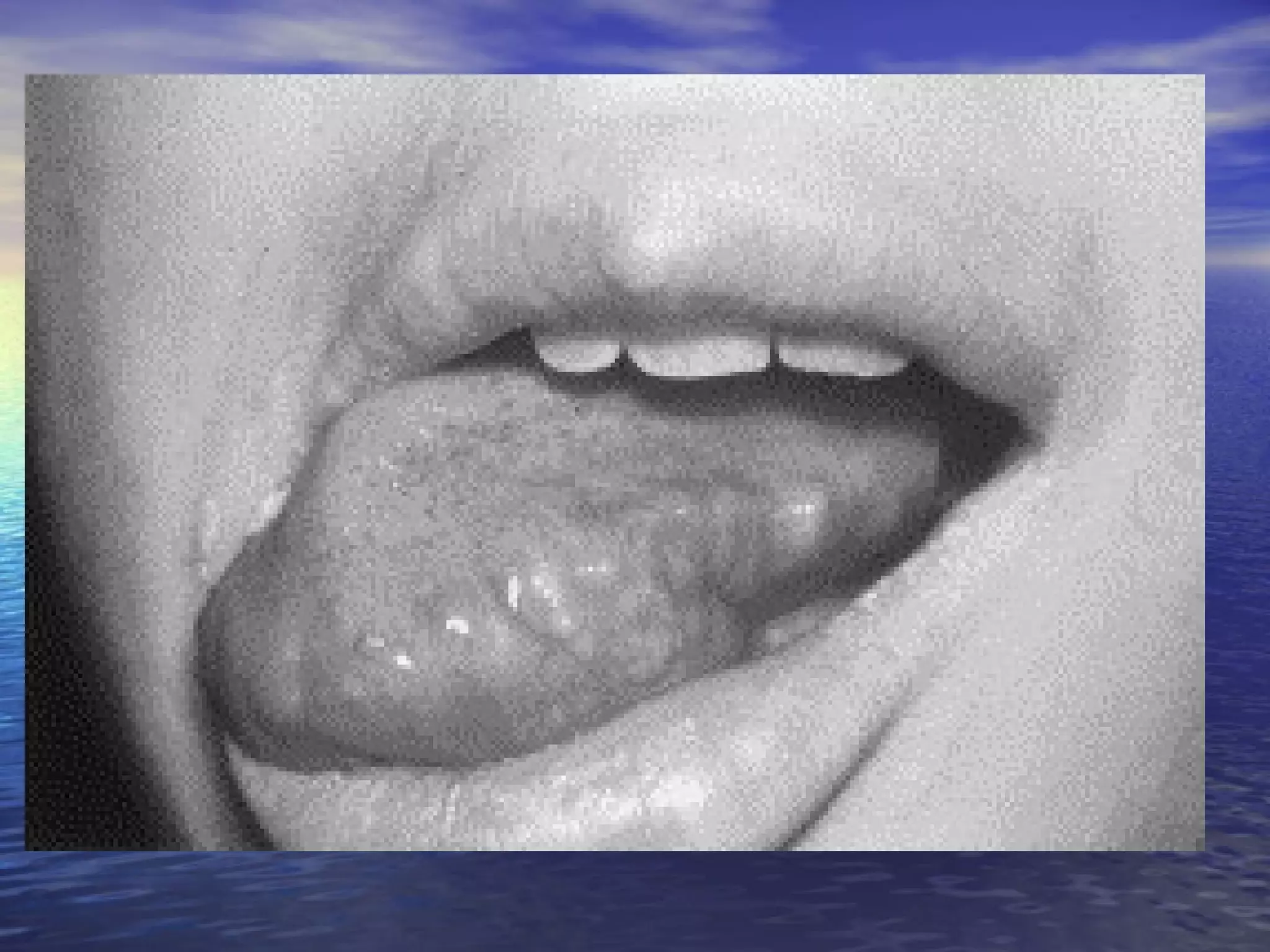
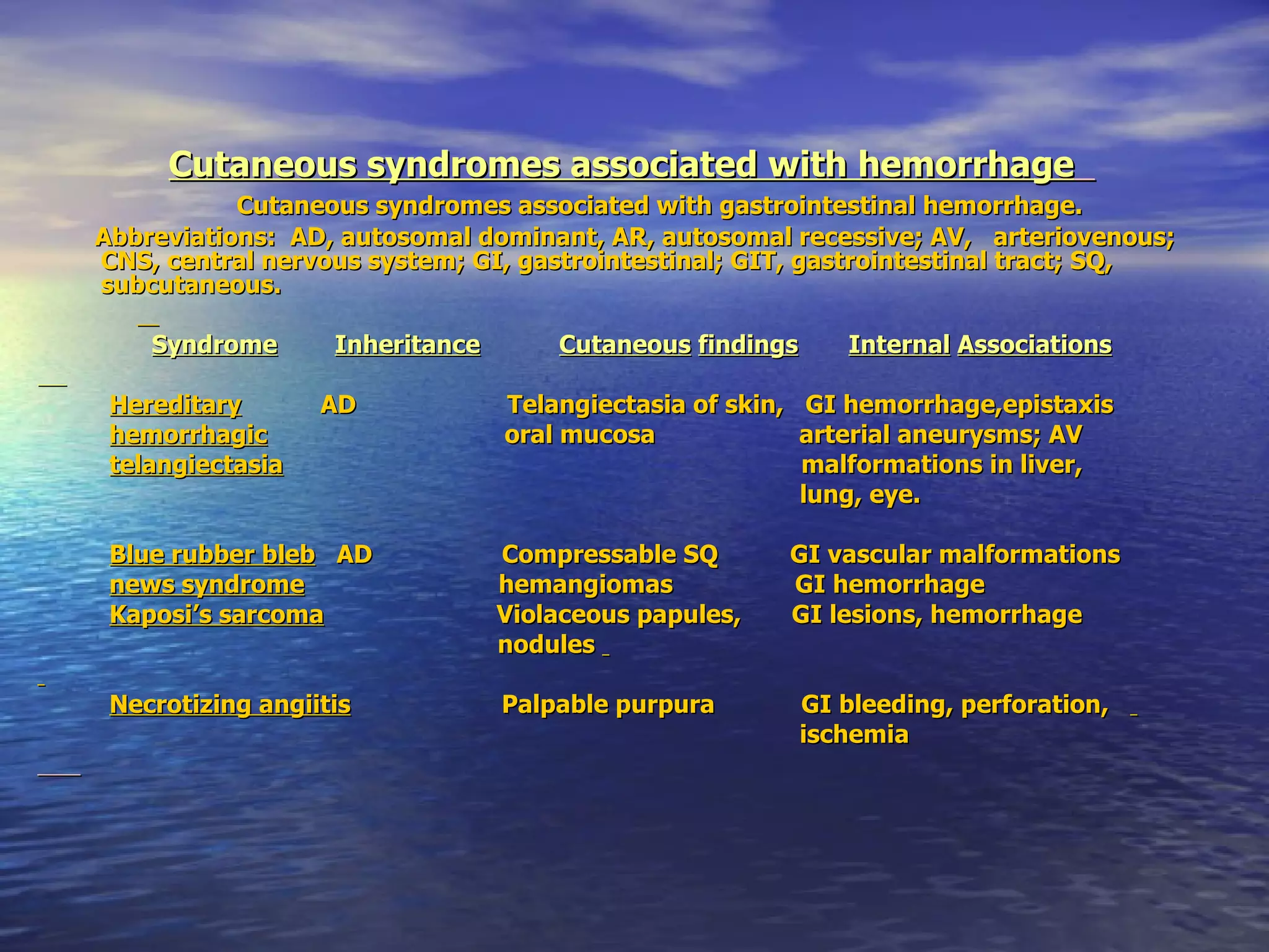
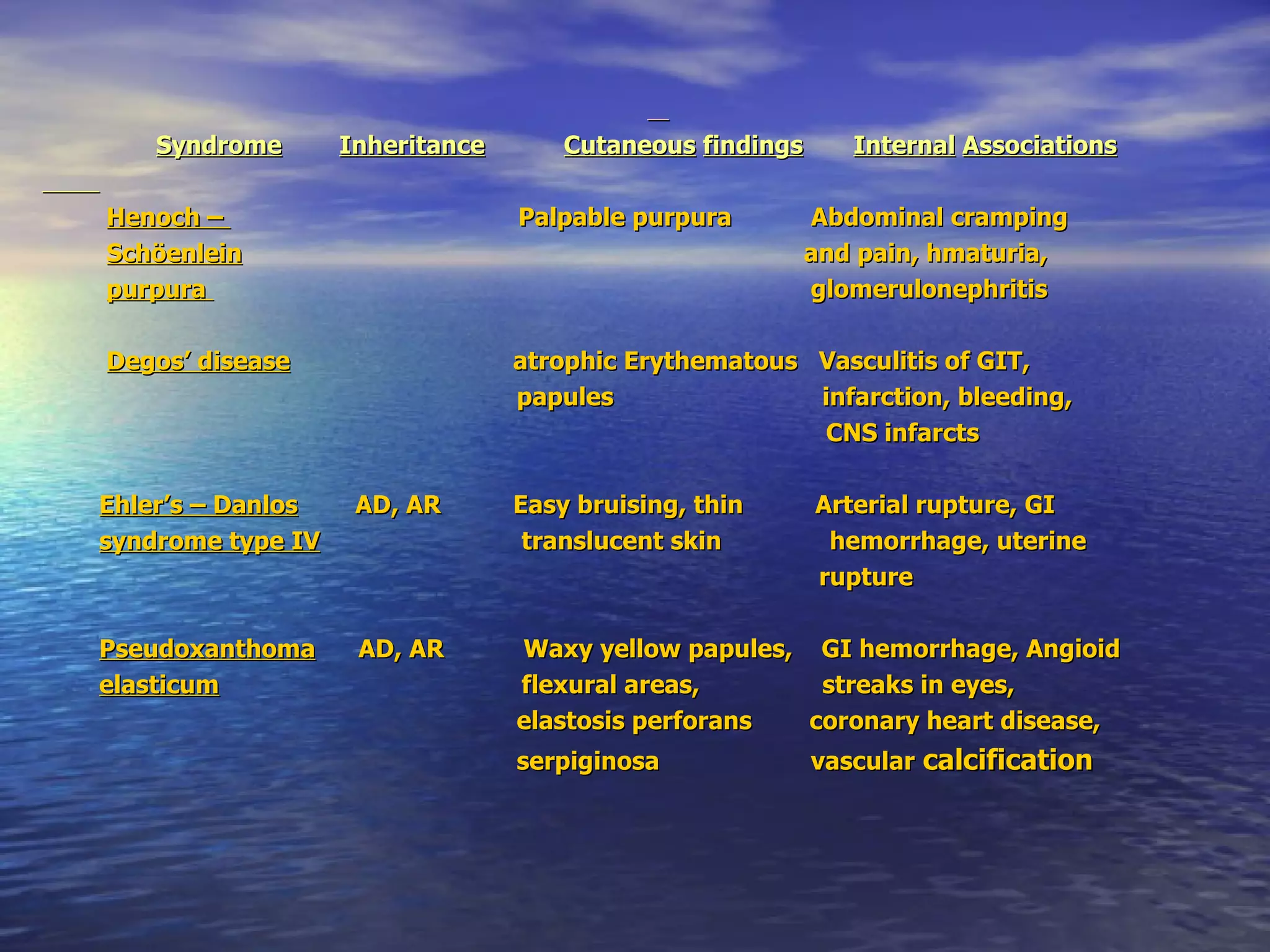
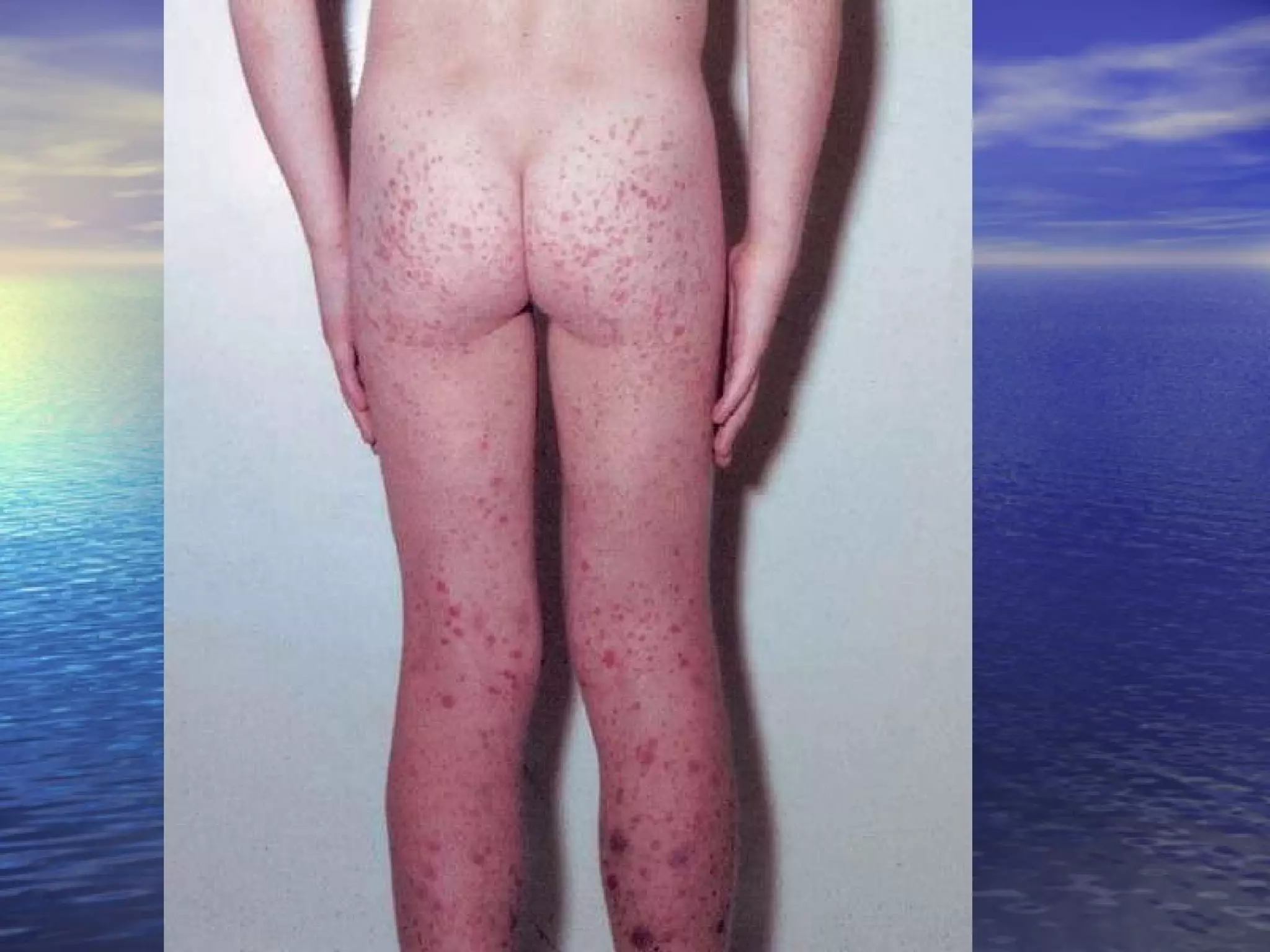
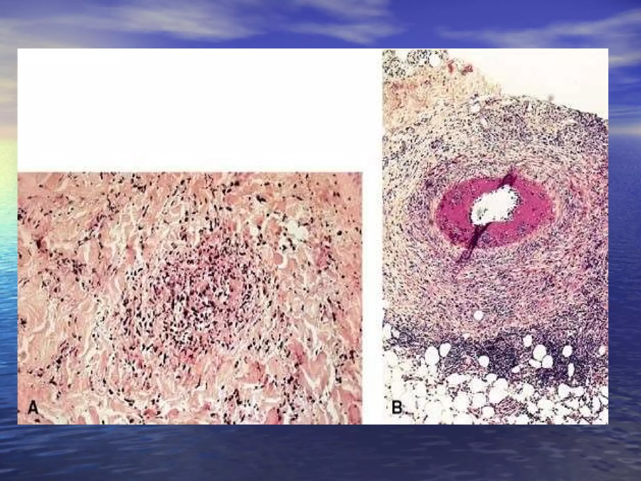
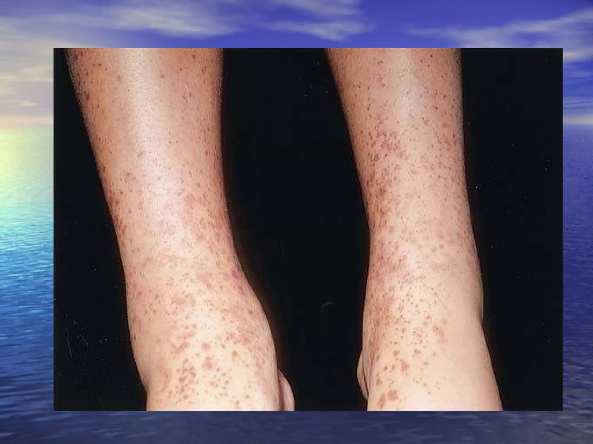
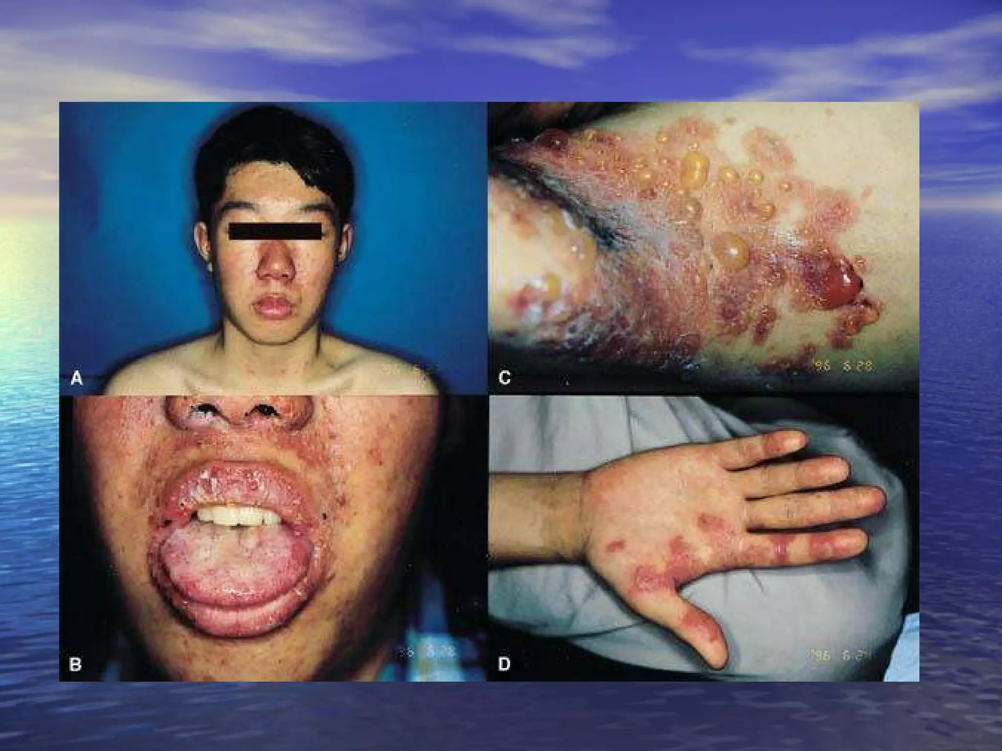
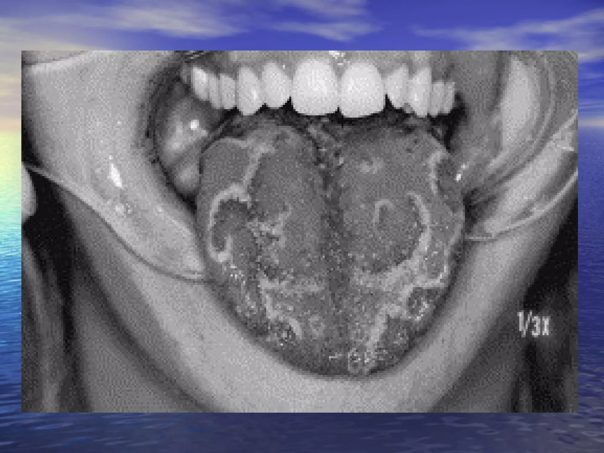
The document discusses many associations between gastrointestinal disorders and skin manifestations. Some key points discussed include inflammatory bowel diseases like ulcerative colitis and Crohn's disease being associated with conditions like erythema nodosum and pyoderma gangrenosum. Liver diseases can cause signs like spider telangiectasias. Viral hepatitis may cause conditions like mixed cryoglobulinemia. Nutritional deficiencies from malabsorption disorders can induce nonspecific eruptions or specific deficiencies like zinc deficiency. Various cutaneous syndromes are also associated with increased cancer risks in the GI tract.
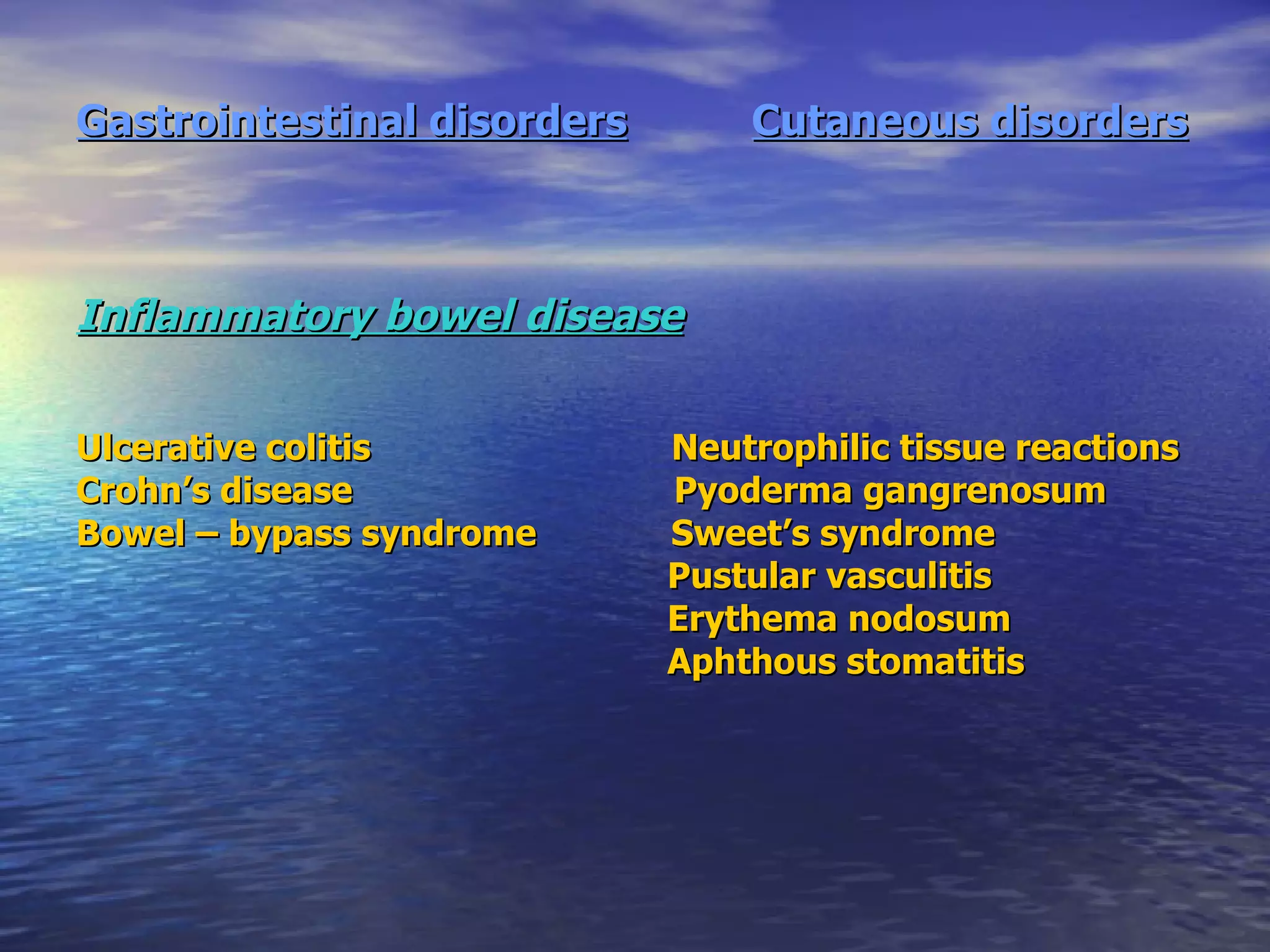
![Inflammatory bowel diseases Inflammatory disorders of the bowel discussed here include ulcerative colitis (UC), Crohn's disease, and bowel bypass syndrome Both UC and Crohn's disease (the traditional inflammatory bowel diseases [IBD]) can present with abdominal pain, GI bleeding, or diarrhea. Bowel bypass syndrome : a bacterial overgrowth in the blind loop associated with a dermatosis-arthritis syndrome](https://image.slidesharecdn.com/gitandskinnn-ppt-100314075600-phpapp02/75/Git-And-Skinnn-Ppt-17-2048.jpg)
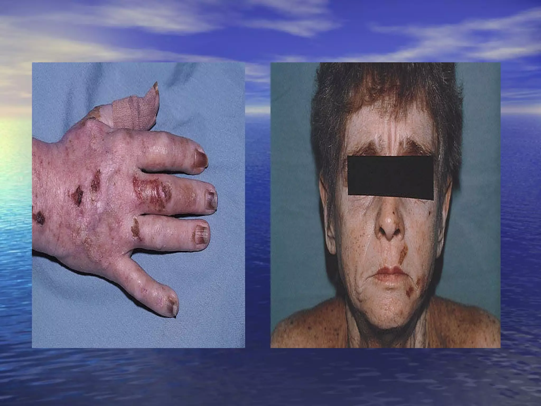
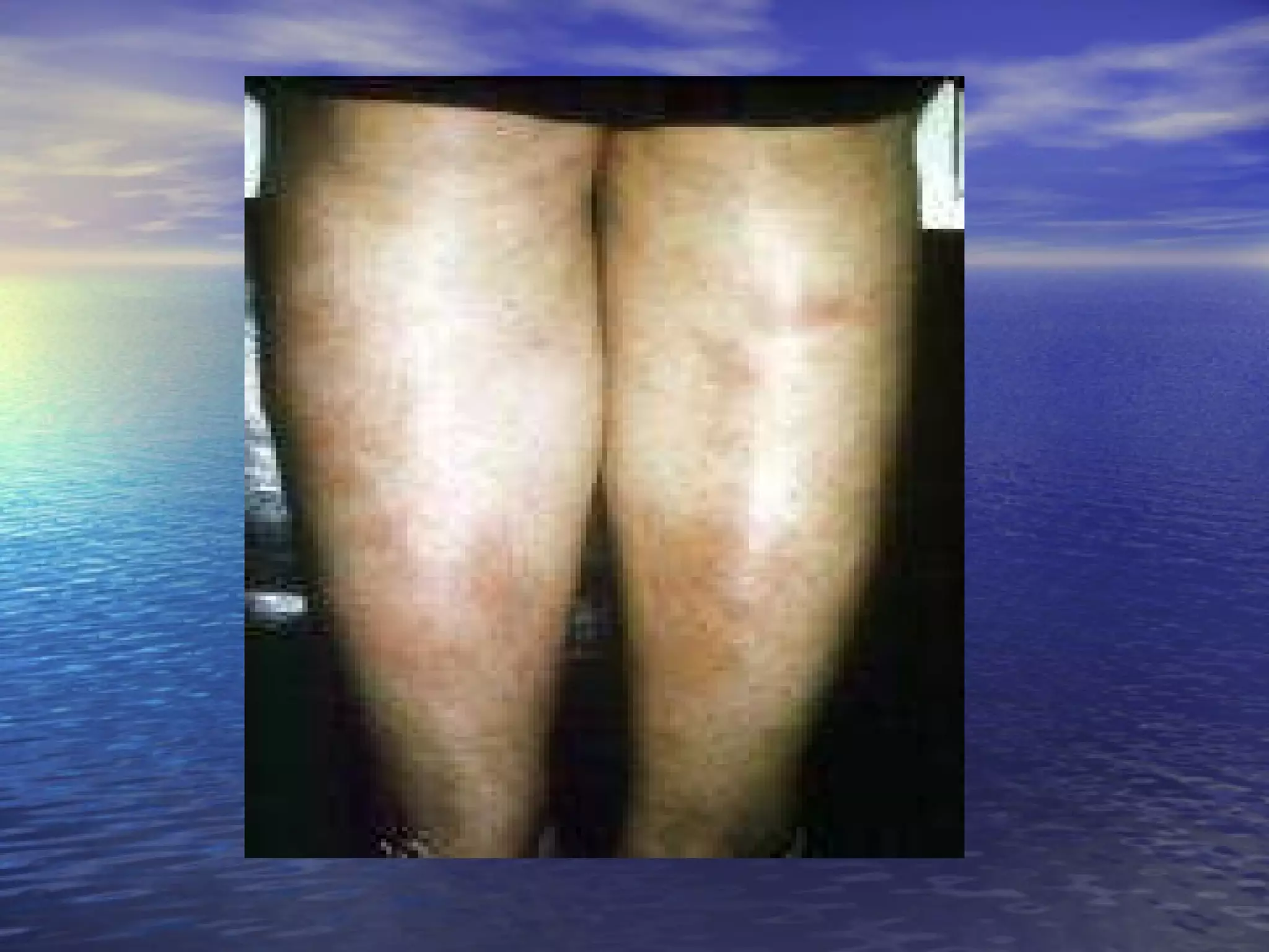
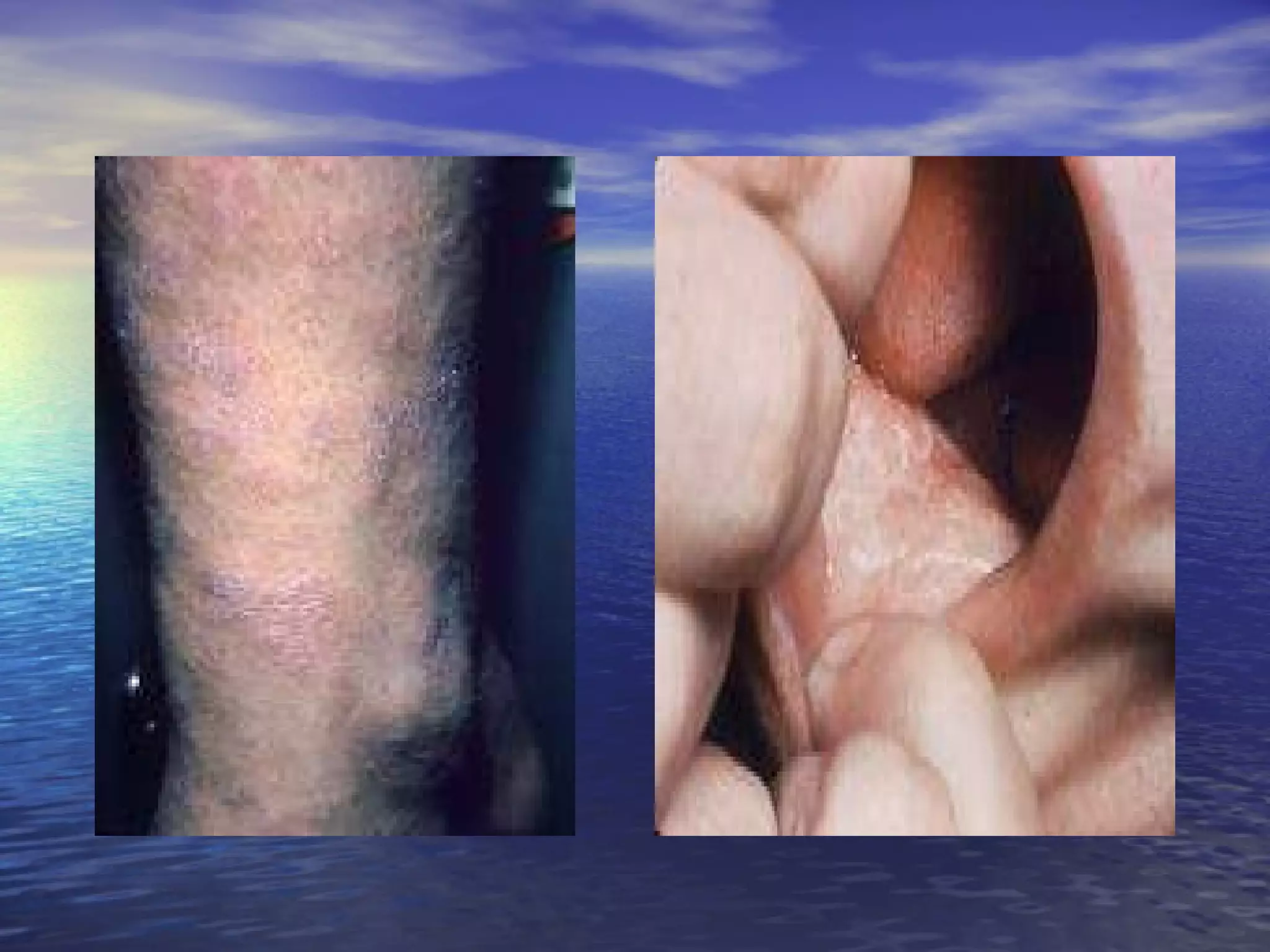
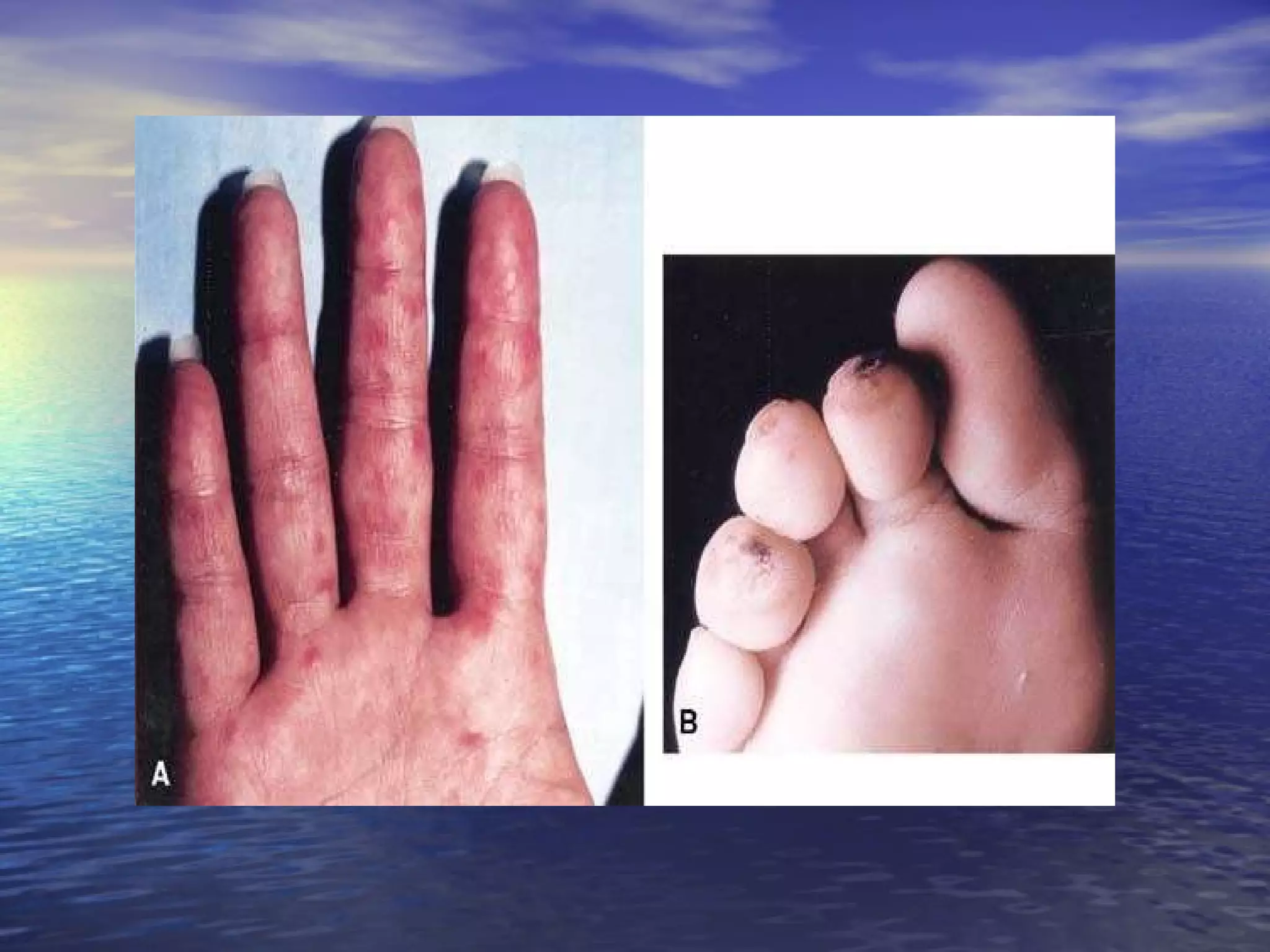

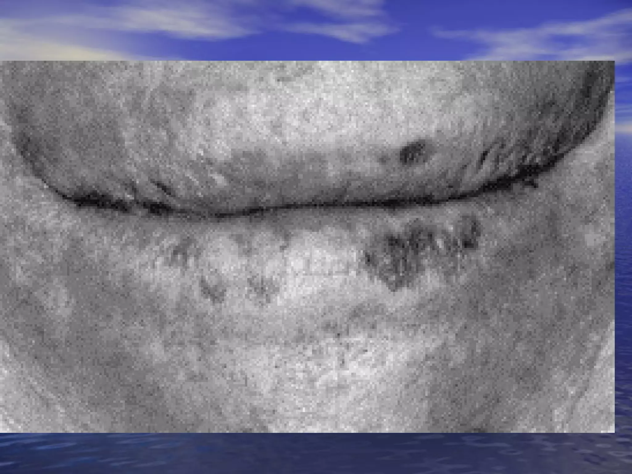
![Inflammatory bowel diseases Inflammatory disorders of the bowel discussed here include ulcerative colitis (UC), Crohn's disease, and bowel bypass syndrome Both UC and Crohn's disease (the traditional inflammatory bowel diseases [IBD]) can present with abdominal pain, GI bleeding, or diarrhea. Bowel bypass syndrome : a bacterial overgrowth in the blind loop associated with a dermatosis-arthritis syndrome](https://image.slidesharecdn.com/gitandskinnn-ppt-100314075600-phpapp02/75/Git-And-Skinnn-Ppt-27-2048.jpg)


![1.1.2. viral infections of skin [compatibility mode]](https://cdn.slidesharecdn.com/ss_thumbnails/1-1-2-viralinfectionsofskincompatibilitymode-120714004456-phpapp02-thumbnail.jpg?width=640&height=640&fit=bounds)


![Apporach to lung biopsy [Auto-saved].pptx latest](https://cdn.slidesharecdn.com/ss_thumbnails/apporachtolungbiopsyauto-saved-251211225655-93258539-thumbnail.jpg?width=640&height=640&fit=bounds)


