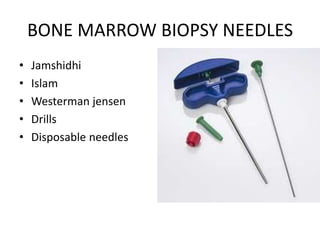
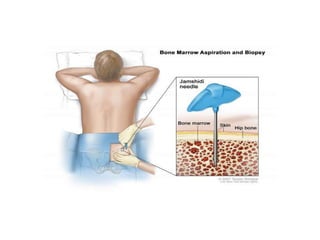
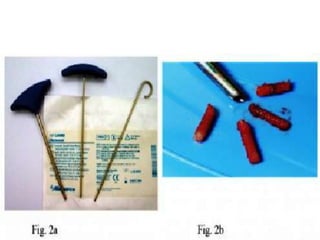
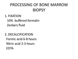
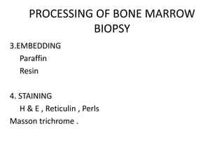
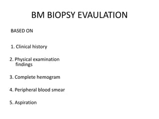
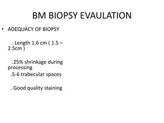
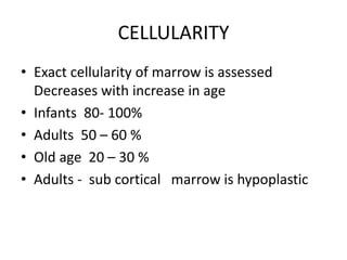
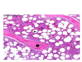
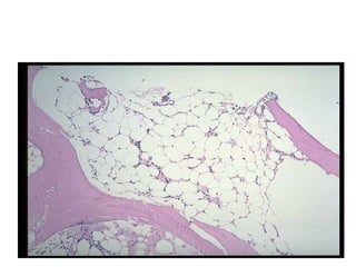
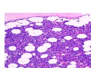
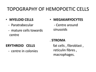
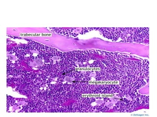
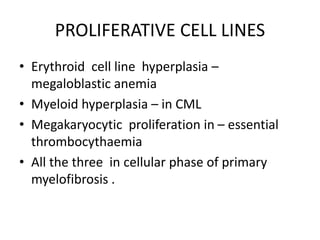
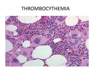
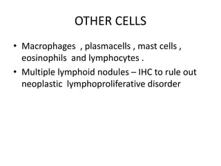
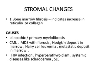
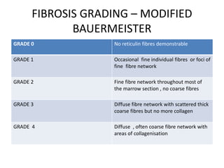
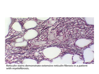
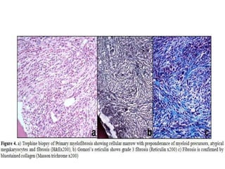
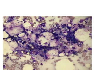
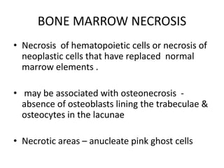
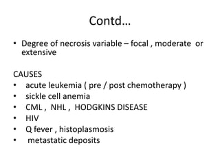
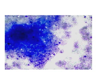
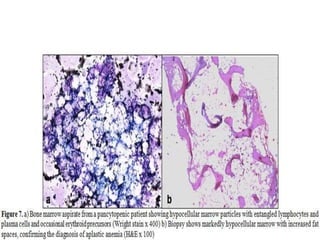
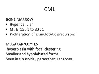
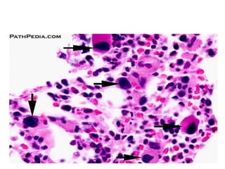
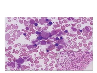
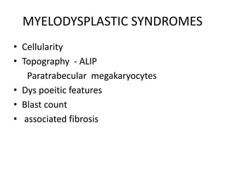
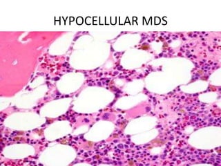
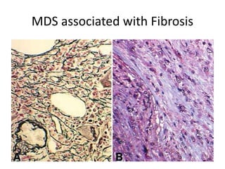
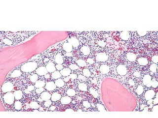
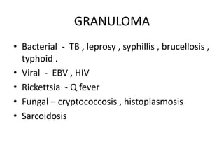
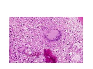
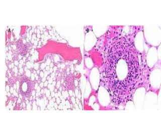
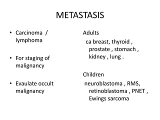
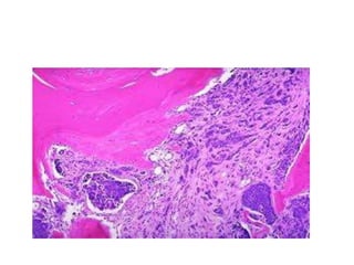
BONE MARROW BIOPSY AND
INTERPRETATION
JULY 2016
STRCTURE OF BONE MARROW
• A) cellular elements
Haematopoetic stem cells , Progenitors ,
precursors .
B) Stroma - unique microenvironment of the
marrow
BONE MARROW BIOPSY NEEDLES
• Jamshidhi
• Islam
• Westerman jensen
• Drills
• Disposable needles
DRILLER
INDICATIONS
• Evaluation of hematologic abnormalities in
peripheral smear
• Evaluation of primary bone marrow disorder
• Staging of bone marrow involvement by
metastatic tumors
• Evaluation of metabolic storage disorders
• PUO.
SITES OF BONE MARROW BIOPSY
• Posterior superior iliac spine
• Anterior superior iliac spine
• Iliac crest
• Tibial tuberosity in children .
PROCESSING OF BONE MARROW
BIOPSY
1. FIXATION
10% buffered formalin
Zenkers fluid
2. DECALCIFICATION
Formic acid 6-8 hours
Nitric acid 2-3 hours
EDTA
PROCESSING OF BONE MARROW
BIOPSY
3.EMBEDDING
Paraffin
Resin
4. STAINING
H & E , Reticulin , Perls
Masson trichrome .
BM BIOPSY EVAULATION
BASED ON
1. Clinical history
2. Physical examination
findings
3. Complete hemogram
4. Peripheral blood smear
5. Aspiration
BM BIOPSY EVAULATION
• ADEQUACY OF BIOPSY
. Length 1.6 cm ( 1.5 –
2.5cm )
.25% shrinkage during
processing
.5-6 trabecular spaces
. Good quality staining
Evaluation
• Cellularity
• Topography of hemopoetic cells
• Proliferation of cell lines
• Fibrosis
• Infections
• Infilterative diseases
CELLULARITY
• Exact cellularity of marrow is assessed
Decreases with increase in age
• Infants 80- 100%
• Adults 50 – 60 %
• Old age 20 – 30 %
• Adults - sub cortical marrow is hypoplastic
TOPOGRAPHY OF HEMOPOETIC CELLS
• MYELOID CELLS
- Paratrabecular
- mature cells towards
centre
ERYTHROID CELLS
- centre in colonies
• MEGAKARYOCYTES
- Centre around
sinusoids
. STROMA
fat cells , fibroblast ,
reticulin fibres ,
macrophages.
PROLIFERATIVE CELL LINES
• Erythroid cell line hyperplasia –
megaloblastic anemia
• Myeloid hyperplasia – in CML
• Megakaryocytic proliferation in – essential
thrombocythaemia
• All the three in cellular phase of primary
myelofibrosis .
THROMBOCYTHEMIA
OTHER CELLS
• Macrophages , plasmacells , mast cells ,
eosinophils and lymphocytes .
• Multiple lymphoid nodules – IHC to rule out
neoplastic lymphoproliferative disorder
STROMAL CHANGES
• 1.Bone marrow fibrosis – indicates increase in
reticulin or collagen
CAUSES
• idiopathic / primary myelofibrosis
• CML , MDS with fibrosis , Hodgkin deposit in
marrow , Hairy cell leukemia , metastatic deposit
in marrow .
• HIV infection , hyperparathyroidism , systemic
diseases like scleroderma , SLE
FIBROSIS GRADING – MODIFIED
BAUERMEISTER
GRADE 0 No reticulin fibres demonstrable
GRADE 1 Occasional fine individual fibres or foci of
fine fibre network
GRADE 2 Fine fibre network throughout most of
the marrow section , no coarse fibres
GRADE 3 Diffuse fibre network with scattered thick
coarse fibres but no more collagen
GRADE 4 Diffuse , often coarse fibre network with
areas of collagenisation
2. GELATINOUS TRANSFORMATION OF
BONE MARROW
• Also known as STARVATION MARROW /
SEROUS ATROPHY .
• Characterised by focal or diffuse extracellular
deposition of gelatinous material in between fat
cells and hypocellular marrow
• CAUSES
post chemotherapy , malnutrition , anorexia
nervosa , HIV , chronic tuberculosis , chronic liver
disease .
acid mucopolysaccharides in the gelatinous
material stain with ALCIAN BLUE
BONE MARROW NECROSIS
• Necrosis of hematopoietic cells or necrosis of
neoplastic cells that have replaced normal
marrow elements .
• may be associated with osteonecrosis -
absence of osteoblasts lining the trabeculae &
osteocytes in the lacunae
• Necrotic areas – anucleate pink ghost cells
Contd…
• Degree of necrosis variable – focal , moderate or
extensive
CAUSES
• acute leukemia ( pre / post chemotherapy )
• sickle cell anemia
• CML , NHL , HODGKINS DISEASE
• HIV
• Q fever , histoplasmosis
• metastatic deposits
APLASTIC ANEMIA
• Progressive pancytopenia , reticulocytopenia
• Bone marrow biopsy < 25 % of normal
cellularity of that age.
CML
BONE MARROW
• Hyper cellular
• M : E 15 : 1 to 30 : 1
• Proliferation of granulocytic precursors
MEGAKARYOCYTES
hyperplasia with focal clustering ,
Smaller and hypolobated forms
Seen in sinusoids , paratrabecular zones
MYELODYSPLASTIC SYNDROMES
• Cellularity
• Topography - ALIP
Paratrabecular megakaryocytes
• Dys poeitic features
• Blast count
• associated fibrosis
HYPOCELLULAR MDS
MDS associated with Fibrosis
HODGKINS LYMHOMA
BONEMARROW BIOPSY
• Staging
• Incidence of bonemarrow involvement
2to 30 %
• Mixed cellularity and lymphocyte depleted
• Manifested as focal or diffuse type
CRITERIA FOR BM INVOLVEMENT
CERTAIN TYPICAL RS CELLS OR MONONUCLEAR
VARIANTS IN CHARACTERISTIC
CELLULAR ENVIRONMENT
SUGGESTIVE ATYPICAL HISTIOCYTE OR
CHARACTERISTIC CELLULAR BACK
GROUND
SUSPICIOUS FIBROSIS / NECROSIS ALONE .
BONE MARROW IN NHL
• Staging
• Marrow involvement is more frequent in low
grade NHL – modest effect on patient
outcome
• Less common in high grade NHL – poor
prognosis .
• Predictor of high risk for CNS involvement
• Pattern of involvement
• nodular , diffuse , paratrabecular , interstitial ,
mixed
• IHC
• tumor cell burden
CLL
• Interstitial / nodular – better prognosis
• Increased fibrosis – bad prognosis
• increased plasma cells – bad prognosis
HAIRY CELL LEUKEMIA
• Pancytopenia , spleenomegaly
• Bone marrow aspiration - DRY TAP
• Biopsy – diffuse / focal / nodular marrow
involvement
• Cell morphology – fried egg appearance /
honey comb appearance
• Web like reticulin meshwork
GRANULOMA
• Bacterial - TB , leprosy , syphillis , brucellosis ,
typhoid .
• Viral - EBV , HIV
• Rickettsia - Q fever
• Fungal – cryptococcosis , histoplasmosis
• Sarcoidosis
METASTASIS
• Carcinoma /
lymphoma
• For staging of
malignancy
• Evaulate occult
malignancy
Adults
ca breast, thyroid ,
prostate , stomach ,
kidney , lung .
Children
neuroblastoma , RMS,
retinoblastoma , PNET ,
Ewings sarcoma
SUMMARY
EVAULATION OF BONE MARROW BIOPSY
• Adequate clinical history
• Better tissue processing
• Cellularity and topography of cellular
elements , their physiological variation .
• Stromal changes – fibrosis , necrosis .
• Infections - granulomas
REFERENCES
• WINTROBES CLINICAL HAEMATOLOGY
13 EDITION
• DAICIE PRACTICAL HAEMATOLOGY
• HOFFBRAND
• INTERNET
THANK YOU

Bone marrow biopsy and interpretation

  • 1.
    BONE MARROW BIOPSYAND INTERPRETATION JULY 2016
  • 2.
    STRCTURE OF BONEMARROW • A) cellular elements Haematopoetic stem cells , Progenitors , precursors . B) Stroma - unique microenvironment of the marrow
  • 3.
    BONE MARROW BIOPSYNEEDLES • Jamshidhi • Islam • Westerman jensen • Drills • Disposable needles
  • 4.
  • 5.
    INDICATIONS • Evaluation ofhematologic abnormalities in peripheral smear • Evaluation of primary bone marrow disorder • Staging of bone marrow involvement by metastatic tumors • Evaluation of metabolic storage disorders • PUO.
  • 6.
    SITES OF BONEMARROW BIOPSY • Posterior superior iliac spine • Anterior superior iliac spine • Iliac crest • Tibial tuberosity in children .
  • 9.
    PROCESSING OF BONEMARROW BIOPSY 1. FIXATION 10% buffered formalin Zenkers fluid 2. DECALCIFICATION Formic acid 6-8 hours Nitric acid 2-3 hours EDTA
  • 10.
    PROCESSING OF BONEMARROW BIOPSY 3.EMBEDDING Paraffin Resin 4. STAINING H & E , Reticulin , Perls Masson trichrome .
  • 11.
    BM BIOPSY EVAULATION BASEDON 1. Clinical history 2. Physical examination findings 3. Complete hemogram 4. Peripheral blood smear 5. Aspiration
  • 12.
    BM BIOPSY EVAULATION •ADEQUACY OF BIOPSY . Length 1.6 cm ( 1.5 – 2.5cm ) .25% shrinkage during processing .5-6 trabecular spaces . Good quality staining
  • 13.
    Evaluation • Cellularity • Topographyof hemopoetic cells • Proliferation of cell lines • Fibrosis • Infections • Infilterative diseases
  • 14.
    CELLULARITY • Exact cellularityof marrow is assessed Decreases with increase in age • Infants 80- 100% • Adults 50 – 60 % • Old age 20 – 30 % • Adults - sub cortical marrow is hypoplastic
  • 18.
    TOPOGRAPHY OF HEMOPOETICCELLS • MYELOID CELLS - Paratrabecular - mature cells towards centre ERYTHROID CELLS - centre in colonies • MEGAKARYOCYTES - Centre around sinusoids . STROMA fat cells , fibroblast , reticulin fibres , macrophages.
  • 20.
    PROLIFERATIVE CELL LINES •Erythroid cell line hyperplasia – megaloblastic anemia • Myeloid hyperplasia – in CML • Megakaryocytic proliferation in – essential thrombocythaemia • All the three in cellular phase of primary myelofibrosis .
  • 21.
  • 22.
    OTHER CELLS • Macrophages, plasmacells , mast cells , eosinophils and lymphocytes . • Multiple lymphoid nodules – IHC to rule out neoplastic lymphoproliferative disorder
  • 23.
    STROMAL CHANGES • 1.Bonemarrow fibrosis – indicates increase in reticulin or collagen CAUSES • idiopathic / primary myelofibrosis • CML , MDS with fibrosis , Hodgkin deposit in marrow , Hairy cell leukemia , metastatic deposit in marrow . • HIV infection , hyperparathyroidism , systemic diseases like scleroderma , SLE
  • 24.
    FIBROSIS GRADING –MODIFIED BAUERMEISTER GRADE 0 No reticulin fibres demonstrable GRADE 1 Occasional fine individual fibres or foci of fine fibre network GRADE 2 Fine fibre network throughout most of the marrow section , no coarse fibres GRADE 3 Diffuse fibre network with scattered thick coarse fibres but no more collagen GRADE 4 Diffuse , often coarse fibre network with areas of collagenisation
  • 27.
    2. GELATINOUS TRANSFORMATIONOF BONE MARROW • Also known as STARVATION MARROW / SEROUS ATROPHY . • Characterised by focal or diffuse extracellular deposition of gelatinous material in between fat cells and hypocellular marrow • CAUSES post chemotherapy , malnutrition , anorexia nervosa , HIV , chronic tuberculosis , chronic liver disease . acid mucopolysaccharides in the gelatinous material stain with ALCIAN BLUE
  • 30.
    BONE MARROW NECROSIS •Necrosis of hematopoietic cells or necrosis of neoplastic cells that have replaced normal marrow elements . • may be associated with osteonecrosis - absence of osteoblasts lining the trabeculae & osteocytes in the lacunae • Necrotic areas – anucleate pink ghost cells
  • 31.
    Contd… • Degree ofnecrosis variable – focal , moderate or extensive CAUSES • acute leukemia ( pre / post chemotherapy ) • sickle cell anemia • CML , NHL , HODGKINS DISEASE • HIV • Q fever , histoplasmosis • metastatic deposits
  • 33.
    APLASTIC ANEMIA • Progressivepancytopenia , reticulocytopenia • Bone marrow biopsy < 25 % of normal cellularity of that age.
  • 35.
    CML BONE MARROW • Hypercellular • M : E 15 : 1 to 30 : 1 • Proliferation of granulocytic precursors MEGAKARYOCYTES hyperplasia with focal clustering , Smaller and hypolobated forms Seen in sinusoids , paratrabecular zones
  • 38.
    MYELODYSPLASTIC SYNDROMES • Cellularity •Topography - ALIP Paratrabecular megakaryocytes • Dys poeitic features • Blast count • associated fibrosis
  • 39.
  • 40.
  • 41.
    HODGKINS LYMHOMA BONEMARROW BIOPSY •Staging • Incidence of bonemarrow involvement 2to 30 % • Mixed cellularity and lymphocyte depleted • Manifested as focal or diffuse type
  • 42.
    CRITERIA FOR BMINVOLVEMENT CERTAIN TYPICAL RS CELLS OR MONONUCLEAR VARIANTS IN CHARACTERISTIC CELLULAR ENVIRONMENT SUGGESTIVE ATYPICAL HISTIOCYTE OR CHARACTERISTIC CELLULAR BACK GROUND SUSPICIOUS FIBROSIS / NECROSIS ALONE .
  • 43.
    BONE MARROW INNHL • Staging • Marrow involvement is more frequent in low grade NHL – modest effect on patient outcome • Less common in high grade NHL – poor prognosis . • Predictor of high risk for CNS involvement
  • 44.
    • Pattern ofinvolvement • nodular , diffuse , paratrabecular , interstitial , mixed • IHC • tumor cell burden
  • 46.
    CLL • Interstitial /nodular – better prognosis • Increased fibrosis – bad prognosis • increased plasma cells – bad prognosis
  • 47.
    HAIRY CELL LEUKEMIA •Pancytopenia , spleenomegaly • Bone marrow aspiration - DRY TAP • Biopsy – diffuse / focal / nodular marrow involvement • Cell morphology – fried egg appearance / honey comb appearance • Web like reticulin meshwork
  • 49.
    GRANULOMA • Bacterial -TB , leprosy , syphillis , brucellosis , typhoid . • Viral - EBV , HIV • Rickettsia - Q fever • Fungal – cryptococcosis , histoplasmosis • Sarcoidosis
  • 52.
    METASTASIS • Carcinoma / lymphoma •For staging of malignancy • Evaulate occult malignancy Adults ca breast, thyroid , prostate , stomach , kidney , lung . Children neuroblastoma , RMS, retinoblastoma , PNET , Ewings sarcoma
  • 54.
    SUMMARY EVAULATION OF BONEMARROW BIOPSY • Adequate clinical history • Better tissue processing • Cellularity and topography of cellular elements , their physiological variation . • Stromal changes – fibrosis , necrosis . • Infections - granulomas
  • 55.
    REFERENCES • WINTROBES CLINICALHAEMATOLOGY 13 EDITION • DAICIE PRACTICAL HAEMATOLOGY • HOFFBRAND • INTERNET
  • 56.