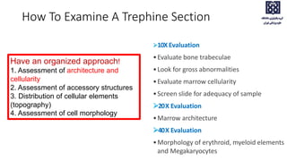
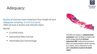
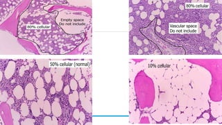
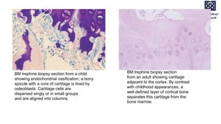
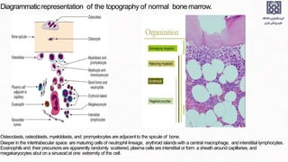
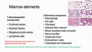
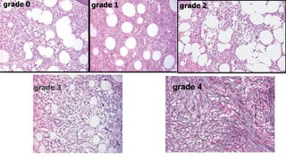
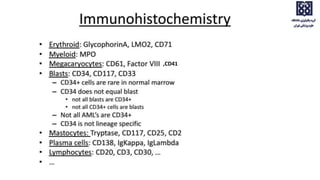
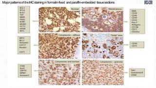
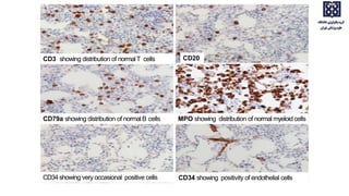
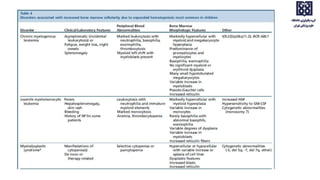
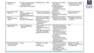
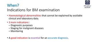
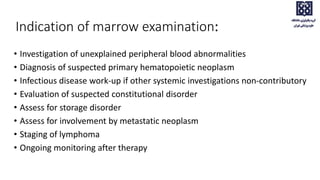
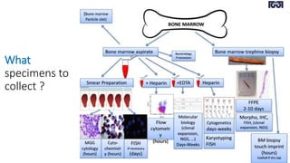
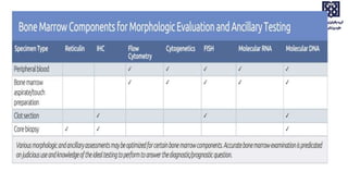
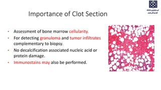
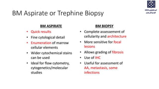
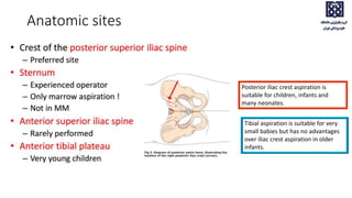
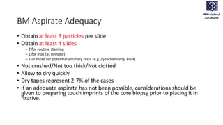
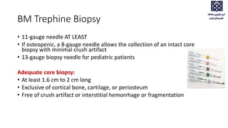
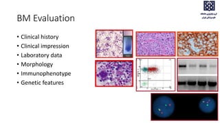
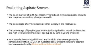
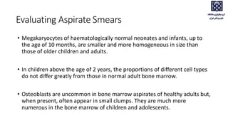
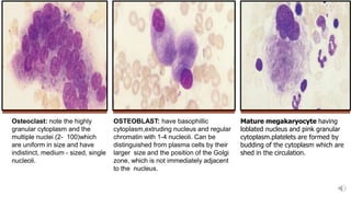
SlideShare a Scribd company logo
1 of 86
BONE MARROW EXAMINATION
When?
Indications for BM examination
• Haematological abnormalities that cannot be explained by available
clinical and laboratory data.
• 3 main indications :
– Diagnostic purposes
– Staging for malignant diseases
– Monitoring
• A good indication is essential for an accurate diagnosis.
Indication of marrow examination:
• Investigation of unexplained peripheral blood abnormalities
• Diagnosis of suspected primary hematopoietic neoplasm
• Infectious disease work-up if other systemic investigations non-contributory
• Evaluation of suspected constitutional disorder
• Assess for storage disorder
• Assess for involvement by metastatic neoplasm
• Staging of lymphoma
• Ongoing monitoring after therapy
What
specimens to
collect ?
Importance of touch/ Imprint smear
• Gives cytological details when aspirate is not
obtained.(fibrotic or inaspirable)
• Shows more neoplastic cells than aspirate.
• Can show marrow infiltration , not seen in
aspirate
• Important for potential ancillary techniques
(e.g., cytochemistry, FISH, iron stains)
• Particularly if BM aspirate is hemodilute/dry tap
• ≥ 3 air-dried imprints
Be gentle, be delicate !
Do NOT CRUSH !
Importance of Clot Section
• Assessment of bone marrow cellularity.
• For detecting granuloma and tumor infiltrates
complementary to biopsy.
• No decalcification associated nucleic acid or
protein damage.
• Immunostains may also be performed.
BM Aspirate or Trephine Biopsy
BM aspirate or trephine biopsy
How to collect these specimens ?
• Anatomic sites
• Collection procedure
• Adequacy
Anatomic sites
Posterior iliac crest aspiration is
suitable for children, infants and
many neonates.
Tibial aspiration is suitable for very
small babies but has no advantages
over iliac crest aspiration in older
infants.
BM Aspirate Adequacy
• Obtain at least 3 particles per slide
• Obtain at least 4 slides
– 2 for routine staining
– 1 for iron (as needed)
– 1 or more for potential ancillary tests (e.g.,cytochemistry, FISH)
• Not crushed/Not too thick/Not clotted
• Allow to dry quickly
• Dry tapes represent 2-7% of the cases
• If an adequate aspirate has not been possible, considerations should be
given to preparing touch imprints of the core biopsy prior to placing it in
fixative.
BM Trephine Biopsy
• 11-gauge needle AT LEAST
• If osteopenic, a 8-gauge needle allows the collection of an intact core
biopsy with minimal crush artifact
• 13-gauge biopsy needle for pediatric patients
Adequate core biopsy:
• At least 1.6 cm to 2 cm long
• Exclusive of cortical bone, cartilage, or periosteum
• Free of crush artifact or interstitial hemorrhage or fragmentation
BM Evaluation
• Clinical history
• Clinical impression
• Laboratory data
• Morphology
• Immunophenotype
• Genetic features
• Erythrocytes remain macrocytic from the first 11 weeks of
gestation until day 5 of postnatal life.
Peripheral blood film for a
normal newborn
demonstrating a normal
lymphocyte, macrocytes,
polychromasia, and one
nucleated red blood cell
(×1000).
• Orthochromic normoblasts frequently are observed in the full-term
infant on the first day of life but disappear within postnatal days 3-5.
• NRBCs may persist longer than a week in immature infants.
• The average number of NRBCs ranges from 3 to 10 per 100 WBCs in a
normal full-term infant to 25 NRBCs per 100 WBCs in a premature
infant.
• The presence of NRBCs for more than 5 days suggests hemolysis,
hypoxic stress, or acute infection.
Bone Marrow Aspiration
Evaluating Aspirate Smears
Screen all slides at low power (4 x)
• Evaluate cellularity, look for abnormal infiltrate
• Find good areas of slides for counting
• Evaluate number of megakaryocytes
Count cells from several areas (100 x)
• If spicules are lacking, accurate counts may be obtained at edges of smears
• If cells are mainly neutrophils and lymphocytes, smears may represent peripeheral
blood rather than marrow
The preparation can be considered satisfactory only when marrow
particles and free marrow cells can be seen in stained films.
Normocellular bone marrow on aspirate High cellularity oaf 95-100%
Cellularity of 40-50% Hypocellular bone marrow (10-20%)
Epidemiology
• Dramatic age-related variations in BM cellularity
-Highest cellularity in neonates: ~ 80-100%
-Gradual decline in cellularity with age
• BM cellularity in elderly patients: ~ 20-30%
Mean values (observed range) for bone marrow
cells in healthy infants and children
• The bone marrow at birth has major erythroid and myeloid components with
few lymphocytes and very few plasma cells.
• The percentage of erythroid cells declines steeply in the first weeks.
• The percentage of lymphocytes increases during the first month and remains
at a high level until 18 months of age (up to 40-50% in young children)
• Numbers decline during childhood and in adults they do not generally
comprise more than 15–25% of nucleated cells, unless the marrow aspirate
has been considerably diluted with peripheral blood.
Evaluating Aspirate Smears
• Megakaryocytes of haematologically normal neonates and infants, up to
the age of 10 months, are smaller and more homogeneous in size than
those of older children and adults.
• In children above the age of 2 years, the proportions of different cell types
do not differ greatly from those in normal adult bone marrow.
• Osteoblasts are uncommon in bone marrow aspirates of healthy adults but,
when present, often appear in small clumps. They are much more
numerous in the bone marrow of children and adolescents.
Evaluating Aspirate Smears
Osteoclast: note the highly
granular cytoplasm and the
multiple nuclei (2- 100)which
are uniform in size and have
indistinct, medium - sized, single
nucleoli.
OSTEOBLAST: have basophillic
cytoplasm,extruding nucleus and regular
chromatin with 1-4 nucleoli. Can be
distinguished from plasma cells by their
larger size and the position of the Golgi
zone, which is not immediately adjacent
to the nucleus.
Mature megakaryocyte having
loblated nucleus and pink granular
cytoplasm.platelets are formed by
budding of the cytoplasm which are
shed in the circulation.
95% Range Mean[12] Mean[11]
Myeloblasts 0–3 1.4 0.4
Promyelocytes 3–12 7.8 13.7[*]
Myelocytes (neutrophil) 2–13 7.6 –
Metamyelocytes 2–6 4.1 –
Neutrophils 22–46 32.1M; 37.4W 35.5
Myelocytes (eosinophil) 0–3 1.3 1.6
Eosinophils 0.3–4 2.2 1.7
Basophils 0–0.5 0.1 0.2
Lymphocytes 5–20 13.1 16.1
Monocytes 0–3 1.3 2.5
Plasma cells 0–3.5 0.6 1.9
Erythroblasts[†] 5–35 28.1M; 22.5W 23.5
Megakaryocytes 0–2 0.5
Macrophages 0–2 0.4 2.0
Normal ranges for differential counts on aspirated bone marrow (500 cells should be counted)
BM Evaluation
Bone Marrow Biopsy
How To Examine A Trephine Section
10X Evaluation
•Evaluate bone trabeculae
•Look for gross abnormalities
•Evaluate marrow cellularity
•Screen slide for adequacy of sample
20X Evaluation
•Marrow architecture
40X Evaluation
•Morphology of erythroid, myeloid elements
and Megakaryocytes
Have an organized approach!
1. Assessment of architecture and
cellularity
2. Assessment of accessory structures
3. Distribution of cellular elements
(topography)
4. Assessment of cell morphology
Adequacy:
Quality of marrow more important than length of core
-Adequate sampling: 1.5 cm (1.6-2cm)
-H&E (at least 2 levels) and reticulin stains
•Avoid
 Crushed areas
 Subcortical fatty marrow
 Intertrabecular hemorrhage
This BM core biopsy is suboptimal for
evaluation with cartilage on both ends
of the biopsy flanking minimal
subcortical BM. Subcortical marrow is
often not representative of the marrow
as a whole, and a statement to this
effect should be issued in the final
report.
• A biopsy specimen containing at least five or six intertrabecular
spaces is desirable, not only for an adequate assessment of cellularity
but also to give a reasonable probability of detecting focal bone
marrow lesions.
• Ideally this requires a core of 2–3 cm in length. A core length of at
least 0.5 cm has been advised in children but one study found 1.0 cm
was necessary to avoid a high rate of non‐interpretable specimens.
Adequacy:
• A section of a trephine biopsy specimen of adequate
size from a patient with Hodgkin lymphoma showing
only a small area of infiltration at one end of the
specimen, illustrating how a small biopsy may miss
focal lesions. at least 16 mm
Adequacy:
Common artifacts in marrow trephine biopsy
• Washed off of marrow space- technical artifact leading to inconclusive findings
• Aspiration artifact
• Crushed artifact
1. ASSESMENT OF ARCHITECTURE AND CELLULARITY
HOW TO EXAMINE A TREPHINE SECTION
• Cellularity varies according to location and age
• Hypocellular in subcortical marrow
(three intertrabecular space)
Evaluation of cellularity
• Average cellularity in the bone marrow of children, assessed on core
biopsy or clot sections:
• 80% at 2 years
• 69% at 2–4 years
• 59% at 5–9 years
• around 60% thereafter
• 20-40% in elderly
Architecture
Myeloid cells
Paratrabecular
Immature myeloid elements
are adjacent to bone
trabeculae
Mature cells towards centre
Erythroid cells
Centre in colonies
Megakaryocytes
Centre around sinusoids
are singletons (<3 together)
2. ASSESMENT OF ACCESSORY STRUCTURES
HOW TO EXAMINE A TREPHINE SECTION
Accessory structures
• Both osteoclasts and osteoblasts normally evident along bony
trabeculae in specimens from pediatric patients.
• Incomplete ossification typical in infants/young children
• Evidence of bony remodeling normal in children, abnormal in middle-
aged to elderly adults.
Trabeculae consists of lamellar bone Osteoblasts along endosteal surface
Ossifying cartilage. Osteocytes within
lacuna (arrow head).
Osteoclast
BM trephine biopsy section from a child
showing endochondrial ossification; a bony
spicule with a core of cartilage is lined by
osteoblasts. Cartilage cells are
dispersed singly or in small groups
and are aligned into columns.
BM trephine biopsy section
from an adult showing cartilage
adjacent to the cortex. By contrast
with childhood appearances, a
well‐defined layer of cortical bone
separates this cartilage from the
bone marrow.
HOW TO EXAMINE A TREPHINE SECTION
3. DISTRIBUTION OF CELLULAR ELEMENTS (TOPOGRAPHY)
4. ASSESMENT OF CELL MORPHOLOGY
Diagrammaticrepresentation of the topography of normal bonemarrow.
Osteoclasts, osteoblasts, myeloblasts, and promyelocytes are adjacent to the spicule of bone.
Deeper in the intertrabecular space are maturing cells of neutrophil lineage, erythroid islands with a central macrophage, and interstitial lymphocytes.
Eosinophils and their precursorsare apparently randomly scattered, plasma cells are interstitial or form a sheath around capillaries, and
megakaryocytes abut on a sinusoidat one extremity of the cell.
Marrow elements
1-Hematopoietic
component
• Erythroid series
• Myeloid Series
• Megakaryocyticseries
• Lymphoid cells
t
2-Stromal componen
• Macrophage
• Fat cells
• Fibroblast
• Delicate fiber network
• Blood vessels(mostly sinusoid)
• Nerve bundles
• Trabecular bone
• Extracellular matrix
• Osteoblast and Osteoclast
Stromal component facilitate maintenance of HSC and support differentiation and maturation of the progenitors
These two components not only coexist but
closely interact with each other
Special Stains on Marrow
-Giemsa(routine)
• Differentiates cytoplasm of erythroid elements and myeloid elements
• Plasma cells, mast cells more obvious
-Reticulin (special)
• Evaluates bone marrow fibrosis
• clues: crush artifact, dry tap, enlarged vascular spaces
-PAS stains highlighting megakaryocytes, fungal organism in institutions with large populations of
immunosuppressed patients
-Silver stain for assessment of reticulin fibrosisIncases of myeloproliferative neoplasms
-Not recommend the routine Perl stain of the core biopsyif a satisfactory marrow aspirate cannot be
obtained, iron stains of the clot or biopsysections>>>potential false-negative results.
Reticulin fibrosis
 Reticulin is normally present in BM
 ‘Normal’ reticulin
• Around blood vessels
• Short wisps
 • Day-to-day variation in silver
• Underdone reticulin stain
• - No staining around blood vessels -> repeat
• Overdone reticulin stain
• - Staining of cell membranes, cell nuceli stained darkly - >
repeat or discount
•
Collagen fibrosis
Most notably seen in association with
myeloproliferative neoplasms
Detected with trichrome stain
Not normally present in BM
Unlikely to be reversible
2 types of BM fibrosis:
- Reticulin fibrosis - Collagen fibrosis
grade 0 grade 1 grade 2,
grade 3 grade 4
Iron Assessment
• Typically assess 2 types of iron stores
• Sideroblastic iron (a.k.a. erythroid iron)
• Macrophage iron (a.k.a. storage iron)
• Assessment of storage iron requires that an adequate number of
fragments are obtained
• A minimum of seven fragments in one or more bone marrow films
need to be examined in order to state with reasonably reliability that
bone marrow iron is absent .
Erythroid iron
Preferred specimen: Spicular, unstained,
air-dried BM aspirate smear/touch
preparation containing adequate red
blood cell precursors
Normal: 20-50% of red blood cell
precursors demonstrate 1-3 small
cytoplasmic granules
Abnormal
– Abnormal size (large), shape (chunky),
or location (perinuclear, ring) of granules
– Ring sideroblast is defined by WHO as
1/3 of red blood cell nucleus tightly
surrounded by 5 or more iron granules.
Storage iron
Preferred specimen: Spicular, unstained, air-dried BMA
smear
Grading may be semiquantitatively assessed (normal,
none, increased, decreased) or assessed by grading scale
(0-6)
In routine practice, grading as absent, scanty, reduced,
normal or increased is also a practical approach.
Need adequate positive control for accuracy
May have heterogeneous iron deposition in BMA
Formalin fixation/histologic processing/decalcification
may interfere with staining of iron stores (ferritin)
Grading of bone marrow storage iron
0 No stainable iron
1+ Small iron particles just visible in
reticulum cells using an oil objective
2+ Small, sparse iron particles in reticulum
cells, visible at lower power
3+ Numerous small particles in reticulum cells
4+ Larger particles with a
tendency to aggregate into clumps
5+ Dense, large clumps
6+ Very large clumps and extracellular iron
Perl’s stain
sideroblast
Perl’s stain
Macrophage with iron
Ringed sideroblast
,CD41
Majorpatternsof theIHCstainingin formalin-fixed andparaffin-embedded tissuesections
MPO showing distribution of normal myeloid cells
CD79a showing distribution of normalB cells
CD20
CD3 showing distribution of normalT cells
CD34 showing positivity of endothelial cells
CD34showing very occasional positive cells
Pathologic Key Features Of Pediatric Bone
Marrow Interpretation
• Interpretation of pediatric bone marrow samples requires that the
pathologist integrate clinical, morphologic, and ancillary study results
to arrive at the correct diagnosis.
• The morphology of normal pediatric bone marrow overlaps
significantly with that of adults.
• The evaluation of pediatric bone marrow poses specific challenges
when compared with the general adult population.
• These challenges stem in part from the higher likelihood of congenital
disorders with hematopoietic manifestations, some of which may give
rise to hematologic malignancies.
Pathologic Key Features Of Pediatric Bone
Marrow Interpretation
• Congenital syndromes and florid reactive processes must always be
considered in the differential diagnosis of hematologic malignancies
in the pediatric population
• Recognition of the general pathologic pattern (hypercellular,
hypocellular, or single-lineage defect) aids in narrowing the
differential diagnosis when interpreting bone marrow samples from
pediatric patients.
Pathologic Key Features Of Pediatric Bone
Marrow Interpretation
• Two features are significantly different and may affect bone marrow
interpretation: the overall cellularity and the presence of benign
lymphoid precursors (hematogones), which may be markedly
increased in some patients.
• Also it is important to realize that a large proportion of the benign
and malignant disorders diagnosed during childhood are likely to be
related to underlying genetic abnormalities, sometimes as the first
manifestation of these defects
Pathologic Key Features Of Pediatric Bone
Marrow Interpretation
• Finally, certain types of malignant neoplasms with partially
overlapping blastic morphologic features (so called “small blue-cell
tumors”), but with vastly different biology requiring distinct
therapeutic approaches, occur relatively frequently in young patients.
Pathologic Key Features Of Pediatric Bone
Marrow Interpretation
Pitfalls Of Pediatric Bone Marrow
Interpretation
• Benign B-cell precursors (hematogones) can occur in significant
numbers in the bone marrow of children with cytopenias of
nonneoplastic causes and may be mistaken for involvement by B-
lymphoblastic leukemia.
Pitfalls Of Pediatric Bone Marrow
Interpretation
• Benign B-cell precursors (hematogones) often represent a significant
proportion of the cells in hypocellular marrow aspirates from acute
megakaryoblastic leukemia (with marrow fibrosis) potentially leading
to a misdiagnosis of B lymphoblastic leukemia.
Pitfalls Of Pediatric Bone Marrow
Interpretation
• B-lymphoblastic leukemias with a CD45-,CD20-, CD99+
immunophenotype may be mistaken for Ewing sarcoma, especially in
small bone or bone marrow biopsies
Pitfalls Of Pediatric Bone Marrow
Interpretation
• Hemophagocytic syndromes may be associated with peripheral T-cell
lymphomas; diagnostic lymphoma cells are sometimes present in
small numbers in the bone marrow aspirates and may be missed.
Pitfalls Of Pediatric Bone Marrow
Interpretation
• Variably prominent populations of polyclonal CD8+ T lymphocytes
with immunophenotypic aberrancies may be present in familial
hemophagocytic histiocytosis; they may be mistaken for peripheral T-
cell lymphoma.
Pitfalls Of Pediatric Bone Marrow
Interpretation
• Parvovirus B19 infections occurring in patients with constitutional red
cell disorders may cause erythroid hyperplasia and significant
dyserythropoiesis, resembling congenital dyserythropoietic anemia.
• Acute lymphoblastic leukaemia can be confused with small cell
tumors of childhood, e.g. neuroblastoma, Ewing’s tumour and other
primitive neuroectodermal tumours (PNET), rhabdomyosarcoma,
medulloblastoma and retinoblastoma.
• It should be noted that leukaemic lymphoblasts may fail to express
CD45, that neuroendocrine tumors can express PAX5 and that blast
cells in a large proportion of ALL patients express CD99, an antigen
commonly expressed in Ewing’s tumour/PNET.
Pitfalls Of Pediatric Bone Marrow
Interpretation
Pitfalls Of Pediatric Bone Marrow
Interpretation
• Terminal deoxynucleotidyl transferase (TDT) in addition to ALL,
positive in up to 15% of AML cases.
• It has been reported to be positive in a proportion of cases of
medulloblastoma and occasionally in other small cell tumors of
childhood
• Small cell tumors of childhood, e.g. those with heavily vacuolated
cells such as alveolar rhabdomyosarcoma, have been misdiagnosed
and even treated as Burkitt lymphoma
Pitfalls Of Pediatric Bone Marrow
Interpretation
Pitfalls Of Pediatric Bone Marrow
Interpretation
• It should be noted that, in children, apparent aplastic anemia may
represent an aplastic presentation of ALL.
Pitfalls Of Pediatric Bone Marrow
Interpretation
• Acute megakaryoblastic leukemia (AMKL), which may yield clusters of
cohesive cells mimicking metastatic solid tumors in some cases
• Some lymphoblastic leukemias with near-tetraploid DNA content may
also show very large blasts reminiscent of solid tumor cells.
Pitfalls Of Pediatric Bone Marrow
Interpretation
• Some leukemias (especially hyperdiploid B-ALL and AMKL) may be
accompanied by significant marrow fibrosis, leading to a paucity of
neoplastic cells available for ancillary studies in the aspirate samples
CONCLUSION
• Integration of clinical, morphologic, immunophenotypic, genetic, and
other biologic features is mandatory to define specific disease entities
• The relative contribution of each feature varies, depending on the
case.
• Make your cytologists/pathologists/geneticians good ! by
providing them relevant clinical information's and optimal samples.
Bone Marrow evaluation EVALUATION (PBS+BMA).pptx

More Related Content

Similar to Bone Marrow evaluation EVALUATION (PBS+BMA).pptx

paris presentation.pptx
paris presentation.pptxparis presentation.pptx
paris presentation.pptxKarishmaBhuyan
 
MALE GENITAL TRACT – TESTICULAR TUMORS
MALE GENITAL TRACT – TESTICULAR TUMORSMALE GENITAL TRACT – TESTICULAR TUMORS
MALE GENITAL TRACT – TESTICULAR TUMORSDr. Roopam Jain
 
Pap Smear: A Bird's Eye View from a Cytopathologist
Pap Smear: A Bird's Eye View from a CytopathologistPap Smear: A Bird's Eye View from a Cytopathologist
Pap Smear: A Bird's Eye View from a CytopathologistDr. Shubhi Saxena
 
Testicular ca [edmond]
Testicular ca [edmond]Testicular ca [edmond]
Testicular ca [edmond]Edmond Wong
 
Deferential diagnosis of mesothelial proliferations and neoplasms
Deferential diagnosis of mesothelial proliferations and neoplasmsDeferential diagnosis of mesothelial proliferations and neoplasms
Deferential diagnosis of mesothelial proliferations and neoplasmsSvetoslav Bardarov
 
Dr shashi bansal approch to bone marrow examination
Dr shashi bansal  approch to bone marrow examinationDr shashi bansal  approch to bone marrow examination
Dr shashi bansal approch to bone marrow examinationShashi Bansal
 
Morphology of erythrocytes
Morphology of erythrocytes Morphology of erythrocytes
Morphology of erythrocytes Amira Shehata
 
Hema II Chapter 7_BM examination.ppt
Hema II Chapter 7_BM examination.pptHema II Chapter 7_BM examination.ppt
Hema II Chapter 7_BM examination.pptPritamGhosh410523
 
Introduction - Cellular Pathology 22-23(1).pptx
Introduction - Cellular Pathology 22-23(1).pptxIntroduction - Cellular Pathology 22-23(1).pptx
Introduction - Cellular Pathology 22-23(1).pptxmehrabrasheed
 
Bone marrow biopsy and interpretation
Bone marrow biopsy and interpretation Bone marrow biopsy and interpretation
Bone marrow biopsy and interpretation Spoorthy Gurajala
 
The cytogenetic study in hematological malignancies
The cytogenetic study in hematological malignanciesThe cytogenetic study in hematological malignancies
The cytogenetic study in hematological malignanciesmohammadjamil37
 

Similar to Bone Marrow evaluation EVALUATION (PBS+BMA).pptx (20)

paris presentation.pptx
paris presentation.pptxparis presentation.pptx
paris presentation.pptx
 
MALE GENITAL TRACT – TESTICULAR TUMORS
MALE GENITAL TRACT – TESTICULAR TUMORSMALE GENITAL TRACT – TESTICULAR TUMORS
MALE GENITAL TRACT – TESTICULAR TUMORS
 
Pap Smear: A Bird's Eye View from a Cytopathologist
Pap Smear: A Bird's Eye View from a CytopathologistPap Smear: A Bird's Eye View from a Cytopathologist
Pap Smear: A Bird's Eye View from a Cytopathologist
 
bonemarrow-
bonemarrow-bonemarrow-
bonemarrow-
 
Testicular ca [edmond]
Testicular ca [edmond]Testicular ca [edmond]
Testicular ca [edmond]
 
Deferential diagnosis of mesothelial proliferations and neoplasms
Deferential diagnosis of mesothelial proliferations and neoplasmsDeferential diagnosis of mesothelial proliferations and neoplasms
Deferential diagnosis of mesothelial proliferations and neoplasms
 
Dr shashi bansal approch to bone marrow examination
Dr shashi bansal  approch to bone marrow examinationDr shashi bansal  approch to bone marrow examination
Dr shashi bansal approch to bone marrow examination
 
solid.pptx
solid.pptxsolid.pptx
solid.pptx
 
Peripheral smear
Peripheral smear Peripheral smear
Peripheral smear
 
bone marrow ppt.pptx
bone marrow ppt.pptxbone marrow ppt.pptx
bone marrow ppt.pptx
 
Morphology of erythrocytes
Morphology of erythrocytes Morphology of erythrocytes
Morphology of erythrocytes
 
Hema II Chapter 7_BM examination.ppt
Hema II Chapter 7_BM examination.pptHema II Chapter 7_BM examination.ppt
Hema II Chapter 7_BM examination.ppt
 
Hema II Chapter 7_BM examination.ppt
Hema II Chapter 7_BM examination.pptHema II Chapter 7_BM examination.ppt
Hema II Chapter 7_BM examination.ppt
 
Introduction - Cellular Pathology 22-23(1).pptx
Introduction - Cellular Pathology 22-23(1).pptxIntroduction - Cellular Pathology 22-23(1).pptx
Introduction - Cellular Pathology 22-23(1).pptx
 
Pindborgs Tumour
Pindborgs TumourPindborgs Tumour
Pindborgs Tumour
 
TESTICULAR TUMOURS
TESTICULAR TUMOURSTESTICULAR TUMOURS
TESTICULAR TUMOURS
 
Overview of Anemia
Overview of AnemiaOverview of Anemia
Overview of Anemia
 
Testicular tumors.pptx
Testicular tumors.pptxTesticular tumors.pptx
Testicular tumors.pptx
 
Bone marrow biopsy and interpretation
Bone marrow biopsy and interpretation Bone marrow biopsy and interpretation
Bone marrow biopsy and interpretation
 
The cytogenetic study in hematological malignancies
The cytogenetic study in hematological malignanciesThe cytogenetic study in hematological malignancies
The cytogenetic study in hematological malignancies
 

Recently uploaded

Bangalore Call Girls Majestic 📞 9907093804 High Profile Service 100% Safe
Bangalore Call Girls Majestic 📞 9907093804 High Profile Service 100% SafeBangalore Call Girls Majestic 📞 9907093804 High Profile Service 100% Safe
Bangalore Call Girls Majestic 📞 9907093804 High Profile Service 100% Safenarwatsonia7
 
Best Rate (Hyderabad) Call Girls Jahanuma ⟟ 8250192130 ⟟ High Class Call Girl...
Best Rate (Hyderabad) Call Girls Jahanuma ⟟ 8250192130 ⟟ High Class Call Girl...Best Rate (Hyderabad) Call Girls Jahanuma ⟟ 8250192130 ⟟ High Class Call Girl...
Best Rate (Hyderabad) Call Girls Jahanuma ⟟ 8250192130 ⟟ High Class Call Girl...astropune
 
Russian Call Girls in Pune Tanvi 9907093804 Short 1500 Night 6000 Best call g...
Russian Call Girls in Pune Tanvi 9907093804 Short 1500 Night 6000 Best call g...Russian Call Girls in Pune Tanvi 9907093804 Short 1500 Night 6000 Best call g...
Russian Call Girls in Pune Tanvi 9907093804 Short 1500 Night 6000 Best call g...Miss joya
 
Vip Call Girls Anna Salai Chennai 👉 8250192130 ❣️💯 Top Class Girls Available
Vip Call Girls Anna Salai Chennai 👉 8250192130 ❣️💯 Top Class Girls AvailableVip Call Girls Anna Salai Chennai 👉 8250192130 ❣️💯 Top Class Girls Available
Vip Call Girls Anna Salai Chennai 👉 8250192130 ❣️💯 Top Class Girls AvailableNehru place Escorts
 
Call Girls Service Jaipur Grishma WhatsApp ❤8445551418 VIP Call Girls Jaipur
Call Girls Service Jaipur Grishma WhatsApp ❤8445551418 VIP Call Girls JaipurCall Girls Service Jaipur Grishma WhatsApp ❤8445551418 VIP Call Girls Jaipur
Call Girls Service Jaipur Grishma WhatsApp ❤8445551418 VIP Call Girls Jaipurparulsinha
 
Call Girls Service In Shyam Nagar Whatsapp 8445551418 Independent Escort Service
Call Girls Service In Shyam Nagar Whatsapp 8445551418 Independent Escort ServiceCall Girls Service In Shyam Nagar Whatsapp 8445551418 Independent Escort Service
Call Girls Service In Shyam Nagar Whatsapp 8445551418 Independent Escort Serviceparulsinha
 
Call Girls Service Navi Mumbai Samaira 8617697112 Independent Escort Service ...
Call Girls Service Navi Mumbai Samaira 8617697112 Independent Escort Service ...Call Girls Service Navi Mumbai Samaira 8617697112 Independent Escort Service ...
Call Girls Service Navi Mumbai Samaira 8617697112 Independent Escort Service ...Call girls in Ahmedabad High profile
 
Call Girls Horamavu WhatsApp Number 7001035870 Meeting With Bangalore Escorts
Call Girls Horamavu WhatsApp Number 7001035870 Meeting With Bangalore EscortsCall Girls Horamavu WhatsApp Number 7001035870 Meeting With Bangalore Escorts
Call Girls Horamavu WhatsApp Number 7001035870 Meeting With Bangalore Escortsvidya singh
 
Call Girls Cuttack Just Call 9907093804 Top Class Call Girl Service Available
Call Girls Cuttack Just Call 9907093804 Top Class Call Girl Service AvailableCall Girls Cuttack Just Call 9907093804 Top Class Call Girl Service Available
Call Girls Cuttack Just Call 9907093804 Top Class Call Girl Service AvailableDipal Arora
 
Call Girl Coimbatore Prisha☎️ 8250192130 Independent Escort Service Coimbatore
Call Girl Coimbatore Prisha☎️  8250192130 Independent Escort Service CoimbatoreCall Girl Coimbatore Prisha☎️  8250192130 Independent Escort Service Coimbatore
Call Girl Coimbatore Prisha☎️ 8250192130 Independent Escort Service Coimbatorenarwatsonia7
 
Low Rate Call Girls Patna Anika 8250192130 Independent Escort Service Patna
Low Rate Call Girls Patna Anika 8250192130 Independent Escort Service PatnaLow Rate Call Girls Patna Anika 8250192130 Independent Escort Service Patna
Low Rate Call Girls Patna Anika 8250192130 Independent Escort Service Patnamakika9823
 
Bangalore Call Girls Hebbal Kempapura Number 7001035870 Meetin With Bangalor...
Bangalore Call Girls Hebbal Kempapura Number 7001035870  Meetin With Bangalor...Bangalore Call Girls Hebbal Kempapura Number 7001035870  Meetin With Bangalor...
Bangalore Call Girls Hebbal Kempapura Number 7001035870 Meetin With Bangalor...narwatsonia7
 
Call Girls Colaba Mumbai ❤️ 9920874524 👈 Cash on Delivery
Call Girls Colaba Mumbai ❤️ 9920874524 👈 Cash on DeliveryCall Girls Colaba Mumbai ❤️ 9920874524 👈 Cash on Delivery
Call Girls Colaba Mumbai ❤️ 9920874524 👈 Cash on Deliverynehamumbai
 
VIP Call Girls Tirunelveli Aaradhya 8250192130 Independent Escort Service Tir...
VIP Call Girls Tirunelveli Aaradhya 8250192130 Independent Escort Service Tir...VIP Call Girls Tirunelveli Aaradhya 8250192130 Independent Escort Service Tir...
VIP Call Girls Tirunelveli Aaradhya 8250192130 Independent Escort Service Tir...narwatsonia7
 
Bangalore Call Girls Marathahalli 📞 9907093804 High Profile Service 100% Safe
Bangalore Call Girls Marathahalli 📞 9907093804 High Profile Service 100% SafeBangalore Call Girls Marathahalli 📞 9907093804 High Profile Service 100% Safe
Bangalore Call Girls Marathahalli 📞 9907093804 High Profile Service 100% Safenarwatsonia7
 
Book Paid Powai Call Girls Mumbai 𖠋 9930245274 𖠋Low Budget Full Independent H...
Book Paid Powai Call Girls Mumbai 𖠋 9930245274 𖠋Low Budget Full Independent H...Book Paid Powai Call Girls Mumbai 𖠋 9930245274 𖠋Low Budget Full Independent H...
Book Paid Powai Call Girls Mumbai 𖠋 9930245274 𖠋Low Budget Full Independent H...Call Girls in Nagpur High Profile
 
Artifacts in Nuclear Medicine with Identifying and resolving artifacts.
Artifacts in Nuclear Medicine with Identifying and resolving artifacts.Artifacts in Nuclear Medicine with Identifying and resolving artifacts.
Artifacts in Nuclear Medicine with Identifying and resolving artifacts.MiadAlsulami
 
Bangalore Call Girl Whatsapp Number 100% Complete Your Sexual Needs
Bangalore Call Girl Whatsapp Number 100% Complete Your Sexual NeedsBangalore Call Girl Whatsapp Number 100% Complete Your Sexual Needs
Bangalore Call Girl Whatsapp Number 100% Complete Your Sexual NeedsGfnyt
 
♛VVIP Hyderabad Call Girls Chintalkunta🖕7001035870🖕Riya Kappor Top Call Girl ...
♛VVIP Hyderabad Call Girls Chintalkunta🖕7001035870🖕Riya Kappor Top Call Girl ...♛VVIP Hyderabad Call Girls Chintalkunta🖕7001035870🖕Riya Kappor Top Call Girl ...
♛VVIP Hyderabad Call Girls Chintalkunta🖕7001035870🖕Riya Kappor Top Call Girl ...astropune
 

Recently uploaded (20)

Bangalore Call Girls Majestic 📞 9907093804 High Profile Service 100% Safe
Bangalore Call Girls Majestic 📞 9907093804 High Profile Service 100% SafeBangalore Call Girls Majestic 📞 9907093804 High Profile Service 100% Safe
Bangalore Call Girls Majestic 📞 9907093804 High Profile Service 100% Safe
 
Best Rate (Hyderabad) Call Girls Jahanuma ⟟ 8250192130 ⟟ High Class Call Girl...
Best Rate (Hyderabad) Call Girls Jahanuma ⟟ 8250192130 ⟟ High Class Call Girl...Best Rate (Hyderabad) Call Girls Jahanuma ⟟ 8250192130 ⟟ High Class Call Girl...
Best Rate (Hyderabad) Call Girls Jahanuma ⟟ 8250192130 ⟟ High Class Call Girl...
 
Russian Call Girls in Pune Tanvi 9907093804 Short 1500 Night 6000 Best call g...
Russian Call Girls in Pune Tanvi 9907093804 Short 1500 Night 6000 Best call g...Russian Call Girls in Pune Tanvi 9907093804 Short 1500 Night 6000 Best call g...
Russian Call Girls in Pune Tanvi 9907093804 Short 1500 Night 6000 Best call g...
 
Vip Call Girls Anna Salai Chennai 👉 8250192130 ❣️💯 Top Class Girls Available
Vip Call Girls Anna Salai Chennai 👉 8250192130 ❣️💯 Top Class Girls AvailableVip Call Girls Anna Salai Chennai 👉 8250192130 ❣️💯 Top Class Girls Available
Vip Call Girls Anna Salai Chennai 👉 8250192130 ❣️💯 Top Class Girls Available
 
Escort Service Call Girls In Sarita Vihar,, 99530°56974 Delhi NCR
Escort Service Call Girls In Sarita Vihar,, 99530°56974 Delhi NCREscort Service Call Girls In Sarita Vihar,, 99530°56974 Delhi NCR
Escort Service Call Girls In Sarita Vihar,, 99530°56974 Delhi NCR
 
Call Girls Service Jaipur Grishma WhatsApp ❤8445551418 VIP Call Girls Jaipur
Call Girls Service Jaipur Grishma WhatsApp ❤8445551418 VIP Call Girls JaipurCall Girls Service Jaipur Grishma WhatsApp ❤8445551418 VIP Call Girls Jaipur
Call Girls Service Jaipur Grishma WhatsApp ❤8445551418 VIP Call Girls Jaipur
 
Call Girls Service In Shyam Nagar Whatsapp 8445551418 Independent Escort Service
Call Girls Service In Shyam Nagar Whatsapp 8445551418 Independent Escort ServiceCall Girls Service In Shyam Nagar Whatsapp 8445551418 Independent Escort Service
Call Girls Service In Shyam Nagar Whatsapp 8445551418 Independent Escort Service
 
Call Girls Service Navi Mumbai Samaira 8617697112 Independent Escort Service ...
Call Girls Service Navi Mumbai Samaira 8617697112 Independent Escort Service ...Call Girls Service Navi Mumbai Samaira 8617697112 Independent Escort Service ...
Call Girls Service Navi Mumbai Samaira 8617697112 Independent Escort Service ...
 
Call Girls Horamavu WhatsApp Number 7001035870 Meeting With Bangalore Escorts
Call Girls Horamavu WhatsApp Number 7001035870 Meeting With Bangalore EscortsCall Girls Horamavu WhatsApp Number 7001035870 Meeting With Bangalore Escorts
Call Girls Horamavu WhatsApp Number 7001035870 Meeting With Bangalore Escorts
 
Call Girls Cuttack Just Call 9907093804 Top Class Call Girl Service Available
Call Girls Cuttack Just Call 9907093804 Top Class Call Girl Service AvailableCall Girls Cuttack Just Call 9907093804 Top Class Call Girl Service Available
Call Girls Cuttack Just Call 9907093804 Top Class Call Girl Service Available
 
Call Girl Coimbatore Prisha☎️ 8250192130 Independent Escort Service Coimbatore
Call Girl Coimbatore Prisha☎️  8250192130 Independent Escort Service CoimbatoreCall Girl Coimbatore Prisha☎️  8250192130 Independent Escort Service Coimbatore
Call Girl Coimbatore Prisha☎️ 8250192130 Independent Escort Service Coimbatore
 
Low Rate Call Girls Patna Anika 8250192130 Independent Escort Service Patna
Low Rate Call Girls Patna Anika 8250192130 Independent Escort Service PatnaLow Rate Call Girls Patna Anika 8250192130 Independent Escort Service Patna
Low Rate Call Girls Patna Anika 8250192130 Independent Escort Service Patna
 
Bangalore Call Girls Hebbal Kempapura Number 7001035870 Meetin With Bangalor...
Bangalore Call Girls Hebbal Kempapura Number 7001035870  Meetin With Bangalor...Bangalore Call Girls Hebbal Kempapura Number 7001035870  Meetin With Bangalor...
Bangalore Call Girls Hebbal Kempapura Number 7001035870 Meetin With Bangalor...
 
Call Girls Colaba Mumbai ❤️ 9920874524 👈 Cash on Delivery
Call Girls Colaba Mumbai ❤️ 9920874524 👈 Cash on DeliveryCall Girls Colaba Mumbai ❤️ 9920874524 👈 Cash on Delivery
Call Girls Colaba Mumbai ❤️ 9920874524 👈 Cash on Delivery
 
VIP Call Girls Tirunelveli Aaradhya 8250192130 Independent Escort Service Tir...
VIP Call Girls Tirunelveli Aaradhya 8250192130 Independent Escort Service Tir...VIP Call Girls Tirunelveli Aaradhya 8250192130 Independent Escort Service Tir...
VIP Call Girls Tirunelveli Aaradhya 8250192130 Independent Escort Service Tir...
 
Bangalore Call Girls Marathahalli 📞 9907093804 High Profile Service 100% Safe
Bangalore Call Girls Marathahalli 📞 9907093804 High Profile Service 100% SafeBangalore Call Girls Marathahalli 📞 9907093804 High Profile Service 100% Safe
Bangalore Call Girls Marathahalli 📞 9907093804 High Profile Service 100% Safe
 
Book Paid Powai Call Girls Mumbai 𖠋 9930245274 𖠋Low Budget Full Independent H...
Book Paid Powai Call Girls Mumbai 𖠋 9930245274 𖠋Low Budget Full Independent H...Book Paid Powai Call Girls Mumbai 𖠋 9930245274 𖠋Low Budget Full Independent H...
Book Paid Powai Call Girls Mumbai 𖠋 9930245274 𖠋Low Budget Full Independent H...
 
Artifacts in Nuclear Medicine with Identifying and resolving artifacts.
Artifacts in Nuclear Medicine with Identifying and resolving artifacts.Artifacts in Nuclear Medicine with Identifying and resolving artifacts.
Artifacts in Nuclear Medicine with Identifying and resolving artifacts.
 
Bangalore Call Girl Whatsapp Number 100% Complete Your Sexual Needs
Bangalore Call Girl Whatsapp Number 100% Complete Your Sexual NeedsBangalore Call Girl Whatsapp Number 100% Complete Your Sexual Needs
Bangalore Call Girl Whatsapp Number 100% Complete Your Sexual Needs
 
♛VVIP Hyderabad Call Girls Chintalkunta🖕7001035870🖕Riya Kappor Top Call Girl ...
♛VVIP Hyderabad Call Girls Chintalkunta🖕7001035870🖕Riya Kappor Top Call Girl ...♛VVIP Hyderabad Call Girls Chintalkunta🖕7001035870🖕Riya Kappor Top Call Girl ...
♛VVIP Hyderabad Call Girls Chintalkunta🖕7001035870🖕Riya Kappor Top Call Girl ...
 

Bone Marrow evaluation EVALUATION (PBS+BMA).pptx

  • 2.
  • 3. When? Indications for BM examination • Haematological abnormalities that cannot be explained by available clinical and laboratory data. • 3 main indications : – Diagnostic purposes – Staging for malignant diseases – Monitoring • A good indication is essential for an accurate diagnosis.
  • 4. Indication of marrow examination: • Investigation of unexplained peripheral blood abnormalities • Diagnosis of suspected primary hematopoietic neoplasm • Infectious disease work-up if other systemic investigations non-contributory • Evaluation of suspected constitutional disorder • Assess for storage disorder • Assess for involvement by metastatic neoplasm • Staging of lymphoma • Ongoing monitoring after therapy
  • 6.
  • 7. Importance of touch/ Imprint smear • Gives cytological details when aspirate is not obtained.(fibrotic or inaspirable) • Shows more neoplastic cells than aspirate. • Can show marrow infiltration , not seen in aspirate • Important for potential ancillary techniques (e.g., cytochemistry, FISH, iron stains) • Particularly if BM aspirate is hemodilute/dry tap • ≥ 3 air-dried imprints Be gentle, be delicate ! Do NOT CRUSH !
  • 8. Importance of Clot Section • Assessment of bone marrow cellularity. • For detecting granuloma and tumor infiltrates complementary to biopsy. • No decalcification associated nucleic acid or protein damage. • Immunostains may also be performed.
  • 9. BM Aspirate or Trephine Biopsy
  • 10. BM aspirate or trephine biopsy
  • 11. How to collect these specimens ? • Anatomic sites • Collection procedure • Adequacy
  • 12. Anatomic sites Posterior iliac crest aspiration is suitable for children, infants and many neonates. Tibial aspiration is suitable for very small babies but has no advantages over iliac crest aspiration in older infants.
  • 13. BM Aspirate Adequacy • Obtain at least 3 particles per slide • Obtain at least 4 slides – 2 for routine staining – 1 for iron (as needed) – 1 or more for potential ancillary tests (e.g.,cytochemistry, FISH) • Not crushed/Not too thick/Not clotted • Allow to dry quickly • Dry tapes represent 2-7% of the cases • If an adequate aspirate has not been possible, considerations should be given to preparing touch imprints of the core biopsy prior to placing it in fixative.
  • 14. BM Trephine Biopsy • 11-gauge needle AT LEAST • If osteopenic, a 8-gauge needle allows the collection of an intact core biopsy with minimal crush artifact • 13-gauge biopsy needle for pediatric patients Adequate core biopsy: • At least 1.6 cm to 2 cm long • Exclusive of cortical bone, cartilage, or periosteum • Free of crush artifact or interstitial hemorrhage or fragmentation
  • 15. BM Evaluation • Clinical history • Clinical impression • Laboratory data • Morphology • Immunophenotype • Genetic features
  • 16.
  • 17.
  • 18. • Erythrocytes remain macrocytic from the first 11 weeks of gestation until day 5 of postnatal life. Peripheral blood film for a normal newborn demonstrating a normal lymphocyte, macrocytes, polychromasia, and one nucleated red blood cell (×1000).
  • 19. • Orthochromic normoblasts frequently are observed in the full-term infant on the first day of life but disappear within postnatal days 3-5. • NRBCs may persist longer than a week in immature infants. • The average number of NRBCs ranges from 3 to 10 per 100 WBCs in a normal full-term infant to 25 NRBCs per 100 WBCs in a premature infant. • The presence of NRBCs for more than 5 days suggests hemolysis, hypoxic stress, or acute infection.
  • 20.
  • 22. Evaluating Aspirate Smears Screen all slides at low power (4 x) • Evaluate cellularity, look for abnormal infiltrate • Find good areas of slides for counting • Evaluate number of megakaryocytes Count cells from several areas (100 x) • If spicules are lacking, accurate counts may be obtained at edges of smears • If cells are mainly neutrophils and lymphocytes, smears may represent peripeheral blood rather than marrow The preparation can be considered satisfactory only when marrow particles and free marrow cells can be seen in stained films.
  • 23. Normocellular bone marrow on aspirate High cellularity oaf 95-100% Cellularity of 40-50% Hypocellular bone marrow (10-20%)
  • 24. Epidemiology • Dramatic age-related variations in BM cellularity -Highest cellularity in neonates: ~ 80-100% -Gradual decline in cellularity with age • BM cellularity in elderly patients: ~ 20-30%
  • 25. Mean values (observed range) for bone marrow cells in healthy infants and children
  • 26. • The bone marrow at birth has major erythroid and myeloid components with few lymphocytes and very few plasma cells. • The percentage of erythroid cells declines steeply in the first weeks. • The percentage of lymphocytes increases during the first month and remains at a high level until 18 months of age (up to 40-50% in young children) • Numbers decline during childhood and in adults they do not generally comprise more than 15–25% of nucleated cells, unless the marrow aspirate has been considerably diluted with peripheral blood. Evaluating Aspirate Smears
  • 27. • Megakaryocytes of haematologically normal neonates and infants, up to the age of 10 months, are smaller and more homogeneous in size than those of older children and adults. • In children above the age of 2 years, the proportions of different cell types do not differ greatly from those in normal adult bone marrow. • Osteoblasts are uncommon in bone marrow aspirates of healthy adults but, when present, often appear in small clumps. They are much more numerous in the bone marrow of children and adolescents. Evaluating Aspirate Smears
  • 28. Osteoclast: note the highly granular cytoplasm and the multiple nuclei (2- 100)which are uniform in size and have indistinct, medium - sized, single nucleoli. OSTEOBLAST: have basophillic cytoplasm,extruding nucleus and regular chromatin with 1-4 nucleoli. Can be distinguished from plasma cells by their larger size and the position of the Golgi zone, which is not immediately adjacent to the nucleus. Mature megakaryocyte having loblated nucleus and pink granular cytoplasm.platelets are formed by budding of the cytoplasm which are shed in the circulation.
  • 29. 95% Range Mean[12] Mean[11] Myeloblasts 0–3 1.4 0.4 Promyelocytes 3–12 7.8 13.7[*] Myelocytes (neutrophil) 2–13 7.6 – Metamyelocytes 2–6 4.1 – Neutrophils 22–46 32.1M; 37.4W 35.5 Myelocytes (eosinophil) 0–3 1.3 1.6 Eosinophils 0.3–4 2.2 1.7 Basophils 0–0.5 0.1 0.2 Lymphocytes 5–20 13.1 16.1 Monocytes 0–3 1.3 2.5 Plasma cells 0–3.5 0.6 1.9 Erythroblasts[†] 5–35 28.1M; 22.5W 23.5 Megakaryocytes 0–2 0.5 Macrophages 0–2 0.4 2.0 Normal ranges for differential counts on aspirated bone marrow (500 cells should be counted)
  • 31. How To Examine A Trephine Section 10X Evaluation •Evaluate bone trabeculae •Look for gross abnormalities •Evaluate marrow cellularity •Screen slide for adequacy of sample 20X Evaluation •Marrow architecture 40X Evaluation •Morphology of erythroid, myeloid elements and Megakaryocytes Have an organized approach! 1. Assessment of architecture and cellularity 2. Assessment of accessory structures 3. Distribution of cellular elements (topography) 4. Assessment of cell morphology
  • 32. Adequacy: Quality of marrow more important than length of core -Adequate sampling: 1.5 cm (1.6-2cm) -H&E (at least 2 levels) and reticulin stains •Avoid  Crushed areas  Subcortical fatty marrow  Intertrabecular hemorrhage This BM core biopsy is suboptimal for evaluation with cartilage on both ends of the biopsy flanking minimal subcortical BM. Subcortical marrow is often not representative of the marrow as a whole, and a statement to this effect should be issued in the final report.
  • 33. • A biopsy specimen containing at least five or six intertrabecular spaces is desirable, not only for an adequate assessment of cellularity but also to give a reasonable probability of detecting focal bone marrow lesions. • Ideally this requires a core of 2–3 cm in length. A core length of at least 0.5 cm has been advised in children but one study found 1.0 cm was necessary to avoid a high rate of non‐interpretable specimens. Adequacy:
  • 34. • A section of a trephine biopsy specimen of adequate size from a patient with Hodgkin lymphoma showing only a small area of infiltration at one end of the specimen, illustrating how a small biopsy may miss focal lesions. at least 16 mm Adequacy:
  • 35. Common artifacts in marrow trephine biopsy • Washed off of marrow space- technical artifact leading to inconclusive findings • Aspiration artifact • Crushed artifact
  • 36. 1. ASSESMENT OF ARCHITECTURE AND CELLULARITY HOW TO EXAMINE A TREPHINE SECTION
  • 37. • Cellularity varies according to location and age • Hypocellular in subcortical marrow (three intertrabecular space) Evaluation of cellularity
  • 38. Evaluation of cellularity • Average cellularity in the bone marrow of children, assessed on core biopsy or clot sections: • 80% at 2 years • 69% at 2–4 years • 59% at 5–9 years • around 60% thereafter • 20-40% in elderly
  • 39.
  • 40. Architecture Myeloid cells Paratrabecular Immature myeloid elements are adjacent to bone trabeculae Mature cells towards centre Erythroid cells Centre in colonies Megakaryocytes Centre around sinusoids are singletons (<3 together)
  • 41.
  • 42. 2. ASSESMENT OF ACCESSORY STRUCTURES HOW TO EXAMINE A TREPHINE SECTION
  • 43. Accessory structures • Both osteoclasts and osteoblasts normally evident along bony trabeculae in specimens from pediatric patients. • Incomplete ossification typical in infants/young children • Evidence of bony remodeling normal in children, abnormal in middle- aged to elderly adults.
  • 44. Trabeculae consists of lamellar bone Osteoblasts along endosteal surface Ossifying cartilage. Osteocytes within lacuna (arrow head). Osteoclast
  • 45. BM trephine biopsy section from a child showing endochondrial ossification; a bony spicule with a core of cartilage is lined by osteoblasts. Cartilage cells are dispersed singly or in small groups and are aligned into columns. BM trephine biopsy section from an adult showing cartilage adjacent to the cortex. By contrast with childhood appearances, a well‐defined layer of cortical bone separates this cartilage from the bone marrow.
  • 46. HOW TO EXAMINE A TREPHINE SECTION 3. DISTRIBUTION OF CELLULAR ELEMENTS (TOPOGRAPHY) 4. ASSESMENT OF CELL MORPHOLOGY
  • 47. Diagrammaticrepresentation of the topography of normal bonemarrow. Osteoclasts, osteoblasts, myeloblasts, and promyelocytes are adjacent to the spicule of bone. Deeper in the intertrabecular space are maturing cells of neutrophil lineage, erythroid islands with a central macrophage, and interstitial lymphocytes. Eosinophils and their precursorsare apparently randomly scattered, plasma cells are interstitial or form a sheath around capillaries, and megakaryocytes abut on a sinusoidat one extremity of the cell.
  • 48. Marrow elements 1-Hematopoietic component • Erythroid series • Myeloid Series • Megakaryocyticseries • Lymphoid cells t 2-Stromal componen • Macrophage • Fat cells • Fibroblast • Delicate fiber network • Blood vessels(mostly sinusoid) • Nerve bundles • Trabecular bone • Extracellular matrix • Osteoblast and Osteoclast Stromal component facilitate maintenance of HSC and support differentiation and maturation of the progenitors These two components not only coexist but closely interact with each other
  • 49. Special Stains on Marrow -Giemsa(routine) • Differentiates cytoplasm of erythroid elements and myeloid elements • Plasma cells, mast cells more obvious -Reticulin (special) • Evaluates bone marrow fibrosis • clues: crush artifact, dry tap, enlarged vascular spaces -PAS stains highlighting megakaryocytes, fungal organism in institutions with large populations of immunosuppressed patients -Silver stain for assessment of reticulin fibrosisIncases of myeloproliferative neoplasms -Not recommend the routine Perl stain of the core biopsyif a satisfactory marrow aspirate cannot be obtained, iron stains of the clot or biopsysections>>>potential false-negative results.
  • 50. Reticulin fibrosis  Reticulin is normally present in BM  ‘Normal’ reticulin • Around blood vessels • Short wisps  • Day-to-day variation in silver • Underdone reticulin stain • - No staining around blood vessels -> repeat • Overdone reticulin stain • - Staining of cell membranes, cell nuceli stained darkly - > repeat or discount • Collagen fibrosis Most notably seen in association with myeloproliferative neoplasms Detected with trichrome stain Not normally present in BM Unlikely to be reversible 2 types of BM fibrosis: - Reticulin fibrosis - Collagen fibrosis
  • 51.
  • 52. grade 0 grade 1 grade 2, grade 3 grade 4
  • 53. Iron Assessment • Typically assess 2 types of iron stores • Sideroblastic iron (a.k.a. erythroid iron) • Macrophage iron (a.k.a. storage iron) • Assessment of storage iron requires that an adequate number of fragments are obtained • A minimum of seven fragments in one or more bone marrow films need to be examined in order to state with reasonably reliability that bone marrow iron is absent .
  • 54. Erythroid iron Preferred specimen: Spicular, unstained, air-dried BM aspirate smear/touch preparation containing adequate red blood cell precursors Normal: 20-50% of red blood cell precursors demonstrate 1-3 small cytoplasmic granules Abnormal – Abnormal size (large), shape (chunky), or location (perinuclear, ring) of granules – Ring sideroblast is defined by WHO as 1/3 of red blood cell nucleus tightly surrounded by 5 or more iron granules. Storage iron Preferred specimen: Spicular, unstained, air-dried BMA smear Grading may be semiquantitatively assessed (normal, none, increased, decreased) or assessed by grading scale (0-6) In routine practice, grading as absent, scanty, reduced, normal or increased is also a practical approach. Need adequate positive control for accuracy May have heterogeneous iron deposition in BMA Formalin fixation/histologic processing/decalcification may interfere with staining of iron stores (ferritin)
  • 55. Grading of bone marrow storage iron 0 No stainable iron 1+ Small iron particles just visible in reticulum cells using an oil objective 2+ Small, sparse iron particles in reticulum cells, visible at lower power 3+ Numerous small particles in reticulum cells 4+ Larger particles with a tendency to aggregate into clumps 5+ Dense, large clumps 6+ Very large clumps and extracellular iron
  • 57. ,CD41
  • 58. Majorpatternsof theIHCstainingin formalin-fixed andparaffin-embedded tissuesections
  • 59. MPO showing distribution of normal myeloid cells CD79a showing distribution of normalB cells CD20 CD3 showing distribution of normalT cells CD34 showing positivity of endothelial cells CD34showing very occasional positive cells
  • 60. Pathologic Key Features Of Pediatric Bone Marrow Interpretation • Interpretation of pediatric bone marrow samples requires that the pathologist integrate clinical, morphologic, and ancillary study results to arrive at the correct diagnosis. • The morphology of normal pediatric bone marrow overlaps significantly with that of adults.
  • 61. • The evaluation of pediatric bone marrow poses specific challenges when compared with the general adult population. • These challenges stem in part from the higher likelihood of congenital disorders with hematopoietic manifestations, some of which may give rise to hematologic malignancies. Pathologic Key Features Of Pediatric Bone Marrow Interpretation
  • 62. • Congenital syndromes and florid reactive processes must always be considered in the differential diagnosis of hematologic malignancies in the pediatric population • Recognition of the general pathologic pattern (hypercellular, hypocellular, or single-lineage defect) aids in narrowing the differential diagnosis when interpreting bone marrow samples from pediatric patients. Pathologic Key Features Of Pediatric Bone Marrow Interpretation
  • 63. • Two features are significantly different and may affect bone marrow interpretation: the overall cellularity and the presence of benign lymphoid precursors (hematogones), which may be markedly increased in some patients. • Also it is important to realize that a large proportion of the benign and malignant disorders diagnosed during childhood are likely to be related to underlying genetic abnormalities, sometimes as the first manifestation of these defects Pathologic Key Features Of Pediatric Bone Marrow Interpretation
  • 64. • Finally, certain types of malignant neoplasms with partially overlapping blastic morphologic features (so called “small blue-cell tumors”), but with vastly different biology requiring distinct therapeutic approaches, occur relatively frequently in young patients. Pathologic Key Features Of Pediatric Bone Marrow Interpretation
  • 65. Pitfalls Of Pediatric Bone Marrow Interpretation • Benign B-cell precursors (hematogones) can occur in significant numbers in the bone marrow of children with cytopenias of nonneoplastic causes and may be mistaken for involvement by B- lymphoblastic leukemia.
  • 66. Pitfalls Of Pediatric Bone Marrow Interpretation • Benign B-cell precursors (hematogones) often represent a significant proportion of the cells in hypocellular marrow aspirates from acute megakaryoblastic leukemia (with marrow fibrosis) potentially leading to a misdiagnosis of B lymphoblastic leukemia.
  • 67. Pitfalls Of Pediatric Bone Marrow Interpretation • B-lymphoblastic leukemias with a CD45-,CD20-, CD99+ immunophenotype may be mistaken for Ewing sarcoma, especially in small bone or bone marrow biopsies
  • 68. Pitfalls Of Pediatric Bone Marrow Interpretation • Hemophagocytic syndromes may be associated with peripheral T-cell lymphomas; diagnostic lymphoma cells are sometimes present in small numbers in the bone marrow aspirates and may be missed.
  • 69. Pitfalls Of Pediatric Bone Marrow Interpretation • Variably prominent populations of polyclonal CD8+ T lymphocytes with immunophenotypic aberrancies may be present in familial hemophagocytic histiocytosis; they may be mistaken for peripheral T- cell lymphoma.
  • 70. Pitfalls Of Pediatric Bone Marrow Interpretation • Parvovirus B19 infections occurring in patients with constitutional red cell disorders may cause erythroid hyperplasia and significant dyserythropoiesis, resembling congenital dyserythropoietic anemia.
  • 71. • Acute lymphoblastic leukaemia can be confused with small cell tumors of childhood, e.g. neuroblastoma, Ewing’s tumour and other primitive neuroectodermal tumours (PNET), rhabdomyosarcoma, medulloblastoma and retinoblastoma. • It should be noted that leukaemic lymphoblasts may fail to express CD45, that neuroendocrine tumors can express PAX5 and that blast cells in a large proportion of ALL patients express CD99, an antigen commonly expressed in Ewing’s tumour/PNET. Pitfalls Of Pediatric Bone Marrow Interpretation
  • 72. Pitfalls Of Pediatric Bone Marrow Interpretation • Terminal deoxynucleotidyl transferase (TDT) in addition to ALL, positive in up to 15% of AML cases. • It has been reported to be positive in a proportion of cases of medulloblastoma and occasionally in other small cell tumors of childhood
  • 73. • Small cell tumors of childhood, e.g. those with heavily vacuolated cells such as alveolar rhabdomyosarcoma, have been misdiagnosed and even treated as Burkitt lymphoma Pitfalls Of Pediatric Bone Marrow Interpretation
  • 74. Pitfalls Of Pediatric Bone Marrow Interpretation • It should be noted that, in children, apparent aplastic anemia may represent an aplastic presentation of ALL.
  • 75. Pitfalls Of Pediatric Bone Marrow Interpretation • Acute megakaryoblastic leukemia (AMKL), which may yield clusters of cohesive cells mimicking metastatic solid tumors in some cases • Some lymphoblastic leukemias with near-tetraploid DNA content may also show very large blasts reminiscent of solid tumor cells.
  • 76. Pitfalls Of Pediatric Bone Marrow Interpretation • Some leukemias (especially hyperdiploid B-ALL and AMKL) may be accompanied by significant marrow fibrosis, leading to a paucity of neoplastic cells available for ancillary studies in the aspirate samples
  • 77.
  • 78.
  • 79.
  • 80.
  • 81.
  • 82.
  • 83.
  • 84.
  • 85. CONCLUSION • Integration of clinical, morphologic, immunophenotypic, genetic, and other biologic features is mandatory to define specific disease entities • The relative contribution of each feature varies, depending on the case. • Make your cytologists/pathologists/geneticians good ! by providing them relevant clinical information's and optimal samples.

Editor's Notes

  1. 32% of carcinomas and 23% of lymphomas were positive on only one side
  2. 4% to 6% at birth. Reticulocytosis persists for about 3 days after birth, then declines abruptly to 0.8% reticulocytes on postnatal day 4 to 7. At 2 months, the number of reticulocytes increases slightly, followed by a slight decline from 3 months to 2 years, when adult levels of 0.5% to 1.5% are attained. 
  3.  Low numbers of blast cells may be present (less than 5% at term, slightly higher earlier in gestation).
  4. It is usual to base assessment of iron stores mainly on intracellular iron since iron stains are prone to artefactual deposits and it can be difficult to distinguish between extracellular iron and artefact.