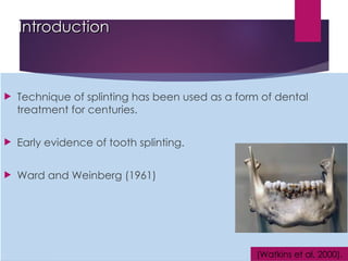
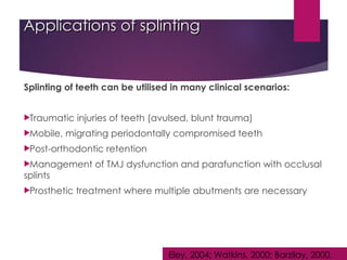
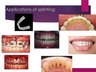
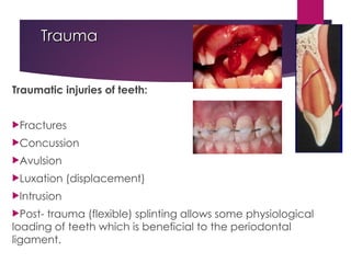
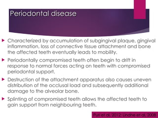
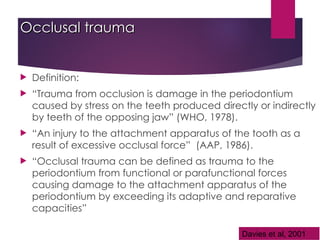
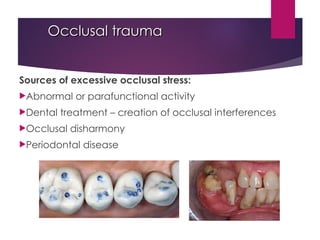
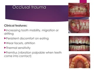
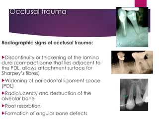
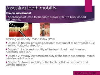
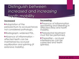
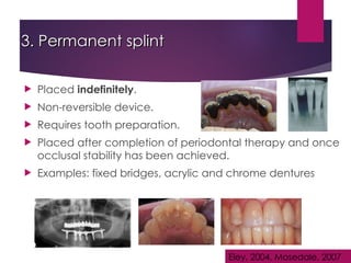
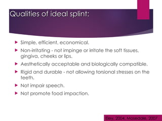
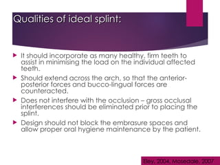
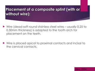
A Boon or Bane?
A Boon or Bane?
Periodontal Splinting
Dr. T Vedan
Dr. T Vedan
(Updated from Dr. G Abrahams)
Department Oral Medicine and
Periodontics
Periodontics
UWC
UWC
Introduction
Introduction
 Technique of splinting has been used as a form of dental
treatment for centuries.
 Early evidence of tooth splinting.
 Ward and Weinberg (1961)
(Watkins et al, 2000).
Definition: Splinting
 “A rigid or flexible material used to protect, immobilize or resist
motion in a part” (Dictionary).
 In dentistry “The joining of two or more teeth into a rigid unit by
means of fixed or removable restorations or devices”(Barzilay,
2000).
 “An appliance designed to stabilize mobile teeth” (Glossary of
Periodontic terms, 1986).
Applications of splinting
Splinting of teeth can be utilised in many clinical scenarios:
Traumatic injuries of teeth (avulsed, blunt trauma)
Mobile, migrating periodontally compromised teeth
Post-orthodontic retention
Management of TMJ dysfunction and parafunction with occlusal
splints
Prosthetic treatment where multiple abutments are necessary
Eley, 2004; Watkins, 2000; Barzilay, 2000.
Principles of splinting:
 Should reduce movement 3 dimensionally
 Centre of rotation of the affected teeth
must be located in the remaining
supporting bone.
 No inflammation
 Minimum one third of bone support
remaining.
 Should allow for oral hygiene methods
 Should not irritate soft tissues
Applications of splinting:
Etiology if mobility
 Trauma
 Pericapical infection
 Periodontal disease
 Occlusal trauma
 Orthodontic treatment
 Root resorption
 Abnormal tooth morphology
Newman et al, 2008; Lindhe et al, 2008
Trauma
Trauma
Traumatic injuries of teeth:
Fractures
Concussion
Avulsion
Luxation (displacement)
Intrusion
Post- trauma (flexible) splinting allows some physiological
loading of teeth which is beneficial to the periodontal
ligament.
Trauma
Trauma
Tooth migration/ mobility may also be associated with
habits:
Pipe smokers
Pencil chewing
Tongue thrusting
Newman et al, 2008; Davies et al, 2001
Periodontal disease
Periodontal disease
 Characterized by accumulation of subgingival plaque, gingival
inflammation, loss of connective tissue attachment and bone
the affected teeth eventually leads to mobility.
 Periodontally compromised teeth often begin to drift in
response to normal forces acting on teeth with compromised
periodontal support.
 Destruction of the attachment apparatus also causes uneven
distribution of the occlusal load and subsequently additional
damage to the alveolar bone.
 Splinting of compromised teeth allows the affected teeth to
gain support from neighbouring teeth.
Puri et al, 2012; Lindhe et al, 2008
Occlusal trauma
Occlusal trauma
 Definition:
 “Trauma from occlusion is damage in the periodontium
caused by stress on the teeth produced directly or indirectly
by teeth of the opposing jaw” (WHO, 1978).
 “An injury to the attachment apparatus of the tooth as a
result of excessive occlusal force” (AAP, 1986).
 “Occlusal trauma can be defined as trauma to the
periodontium from functional or parafunctional forces
causing damage to the attachment apparatus of the
periodontium by exceeding its adaptive and reparative
capacities”
Davies et al, 2001
Occlusal trauma
Occlusal trauma
Sources of excessive occlusal stress:
Abnormal or parafunctional activity
Dental treatment – creation of occlusal interferences
Occlusal disharmony
Periodontal disease
Occlusal trauma
Occlusal trauma
Clinical features:
Increasing tooth mobility, migration or
drifting
Persistent discomfort on eating
Wear facets, attrition
Thermal sensitivity
Fremitus (vibration palpable when teeth
come into contact)
Occlusal trauma
Occlusal trauma
Radiographic signs of occlusal trauma:
Discontinuity or thickening of the lamina
dura (compact bone that lies adjacent to
the PDL, allows attachment surface for
Sharpey’s fibres)
Widening of periodontal ligament space
(PDL)
Radiolucency and destruction of the
alveolar bone
Root resorbtion
Formation of angular bone defects
Assessing tooth mobility
Clinical assessment :
Application of force to the tooth crown with two blunt ended
instruments.
Grading of mobility: Millers Index (1950)
Degree 0: Normal physiological tooth movement of between 0,1-0,2
mm in a horizontal direction.
Degree 1: Increased mobility of the tooth to at most 1mm in a
horizontal direction.
Degree 2: Visually increased mobility of the tooth exceeding 1mm in
a horizontal direction.
Degree 3: Severe mobility of the tooth both in a horizontal and
vertical direction
Lindhe et al, 2008
Distinguish between
Distinguish between
increased
increased and
and increasing
increasing
tooth mobility
tooth mobility
Increased
Adaptation of the
periodontium to occlusal forces
not considered pathologic.
Radiograph: widened PDL.
Absence of inflammation –
affected teeth can be
maintained by occlusal
equilibration and splinting (if
extensive mobility).
Increasing:
Presence of inflammation
(pocketing and bleeding in
affected sites) and tooth
mobility.
Periodontal treatment
must first be performed.
Thereafter – occlusal
interferences can be
corrected and teeth
splinted.
Puri et al, 2012.
Splinting as a treatment modality
 Rationale: joining mobile teeth to less mobile teeth creating a
“Multi-rooted unit”,
 This increases the total area of root resistance thus preventing a
local gross overload of force on the compromised tooth
 Resulting in a force distribution among a number of teeth.
 Involve individual or multiple teeth – depending on the clinical
situation.
 Possible alternative in situations where extractions and implant
therapy is not a viable treatment option for the patient.
 Splinting of mobile teeth should only be viewed as a short-term or
transitional treatment modality.
Watkins et al, 2000; Mosedale, 2007; Griffin, 2005
Indications splinting:
Only be applied once periodontal treatment has been
performed, local factors eliminated and occlusal interferences
have been corrected.
Mobility is progressively increasing – the teeth are exposed to
extraction forces during function.
Multiple teeth that have become mobile and are drifting as a
result of gradual alveolar bone loss.
Restore proximal contacts that have been disrupted by the
migration of teeth.
Puri et al, 2012; Eley, 2004
Indications splinting:
 Stabilize moderate to advanced tooth mobility (teeth that
have not responded to occlusal adjustment and periodontal
therapy).
 It permits and improves masticatory function and comfort.
 To facilitate mechanical debridement and surgical
procedures of mobile teeth.
 To protect, provide stability and rest for the tooth-supporting
tissues during the healing period after periodontal treatment.
Puri et al, 2012; Eley, 2004
Indications for splinting:
 Redirection of forces - forces of occlusion can be
redirected in a more axial direction over the teeth in the
splint.
 Redistribution of forces - there is stabilization in response to
the applied force. This ensures that forces do not exceed
the adaptive capacity of compromised teeth.
 Psychological well-being - provides some psychological
comfort from the fear of the loss of loose teeth.
Puri et al, 2012; Eley, 2004
Contra-indications for splinting
 In situations where occlusal stability and optimal
periodontal conditions cannot be obtained.
 Occlusal or masticatory forces exceed the resistance
provided by the splinted teeth.
Puri et al, 2012; Eley, 2004
Classification of splints
Eley, 2004, Mosedale, 2007
1. Temporary splint
1. Temporary splint
 Indicated to stabilize teeth and absorb occlusal forces for
a limited period.
 Usually not longer than 2 months.
 Clinical scenarios : stabilize teeth during periodontal tx
/regenerative therapy/ after trauma.
 NO tooth structure removed with placement.
 Examples: composite resins (with or without wire)
Eley, 2004, Mosedale, 2007
2. Provisional splint
 Usually used for 4-6 months.
 Used for diagnostic information.
 Allows clinician to observe the healing response to treatment
and make adjustments based on patient response to
treatment.
 At a later stage the clinician can design a more permanent
and biologically acceptable form of treatment.
 Examples: acrylic splints, vacuum-formed splints.
Eley, 2004, Mosedale, 2007
3. Permanent splint
3. Permanent splint
 Placed indefinitely.
 Non-reversible device.
 Requires tooth preparation.
 Placed after completion of periodontal therapy and once
occlusal stability has been achieved.
 Examples: fixed bridges, acrylic and chrome dentures
Eley, 2004, Mosedale, 2007
Qualities of ideal splint:
 Simple, efficient, economical.
 Non-irritating - not impinge or irritate the soft tissues,
gingiva, cheeks or lips.
 Aesthetically acceptable and biologically compatible.
 Rigid and durable - not allowing torsional stresses on the
teeth.
 Not impair speech.
 Not promote food impaction.
Eley, 2004, Mosedale, 2007
Qualities of ideal splint:
 It should incorporate as many healthy, firm teeth to
assist in minimising the load on the individual affected
teeth.
 Should extend across the arch, so that the anterior-
posterior forces and bucco-lingual forces are
counteracted.
 Does not interfere with the occlusion – gross occlusal
interferences should be eliminated prior to placing the
splint.
 Design should not block the embrasure spaces and
allow proper oral hygiene maintenance by the patient.
Eley, 2004, Mosedale, 2007
Placement of a composite splint (with or
without wire):
without wire):
 Wire (dead-soft round stainless steel wires – usually 0,25 to
0,30mm thickness) is adapted to the tooth arch for
placement on the teeth.
 Wire is placed apical to proximal contacts and incisal to
the cervical contacts.
Eley, 2004
Materials needed:
Materials needed:
1
2
3
Placement of a composite splint (with or
without wire):
without wire):
 Moisture control is essential during splinting and
the use of a rubberdam is advocated.
 Enamel surfaces must be properly cleaned to
achieve an adequate etching of the area.
 The enamel is etched for 15-20 seconds with
35% phosphoric acid, rinsed, dried and a light
curable dentin-bonding agent is applied.
 Composite filling material (with or without a
wire) is placed and cured.
Disadvantages of splinting
Plaque control
Removable splints allow the patient to practise normal plaque
control measures.
Fixed splinting modalities may compromise the ability of
patients to maintain adequate plaque control.
Periodontal monitoring
Fixed splints prevent proper clinical assessment of teeth and
also reduces the patients awareness of increasing tooth
mobility.
Effective recall systems need to be established with regular
clinical and radiographic review. Watkins, 2000
Disadvantages of splinting
Dental caries
In the presence of poor plaque control associated with poor
splint design, there is a risk for caries development.
Thus regular review, plaque control and proper splint design is
essential to prevent detrimental effects.
Maintenance of splints
Biological failure of splints can be the result of dental caries,
progressing periodontal disease, endodontic failures and
mechanical splint failure.
Occlusion needs to be meticulously monitored and finely
adjusted after splint placement . Watkins, 2000
Conclusion
Conclusion
 Periodontal splinting can be a useful tool in specific
situations.
 However if applied incorrectly, it can have a further
detrimental effect.
 After placement of a splint, vigilant periodontal
maintenance and monitoring is essential.
 Splinting is never a substitute for thorough diagnosis and
effective periodontal treatment.
 Decision to splint should not be taken lightly and should
only be considered following appropriate periodontal
and occlusal management.
Watkins, 2000; Mosedale, 2007
References
Barzilay, I. (2000). Splinting teeth – A review of the methodology and clinical case reports. J
Can Dent Asso; 66:440-443.
Bernal,G. Carvajal,JC. & Munoz, CA.(2002). A review of the clinical management of mobile teeth. The Journal of
Contemporary Dental Practice; Volume 3, No4.
Davies, SJ. Gray, RJM. Linden, GJ. & James, JA. (2001). Occlusal considerations in periodontics. British Dental Journal;
191(11): 597-604.
Eley, BM. (2004). Periodontics. Fifth edition. Wright; pg: 361-368.
Griffin, EC. (2005). To splint or not to splint: Treating periodontally compromised teeth by improving occlusion.
Contemporary Aesthetics and Restorative Practice. Continuing Education.
Lindhe, J. Lang, NP. & Karring, T. (2008). Clinical periodontology and Implant Dentistry. Blackwell Munksgaard.Volume
2: 183-249.
Mosedale, RF. (2007). Current indications and methods of periodontal splinting. Dental Update; 34: 168-180.
Newman,M. Takei, HH. Klokkevold, PR. Carranza, FA. (2006). Caranza’s Clinical Periodontology –Tenth Edition.
Saunders.
Puri, MS. Grover, HS. Gupta, A. Puri,N. & Luthra, S. (2012). Splinting – A healing touch for an ailing periodontium.
Journal of Oral health and Community Dentistry; 8: 145-148.
Watkins, SJ. & Hemmings, KW. (2000). Periodontal splinting in general dental practice. Dental Update; 27: 278-285.

Splinting procedure and steps in dentistry.

  • 1.
    A Boon orBane? A Boon or Bane? Periodontal Splinting Periodontal Splinting Dr. T Vedan Dr. T Vedan (Updated from Dr. G Abrahams) (Updated from Dr. G Abrahams) Department Oral Medicine and Department Oral Medicine and Periodontics Periodontics UWC UWC
  • 3.
    Introduction Introduction  Technique ofsplinting has been used as a form of dental treatment for centuries.  Early evidence of tooth splinting.  Ward and Weinberg (1961) (Watkins et al, 2000).
  • 4.
    Definition: Splinting Definition: Splinting “A rigid or flexible material used to protect, immobilize or resist motion in a part” (Dictionary).  In dentistry “The joining of two or more teeth into a rigid unit by means of fixed or removable restorations or devices”(Barzilay, 2000).  “An appliance designed to stabilize mobile teeth” (Glossary of Periodontic terms, 1986).
  • 5.
    Applications of splinting Applicationsof splinting Splinting of teeth can be utilised in many clinical scenarios: Traumatic injuries of teeth (avulsed, blunt trauma) Mobile, migrating periodontally compromised teeth Post-orthodontic retention Management of TMJ dysfunction and parafunction with occlusal splints Prosthetic treatment where multiple abutments are necessary Eley, 2004; Watkins, 2000; Barzilay, 2000.
  • 6.
    Principles of splinting: Principlesof splinting:  Should reduce movement 3 dimensionally  Centre of rotation of the affected teeth must be located in the remaining supporting bone.  No inflammation  Minimum one third of bone support remaining.  Should allow for oral hygiene methods  Should not irritate soft tissues
  • 7.
  • 8.
    Etiology if mobility Trauma  Pericapical infection  Periodontal disease  Occlusal trauma  Orthodontic treatment  Root resorption  Abnormal tooth morphology Newman et al, 2008; Lindhe et al, 2008
  • 9.
    Trauma Trauma Traumatic injuries ofteeth: Fractures Concussion Avulsion Luxation (displacement) Intrusion Post- trauma (flexible) splinting allows some physiological loading of teeth which is beneficial to the periodontal ligament.
  • 10.
    Trauma Trauma Tooth migration/ mobilitymay also be associated with habits: Pipe smokers Pencil chewing Tongue thrusting Newman et al, 2008; Davies et al, 2001
  • 11.
    Periodontal disease Periodontal disease Characterized by accumulation of subgingival plaque, gingival inflammation, loss of connective tissue attachment and bone the affected teeth eventually leads to mobility.  Periodontally compromised teeth often begin to drift in response to normal forces acting on teeth with compromised periodontal support.  Destruction of the attachment apparatus also causes uneven distribution of the occlusal load and subsequently additional damage to the alveolar bone.  Splinting of compromised teeth allows the affected teeth to gain support from neighbouring teeth. Puri et al, 2012; Lindhe et al, 2008
  • 12.
    Occlusal trauma Occlusal trauma Definition:  “Trauma from occlusion is damage in the periodontium caused by stress on the teeth produced directly or indirectly by teeth of the opposing jaw” (WHO, 1978).  “An injury to the attachment apparatus of the tooth as a result of excessive occlusal force” (AAP, 1986).  “Occlusal trauma can be defined as trauma to the periodontium from functional or parafunctional forces causing damage to the attachment apparatus of the periodontium by exceeding its adaptive and reparative capacities” Davies et al, 2001
  • 13.
    Occlusal trauma Occlusal trauma Sourcesof excessive occlusal stress: Abnormal or parafunctional activity Dental treatment – creation of occlusal interferences Occlusal disharmony Periodontal disease
  • 14.
    Occlusal trauma Occlusal trauma Clinicalfeatures: Increasing tooth mobility, migration or drifting Persistent discomfort on eating Wear facets, attrition Thermal sensitivity Fremitus (vibration palpable when teeth come into contact)
  • 15.
    Occlusal trauma Occlusal trauma Radiographicsigns of occlusal trauma: Discontinuity or thickening of the lamina dura (compact bone that lies adjacent to the PDL, allows attachment surface for Sharpey’s fibres) Widening of periodontal ligament space (PDL) Radiolucency and destruction of the alveolar bone Root resorbtion Formation of angular bone defects
  • 16.
    Assessing tooth mobility Assessingtooth mobility Clinical assessment : Application of force to the tooth crown with two blunt ended instruments. Grading of mobility: Millers Index (1950) Degree 0: Normal physiological tooth movement of between 0,1-0,2 mm in a horizontal direction. Degree 1: Increased mobility of the tooth to at most 1mm in a horizontal direction. Degree 2: Visually increased mobility of the tooth exceeding 1mm in a horizontal direction. Degree 3: Severe mobility of the tooth both in a horizontal and vertical direction Lindhe et al, 2008
  • 17.
    Distinguish between Distinguish between increased increasedand and increasing increasing tooth mobility tooth mobility Increased Adaptation of the periodontium to occlusal forces not considered pathologic. Radiograph: widened PDL. Absence of inflammation – affected teeth can be maintained by occlusal equilibration and splinting (if extensive mobility). Increasing: Presence of inflammation (pocketing and bleeding in affected sites) and tooth mobility. Periodontal treatment must first be performed. Thereafter – occlusal interferences can be corrected and teeth splinted. Puri et al, 2012.
  • 18.
    Splinting as atreatment modality Splinting as a treatment modality  Rationale: joining mobile teeth to less mobile teeth creating a “Multi-rooted unit”,  This increases the total area of root resistance thus preventing a local gross overload of force on the compromised tooth  Resulting in a force distribution among a number of teeth.  Involve individual or multiple teeth – depending on the clinical situation.  Possible alternative in situations where extractions and implant therapy is not a viable treatment option for the patient.  Splinting of mobile teeth should only be viewed as a short-term or transitional treatment modality. Watkins et al, 2000; Mosedale, 2007; Griffin, 2005
  • 19.
    Indications splinting: Indications splinting: Onlybe applied once periodontal treatment has been performed, local factors eliminated and occlusal interferences have been corrected. Mobility is progressively increasing – the teeth are exposed to extraction forces during function. Multiple teeth that have become mobile and are drifting as a result of gradual alveolar bone loss. Restore proximal contacts that have been disrupted by the migration of teeth. Puri et al, 2012; Eley, 2004
  • 20.
    Indications splinting: Indications splinting: Stabilize moderate to advanced tooth mobility (teeth that have not responded to occlusal adjustment and periodontal therapy).  It permits and improves masticatory function and comfort.  To facilitate mechanical debridement and surgical procedures of mobile teeth.  To protect, provide stability and rest for the tooth-supporting tissues during the healing period after periodontal treatment. Puri et al, 2012; Eley, 2004
  • 21.
    Indications for splinting: Indicationsfor splinting:  Redirection of forces - forces of occlusion can be redirected in a more axial direction over the teeth in the splint.  Redistribution of forces - there is stabilization in response to the applied force. This ensures that forces do not exceed the adaptive capacity of compromised teeth.  Psychological well-being - provides some psychological comfort from the fear of the loss of loose teeth. Puri et al, 2012; Eley, 2004
  • 22.
    Contra-indications for splinting Contra-indicationsfor splinting  In situations where occlusal stability and optimal periodontal conditions cannot be obtained.  Occlusal or masticatory forces exceed the resistance provided by the splinted teeth. Puri et al, 2012; Eley, 2004
  • 23.
    Classification of splints Classificationof splints Eley, 2004, Mosedale, 2007
  • 24.
    1. Temporary splint 1.Temporary splint  Indicated to stabilize teeth and absorb occlusal forces for a limited period.  Usually not longer than 2 months.  Clinical scenarios : stabilize teeth during periodontal tx /regenerative therapy/ after trauma.  NO tooth structure removed with placement.  Examples: composite resins (with or without wire) Eley, 2004, Mosedale, 2007
  • 25.
    2. Provisional splint 2.Provisional splint  Usually used for 4-6 months.  Used for diagnostic information.  Allows clinician to observe the healing response to treatment and make adjustments based on patient response to treatment.  At a later stage the clinician can design a more permanent and biologically acceptable form of treatment.  Examples: acrylic splints, vacuum-formed splints. Eley, 2004, Mosedale, 2007
  • 26.
    3. Permanent splint 3.Permanent splint  Placed indefinitely.  Non-reversible device.  Requires tooth preparation.  Placed after completion of periodontal therapy and once occlusal stability has been achieved.  Examples: fixed bridges, acrylic and chrome dentures Eley, 2004, Mosedale, 2007
  • 27.
    Qualities of idealsplint: Qualities of ideal splint:  Simple, efficient, economical.  Non-irritating - not impinge or irritate the soft tissues, gingiva, cheeks or lips.  Aesthetically acceptable and biologically compatible.  Rigid and durable - not allowing torsional stresses on the teeth.  Not impair speech.  Not promote food impaction. Eley, 2004, Mosedale, 2007
  • 28.
    Qualities of idealsplint: Qualities of ideal splint:  It should incorporate as many healthy, firm teeth to assist in minimising the load on the individual affected teeth.  Should extend across the arch, so that the anterior- posterior forces and bucco-lingual forces are counteracted.  Does not interfere with the occlusion – gross occlusal interferences should be eliminated prior to placing the splint.  Design should not block the embrasure spaces and allow proper oral hygiene maintenance by the patient. Eley, 2004, Mosedale, 2007
  • 29.
    Placement of acomposite splint (with or Placement of a composite splint (with or without wire): without wire):  Wire (dead-soft round stainless steel wires – usually 0,25 to 0,30mm thickness) is adapted to the tooth arch for placement on the teeth.  Wire is placed apical to proximal contacts and incisal to the cervical contacts. Eley, 2004
  • 30.
  • 31.
    Placement of acomposite splint (with or Placement of a composite splint (with or without wire): without wire):  Moisture control is essential during splinting and the use of a rubberdam is advocated.  Enamel surfaces must be properly cleaned to achieve an adequate etching of the area.  The enamel is etched for 15-20 seconds with 35% phosphoric acid, rinsed, dried and a light curable dentin-bonding agent is applied.  Composite filling material (with or without a wire) is placed and cured.
  • 38.
    Disadvantages of splinting Disadvantagesof splinting Plaque control Removable splints allow the patient to practise normal plaque control measures. Fixed splinting modalities may compromise the ability of patients to maintain adequate plaque control. Periodontal monitoring Fixed splints prevent proper clinical assessment of teeth and also reduces the patients awareness of increasing tooth mobility. Effective recall systems need to be established with regular clinical and radiographic review. Watkins, 2000
  • 39.
    Disadvantages of splinting Disadvantagesof splinting Dental caries In the presence of poor plaque control associated with poor splint design, there is a risk for caries development. Thus regular review, plaque control and proper splint design is essential to prevent detrimental effects. Maintenance of splints Biological failure of splints can be the result of dental caries, progressing periodontal disease, endodontic failures and mechanical splint failure. Occlusion needs to be meticulously monitored and finely adjusted after splint placement . Watkins, 2000
  • 40.
    Conclusion Conclusion  Periodontal splintingcan be a useful tool in specific situations.  However if applied incorrectly, it can have a further detrimental effect.  After placement of a splint, vigilant periodontal maintenance and monitoring is essential.  Splinting is never a substitute for thorough diagnosis and effective periodontal treatment.  Decision to splint should not be taken lightly and should only be considered following appropriate periodontal and occlusal management. Watkins, 2000; Mosedale, 2007
  • 41.
    References Barzilay, I. (2000).Splinting teeth – A review of the methodology and clinical case reports. J Can Dent Asso; 66:440-443. Bernal,G. Carvajal,JC. & Munoz, CA.(2002). A review of the clinical management of mobile teeth. The Journal of Contemporary Dental Practice; Volume 3, No4. Davies, SJ. Gray, RJM. Linden, GJ. & James, JA. (2001). Occlusal considerations in periodontics. British Dental Journal; 191(11): 597-604. Eley, BM. (2004). Periodontics. Fifth edition. Wright; pg: 361-368. Griffin, EC. (2005). To splint or not to splint: Treating periodontally compromised teeth by improving occlusion. Contemporary Aesthetics and Restorative Practice. Continuing Education. Lindhe, J. Lang, NP. & Karring, T. (2008). Clinical periodontology and Implant Dentistry. Blackwell Munksgaard.Volume 2: 183-249. Mosedale, RF. (2007). Current indications and methods of periodontal splinting. Dental Update; 34: 168-180. Newman,M. Takei, HH. Klokkevold, PR. Carranza, FA. (2006). Caranza’s Clinical Periodontology –Tenth Edition. Saunders. Puri, MS. Grover, HS. Gupta, A. Puri,N. & Luthra, S. (2012). Splinting – A healing touch for an ailing periodontium. Journal of Oral health and Community Dentistry; 8: 145-148. Watkins, SJ. & Hemmings, KW. (2000). Periodontal splinting in general dental practice. Dental Update; 27: 278-285.

Editor's Notes

  • #3 History..!!! A Phoenician mandible from 500BC found in modern day Lebanon which has two carved ivory teeth attached to four natural teeth by gold wire. Obin and Arvin's (1951) -self curing internal splint. Harrington(1957) modified the splint by incorporating cemented stainless steel wire. Ward & Weinberg (1961) – developed new techniques using a plastic matrix or using wire reinforcement. Splints have thus come a far way to INTRACORONAL BONDABLE FIBER SPLINTING of the present.
  • #5 Objectives: To provide rest  For redirection of forces  For redistribution of forces  To preserve arch integrity  Restore of functional stability  Psychological well being  To stabilize mobile teeth during surgery , especially during regenerative therapy  To prevent the eruption of unopposed teeth