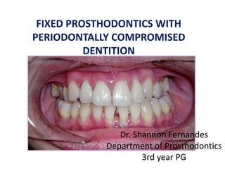
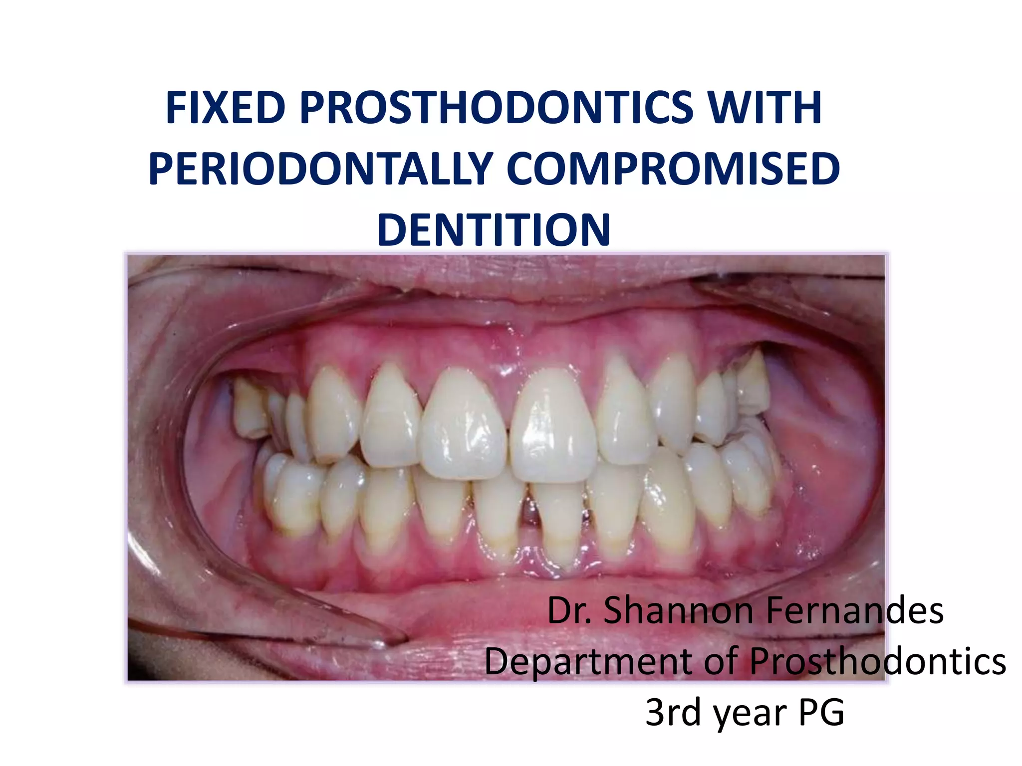
This document discusses considerations for fixed prosthodontics in patients with compromised periodontal health. Key points include:
- Periodontal health plays an important role in the longevity of restorations, and defective prostheses can contribute to periodontal disease progression. Successful treatment requires cooperation between periodontists and prosthodontists.
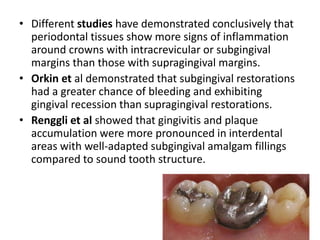
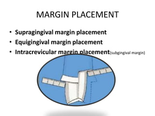
- Periodontal issues must be resolved before restorative treatment to avoid tensions on the periodontium from tooth movement. Supragingival margins and open embrasures are preferred for periodontal health.
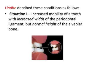
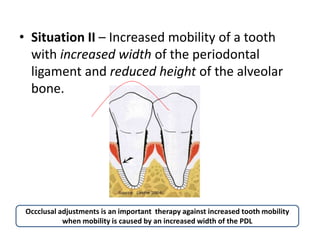
- Temporary splinting can help determine the prognosis of a permanent restoration in periodontally compromised patients. Occlusion should not interfere with plaque control.
This seminar deals with implant-related complications that lead to implant failure.this also discus diagnostic criteria and preventive methods for an implant failure.
Fixed prosthodontic treatment can offer exceptional satisfaction for both patient and the dentist. Fixed Prosthodontics can transform an unhealthy, unattractive dentition with poor function into a comfortable, healthy occlusion capable of giving years of further service while greatly enhancing esthetics.
Nothing is more important in the construction of fixed partial dentures than an adequate diagnosis and a well-devised treatment plan. Although these two subjects are usually considered together. Astute dentists must recognize their subtle differences. Diagnosis is an evaluation of the condition of the patient when he presents for treatment. Treatment planning concerns the treatment procedures by which the dentist will restore the patient to an optimum state of dental health.
The patient who requires a single denture opposing a natural or restored dentition challenges the clinician even more than the completely edentulous patient does. This is due to the biomechanical differences in the supporting tissues of the opposing arches. So a proper evaluation, correction of the existing factors and proper sequence of denture construction is necessary to give a more stable prosthesis.
Indian Dental Academy: will be one of the most relevant and exciting
training center with best faculty and flexible training programs
for dental professionals who wish to advance in their dental
practice,Offers certified courses in Dental
implants,Orthodontics,Endodontics,Cosmetic Dentistry, Prosthetic
Dentistry, Periodontics and General Dentistry.
A detailed description about endo perio interrelationship, including introduction, development and etiology, historical aspects, definition, classification, diagnosis, differential diagnosis, management, special consideration in management,controversies prognosis, conclusion.
Failures in FPDs and its management is very well described in this seminar and done according to the main books.
Described under classification of biologic, mechanical, aesthetics, Psychogenic, Maintenances Failures
Review of literature is also given in this presentation
Indian Dental Academy: will be one of the most relevant and exciting training center with best faculty and flexible training programs for dental professionals who wish to advance in their dental practice,Offers certified courses in Dental implants,Orthodontics,Endodontics,Cosmetic Dentistry, Prosthetic Dentistry, Periodontics and General Dentistry.
Orthodontic-periodontic interactions are mutually beneficial. Orthodontic treatment can be justified as a part of periodontal therapy if it is used to reduce plaque accumulation, correct abnormal gingival and osseous forms, improve aesthetics, and facilitate prosthetic replacement.
Overdentures are a useful treatment option in many clinical situations. A simple complete lower overdenture which encloses the roots of two root-treated canines has been shown above (Fig. 12.51). Cases can be more complicated than this. The reduction in the crowns of the teeth may have occurred due to tooth wear from a combination of erosion and attrition. In the elderly, where such tooth reduction has occurred, root canal treatment may not be necessary. The removal of the roots will not benefit the patient and the overdenture is the best form of treatment.
Less common situations, such as partial anodontia, cleft palate or loss of tooth crown substance in dentinogenesis imperfecta, may also require restoration using overdentures. The distinction between an onlay and an overdenture is not clear-cut and a potentially difficult partial denture treatment, such as the restoration of a free end saddle, may be helped by the coverage of a canine or molar tooth with a reduced crown rather than a more involved crown restoration.
In the case illustrated in Figure 12.53, an elderly patient has severe tooth surface loss. The aetiology of this wear must be diagnosed before treatment is commenced. For instance, is this wear a result of parafunction or erosion from the consumption of acidic drinks? The remaining dentition has been restored and a definitive overdenture placed.
Overdenture is a favored treatment modality for elderly patients with few remaining teeth. Roots maintained under the denture base preserve the alveolar ridge, provide sensory feedback and improve the stability of the dentures. Furthermore, the use of copings and precision attachments on the remaining teeth enhances the retention of the denture. This clinical report describes a novel method of fabricating a tooth supported overdenture retained with custom made ball attachments using orthodontic separators as a female component. Customized ball attachments with orthodontic separators are a simple and cost effective alternative treatment to the use of prefabricated attachments for enhancing the retention of tooth supported overdentures.
This presentation describe the evaluation of badly damaged teeth for crowning before starting RCT treatment, because the treatment of such teeth is always achieved by crowning otherwise they will end for extraction. All the necessary procedures to save the damaged teeth are discussed in the context of restoring function, aesthetic and mechanical qualities. Evaluation of any case based on scientific data will insure durability and patient satisfaction.
Indian Dental Academy: will be one of the most relevant and exciting training center with best faculty and flexible training programs for dental professionals who wish to advance in their dental practice,Offers certified courses in Dental implants,Orthodontics,Endodontics,Cosmetic Dentistry, Prosthetic Dentistry, Periodontics and General Dentistry.
micro teaching on communication m.sc nursing.pdfAnurag Sharma
Microteaching is a unique model of practice teaching. It is a viable instrument for the. desired change in the teaching behavior or the behavior potential which, in specified types of real. classroom situations, tends to facilitate the achievement of specified types of objectives.
Acute scrotum is a general term referring to an emergency condition affecting the contents or the wall of the scrotum.
There are a number of conditions that present acutely, predominantly with pain and/or swelling
A careful and detailed history and examination, and in some cases, investigations allow differentiation between these diagnoses. A prompt diagnosis is essential as the patient may require urgent surgical intervention
Testicular torsion refers to twisting of the spermatic cord, causing ischaemia of the testicle.
Testicular torsion results from inadequate fixation of the testis to the tunica vaginalis producing ischemia from reduced arterial inflow and venous outflow obstruction.
The prevalence of testicular torsion in adult patients hospitalized with acute scrotal pain is approximately 25 to 50 percent
Title: Sense of Taste
Presenter: Dr. Faiza, Assistant Professor of Physiology
Qualifications:
MBBS (Best Graduate, AIMC Lahore)
FCPS Physiology
ICMT, CHPE, DHPE (STMU)
MPH (GC University, Faisalabad)
MBA (Virtual University of Pakistan)
Learning Objectives:
Describe the structure and function of taste buds.
Describe the relationship between the taste threshold and taste index of common substances.
Explain the chemical basis and signal transduction of taste perception for each type of primary taste sensation.
Recognize different abnormalities of taste perception and their causes.
Key Topics:
Significance of Taste Sensation:
Differentiation between pleasant and harmful food
Influence on behavior
Selection of food based on metabolic needs
Receptors of Taste:
Taste buds on the tongue
Influence of sense of smell, texture of food, and pain stimulation (e.g., by pepper)
Primary and Secondary Taste Sensations:
Primary taste sensations: Sweet, Sour, Salty, Bitter, Umami
Chemical basis and signal transduction mechanisms for each taste
Taste Threshold and Index:
Taste threshold values for Sweet (sucrose), Salty (NaCl), Sour (HCl), and Bitter (Quinine)
Taste index relationship: Inversely proportional to taste threshold
Taste Blindness:
Inability to taste certain substances, particularly thiourea compounds
Example: Phenylthiocarbamide
Structure and Function of Taste Buds:
Composition: Epithelial cells, Sustentacular/Supporting cells, Taste cells, Basal cells
Features: Taste pores, Taste hairs/microvilli, and Taste nerve fibers
Location of Taste Buds:
Found in papillae of the tongue (Fungiform, Circumvallate, Foliate)
Also present on the palate, tonsillar pillars, epiglottis, and proximal esophagus
Mechanism of Taste Stimulation:
Interaction of taste substances with receptors on microvilli
Signal transduction pathways for Umami, Sweet, Bitter, Sour, and Salty tastes
Taste Sensitivity and Adaptation:
Decrease in sensitivity with age
Rapid adaptation of taste sensation
Role of Saliva in Taste:
Dissolution of tastants to reach receptors
Washing away the stimulus
Taste Preferences and Aversions:
Mechanisms behind taste preference and aversion
Influence of receptors and neural pathways
Impact of Sensory Nerve Damage:
Degeneration of taste buds if the sensory nerve fiber is cut
Abnormalities of Taste Detection:
Conditions: Ageusia, Hypogeusia, Dysgeusia (parageusia)
Causes: Nerve damage, neurological disorders, infections, poor oral hygiene, adverse drug effects, deficiencies, aging, tobacco use, altered neurotransmitter levels
Neurotransmitters and Taste Threshold:
Effects of serotonin (5-HT) and norepinephrine (NE) on taste sensitivity
Supertasters:
25% of the population with heightened sensitivity to taste, especially bitterness
Increased number of fungiform papillae
Pulmonary Thromboembolism - etilogy, types, medical- Surgical and nursing man...VarunMahajani
Disruption of blood supply to lung alveoli due to blockage of one or more pulmonary blood vessels is called as Pulmonary thromboembolism. In this presentation we will discuss its causes, types and its management in depth.
TEST BANK for Operations Management, 14th Edition by William J. Stevenson, Ve...kevinkariuki227
TEST BANK for Operations Management, 14th Edition by William J. Stevenson, Verified Chapters 1 - 19, Complete Newest Version.pdf
Recomendações da OMS sobre cuidados maternos e neonatais para uma experiência pós-natal positiva.
Em consonância com os ODS – Objetivos do Desenvolvimento Sustentável e a Estratégia Global para a Saúde das Mulheres, Crianças e Adolescentes, e aplicando uma abordagem baseada nos direitos humanos, os esforços de cuidados pós-natais devem expandir-se para além da cobertura e da simples sobrevivência, de modo a incluir cuidados de qualidade.
Estas diretrizes visam melhorar a qualidade dos cuidados pós-natais essenciais e de rotina prestados às mulheres e aos recém-nascidos, com o objetivo final de melhorar a saúde e o bem-estar materno e neonatal.
Uma “experiência pós-natal positiva” é um resultado importante para todas as mulheres que dão à luz e para os seus recém-nascidos, estabelecendo as bases para a melhoria da saúde e do bem-estar a curto e longo prazo. Uma experiência pós-natal positiva é definida como aquela em que as mulheres, pessoas que gestam, os recém-nascidos, os casais, os pais, os cuidadores e as famílias recebem informação consistente, garantia e apoio de profissionais de saúde motivados; e onde um sistema de saúde flexível e com recursos reconheça as necessidades das mulheres e dos bebês e respeite o seu contexto cultural.
Estas diretrizes consolidadas apresentam algumas recomendações novas e já bem fundamentadas sobre cuidados pós-natais de rotina para mulheres e neonatos que recebem cuidados no pós-parto em unidades de saúde ou na comunidade, independentemente dos recursos disponíveis.
É fornecido um conjunto abrangente de recomendações para cuidados durante o período puerperal, com ênfase nos cuidados essenciais que todas as mulheres e recém-nascidos devem receber, e com a devida atenção à qualidade dos cuidados; isto é, a entrega e a experiência do cuidado recebido. Estas diretrizes atualizam e ampliam as recomendações da OMS de 2014 sobre cuidados pós-natais da mãe e do recém-nascido e complementam as atuais diretrizes da OMS sobre a gestão de complicações pós-natais.
O estabelecimento da amamentação e o manejo das principais intercorrências é contemplada.
Recomendamos muito.
Vamos discutir essas recomendações no nosso curso de pós-graduação em Aleitamento no Instituto Ciclos.
Esta publicação só está disponível em inglês até o momento.
Prof. Marcus Renato de Carvalho
www.agostodourado.com
Lung Cancer: Artificial Intelligence, Synergetics, Complex System Analysis, S...Oleg Kshivets
RESULTS: Overall life span (LS) was 2252.1±1742.5 days and cumulative 5-year survival (5YS) reached 73.2%, 10 years – 64.8%, 20 years – 42.5%. 513 LCP lived more than 5 years (LS=3124.6±1525.6 days), 148 LCP – more than 10 years (LS=5054.4±1504.1 days).199 LCP died because of LC (LS=562.7±374.5 days). 5YS of LCP after bi/lobectomies was significantly superior in comparison with LCP after pneumonectomies (78.1% vs.63.7%, P=0.00001 by log-rank test). AT significantly improved 5YS (66.3% vs. 34.8%) (P=0.00000 by log-rank test) only for LCP with N1-2. Cox modeling displayed that 5YS of LCP significantly depended on: phase transition (PT) early-invasive LC in terms of synergetics, PT N0—N12, cell ratio factors (ratio between cancer cells- CC and blood cells subpopulations), G1-3, histology, glucose, AT, blood cell circuit, prothrombin index, heparin tolerance, recalcification time (P=0.000-0.038). Neural networks, genetic algorithm selection and bootstrap simulation revealed relationships between 5YS and PT early-invasive LC (rank=1), PT N0—N12 (rank=2), thrombocytes/CC (3), erythrocytes/CC (4), eosinophils/CC (5), healthy cells/CC (6), lymphocytes/CC (7), segmented neutrophils/CC (8), stick neutrophils/CC (9), monocytes/CC (10); leucocytes/CC (11). Correct prediction of 5YS was 100% by neural networks computing (area under ROC curve=1.0; error=0.0).
CONCLUSIONS: 5YS of LCP after radical procedures significantly depended on: 1) PT early-invasive cancer; 2) PT N0--N12; 3) cell ratio factors; 4) blood cell circuit; 5) biochemical factors; 6) hemostasis system; 7) AT; 8) LC characteristics; 9) LC cell dynamics; 10) surgery type: lobectomy/pneumonectomy; 11) anthropometric data. Optimal diagnosis and treatment strategies for LC are: 1) screening and early detection of LC; 2) availability of experienced thoracic surgeons because of complexity of radical procedures; 3) aggressive en block surgery and adequate lymph node dissection for completeness; 4) precise prediction; 5) adjuvant chemoimmunoradiotherapy for LCP with unfavorable prognosis.
Title: Sense of Smell
Presenter: Dr. Faiza, Assistant Professor of Physiology
Qualifications:
MBBS (Best Graduate, AIMC Lahore)
FCPS Physiology
ICMT, CHPE, DHPE (STMU)
MPH (GC University, Faisalabad)
MBA (Virtual University of Pakistan)
Learning Objectives:
Describe the primary categories of smells and the concept of odor blindness.
Explain the structure and location of the olfactory membrane and mucosa, including the types and roles of cells involved in olfaction.
Describe the pathway and mechanisms of olfactory signal transmission from the olfactory receptors to the brain.
Illustrate the biochemical cascade triggered by odorant binding to olfactory receptors, including the role of G-proteins and second messengers in generating an action potential.
Identify different types of olfactory disorders such as anosmia, hyposmia, hyperosmia, and dysosmia, including their potential causes.
Key Topics:
Olfactory Genes:
3% of the human genome accounts for olfactory genes.
400 genes for odorant receptors.
Olfactory Membrane:
Located in the superior part of the nasal cavity.
Medially: Folds downward along the superior septum.
Laterally: Folds over the superior turbinate and upper surface of the middle turbinate.
Total surface area: 5-10 square centimeters.
Olfactory Mucosa:
Olfactory Cells: Bipolar nerve cells derived from the CNS (100 million), with 4-25 olfactory cilia per cell.
Sustentacular Cells: Produce mucus and maintain ionic and molecular environment.
Basal Cells: Replace worn-out olfactory cells with an average lifespan of 1-2 months.
Bowman’s Gland: Secretes mucus.
Stimulation of Olfactory Cells:
Odorant dissolves in mucus and attaches to receptors on olfactory cilia.
Involves a cascade effect through G-proteins and second messengers, leading to depolarization and action potential generation in the olfactory nerve.
Quality of a Good Odorant:
Small (3-20 Carbon atoms), volatile, water-soluble, and lipid-soluble.
Facilitated by odorant-binding proteins in mucus.
Membrane Potential and Action Potential:
Resting membrane potential: -55mV.
Action potential frequency in the olfactory nerve increases with odorant strength.
Adaptation Towards the Sense of Smell:
Rapid adaptation within the first second, with further slow adaptation.
Psychological adaptation greater than receptor adaptation, involving feedback inhibition from the central nervous system.
Primary Sensations of Smell:
Camphoraceous, Musky, Floral, Pepperminty, Ethereal, Pungent, Putrid.
Odor Detection Threshold:
Examples: Hydrogen sulfide (0.0005 ppm), Methyl-mercaptan (0.002 ppm).
Some toxic substances are odorless at lethal concentrations.
Characteristics of Smell:
Odor blindness for single substances due to lack of appropriate receptor protein.
Behavioral and emotional influences of smell.
Transmission of Olfactory Signals:
From olfactory cells to glomeruli in the olfactory bulb, involving lateral inhibition.
Primitive, less old, and new olfactory systems with different path
Flu Vaccine Alert in Bangalore Karnatakaaddon Scans
As flu season approaches, health officials in Bangalore, Karnataka, are urging residents to get their flu vaccinations. The seasonal flu, while common, can lead to severe health complications, particularly for vulnerable populations such as young children, the elderly, and those with underlying health conditions.
Dr. Vidisha Kumari, a leading epidemiologist in Bangalore, emphasizes the importance of getting vaccinated. "The flu vaccine is our best defense against the influenza virus. It not only protects individuals but also helps prevent the spread of the virus in our communities," he says.
This year, the flu season is expected to coincide with a potential increase in other respiratory illnesses. The Karnataka Health Department has launched an awareness campaign highlighting the significance of flu vaccinations. They have set up multiple vaccination centers across Bangalore, making it convenient for residents to receive their shots.
To encourage widespread vaccination, the government is also collaborating with local schools, workplaces, and community centers to facilitate vaccination drives. Special attention is being given to ensuring that the vaccine is accessible to all, including marginalized communities who may have limited access to healthcare.
Residents are reminded that the flu vaccine is safe and effective. Common side effects are mild and may include soreness at the injection site, mild fever, or muscle aches. These side effects are generally short-lived and far less severe than the flu itself.
Healthcare providers are also stressing the importance of continuing COVID-19 precautions. Wearing masks, practicing good hand hygiene, and maintaining social distancing are still crucial, especially in crowded places.
Protect yourself and your loved ones by getting vaccinated. Together, we can help keep Bangalore healthy and safe this flu season. For more information on vaccination centers and schedules, residents can visit the Karnataka Health Department’s official website or follow their social media pages.
Stay informed, stay safe, and get your flu shot today!
These simplified slides by Dr. Sidra Arshad present an overview of the non-respiratory functions of the respiratory tract.
Learning objectives:
1. Enlist the non-respiratory functions of the respiratory tract
2. Briefly explain how these functions are carried out
3. Discuss the significance of dead space
4. Differentiate between minute ventilation and alveolar ventilation
5. Describe the cough and sneeze reflexes
Study Resources:
1. Chapter 39, Guyton and Hall Textbook of Medical Physiology, 14th edition
2. Chapter 34, Ganong’s Review of Medical Physiology, 26th edition
3. Chapter 17, Human Physiology by Lauralee Sherwood, 9th edition
4. Non-respiratory functions of the lungs https://academic.oup.com/bjaed/article/13/3/98/278874
The prostate is an exocrine gland of the male mammalian reproductive system
It is a walnut-sized gland that forms part of the male reproductive system and is located in front of the rectum and just below the urinary bladder
Function is to store and secrete a clear, slightly alkaline fluid that constitutes 10-30% of the volume of the seminal fluid that along with the spermatozoa, constitutes semen
A healthy human prostate measures (4cm-vertical, by 3cm-horizontal, 2cm ant-post ).
It surrounds the urethra just below the urinary bladder. It has anterior, median, posterior and two lateral lobes
It’s work is regulated by androgens which are responsible for male sex characteristics
Generalised disease of the prostate due to hormonal derangement which leads to non malignant enlargement of the gland (increase in the number of epithelial cells and stromal tissue)to cause compression of the urethra leading to symptoms (LUTS
NVBDCP.pptx Nation vector borne disease control programSapna Thakur
NVBDCP was launched in 2003-2004 . Vector-Borne Disease: Disease that results from an infection transmitted to humans and other animals by blood-feeding arthropods, such as mosquitoes, ticks, and fleas. Examples of vector-borne diseases include Dengue fever, West Nile Virus, Lyme disease, and malaria.
2. INTRODUCTION
• Dentists face special problems in patients with
history of periodontitis requiring crowns or
FPD’s to restore carious or missing teeth.
• These problems include poor crown-root ratio,
esthetic compromise, furcation invasion,
progressive tooth mobility, migration,
inadequate zone of attached gingiva and
prominent root concavities.
3. • Of all disciplines within modern dentistry,
periodontics and prosthodontics have the
strongest and the most intimate connections.
• For prosthodontics, periodontal health plays an
important role on the longevity of restorations.
On the other hand, defective prostheses may
contribute to progression of periodontal diseases.
• To achieve successful treatment outcomes,
periodontists and prosthodontist should
cooperate in treatment plan and maintenance.
4. Importance of Preparation of the
Periodontium for Restorative Dentistry
• The reasons why periodontal disease must be
eliminated prior to restorative dentistry are:
1. Gingiva shrinks after periodontal treatment.
2. The position of teeth is frequently altered in
periodontal disease. Resolution of inflammation after
treatment causes the teeth to move again, often back
to their original position. Restorations designed for
teeth before the periodontium is treated, may
produce injurious tensions and pressures on the
treated periodontium.
3. Inflammation of the periodontium impairs the
capacity of abutment teeth.
5. 4. Discomfort from tooth mobility interferes
with mastication and function.
5. It is easy to obtain accurate impressions and
make precise preparations on healthy gingiva
than inflamed one.
6. To minimize the risk of trauma to the gingival
tissues during preparation and impression
procedures.
6. • Different studies have demonstrated conclusively that
periodontal tissues show more signs of inflammation
around crowns with intracrevicular or subgingival
margins than those with supragingival margins.
• Orkin et al demonstrated that subgingival restorations
had a greater chance of bleeding and exhibiting
gingival recession than supragingival restorations.
• Renggli et al showed that gingivitis and plaque
accumulation were more pronounced in interdental
areas with well-adapted subgingival amalgam fillings
compared to sound tooth structure.
7. • Flores-de-Jacoby et al studied the effects of
crown margin location on periodontal health and
bacterial morphotypes in human 6-8 weeks and 1
year postinsertion. Subgingival margins
demonstrated increased plaque, gingival index
score and probing depths. Furthermore, more
spirochetes, fusiforms, rods and filamentous
bacteria were found to be associated with
subgingival margins.
• Silness evaluated the periodontal condition of
the lingual surfaces of 385 fixed partial denture
abutment teeth. He found that a supragingival
position of the crown margin was the most
favorable, whereas margins below the gingival
margin significantly compromised gingival health.
8. PERIODONTAL SPLINTS
• Joining together of two
or more teeth for
stabilization.
• Purposes:
i. To protect loose teeth from
injury during stabilization
in a favourable occlusal
relationship.
ii. To distribute occlusal forces
for teeth weakened by loss
of periodontal support.
iii. To prevent a natural tooth
from migrating.
9. PERIODONTAL SPLINTS
Contraindications
1. Early and moderate periodontitis: most
patients with moderate periodontitis with
slight or no mobility after periodontal
treatment do not require fixed splints.
2. Mobility is considered physiologic if it is
increased but not increasing and does not
impair function or cause patient discomfort.
10. Lindhe decribed these conditions as follow:
• Situation I – Increased mobility of a tooth
with increased width of the periodontal
ligament, but normal height of the alveolar
bone.
11. • Situation II – Increased mobility of a tooth
with increased width of the periodontal
ligament and reduced height of the alveolar
bone.
Occclusal adjustments is an important therapy against increased tooth mobility
when mobility is caused by an increased width of the PDL
12. 2. Advanced periodontitis:
clinical crown to clinical root
ratio and root morphology
are important determinants
of mobility.
Situation III – increased
mobility of a tooth with
reduced height of the
alveolar bone and normal
width of the periodontal
ligament (residual mobility
of tooth with bone loss but
not increasing ). This cannot
be reduced by occlusal
adjustments.
Splinting is indicated only if the mobility affects the
patients chewing ability and not otherwise.
13. INDICATIONS
• Situation IV – Progressive (increasing)
mobility of a tooth (teeth) as a result of
gradually increasing width of the periodontal
ligament in teeth with a reduced height of the
alveolar bone. (unilateral splinting)
• Situation V – Increased bridge mobility
despite splinting.(cross arch splinting)
14. TEMPORARY AND PROVISIONAL
SPLINTS
• In patients with advanced periodontitis, it is
difficult to predict in the early stages if an FPD or
splint will exhibit increasing mobility after
insertion. Therefore provisional splints are made
to gain insight into the prognosis.
• They are also beneficial into acceptance of
complex treatment by the patients.
• Scaling and root planning, plaque control,
occlusal therapy, provisional splinting are
nonthreatening.
15. Types of treatment restoration (Stern):-
1. External devices ligated or fixed to intact
tooth surface.
2. Intracoronal internal devices that are bonded
to cavity preparation within enamel or
dentin.
3. Circumcoronal internal devices bonded to
surface of crown preparations.
16. • Permanent Splints - are commonly fabricated
after the completion of definitive periodontal
therapy.
• Removable splints - They do not provide the
rigidity or as favorable force distribution as
fixed partial dentures. They have shown to
increase tooth mobility; this mobility can
return to presplint levels in 2-3 years.
18. • Advantages:-
1. Increased Retention on short clinical crown or
over tapered preparations.
(not used in short abutment teeth in FPDs due
to space occupied by additional casting )
2. Paralleling of the severely tilted abutment
3. Full arch Periodontal Splinting in multiple
smaller segments
19. 4. Protection to the abutment tooth (Cemented
Coping)
5. Superstructures can be easily removed and
converted to pontic.
6. Additional retention can be included on terminal
abutments in long span splints with tooth
preparations for rods on inside of copings. Outer
surface of copings internal grooves or external
rod interlocking with opposing rods on grooves
on inner surface of superstructure.
20. • Disadvantages:-
1. Retention between coping and tooth will be
more.
2. Fit is difficult
3. Esthetic limitation in the anterior region.
4. Short abutment tooth or narrow embrasures
5. Expensive
21. ORTHODONTIC THERAPY
• Telescopic prostheses for malaligned
abutment teeth are circumvented by
orthodontics.
• After tooth realignment, conditions improve.
Pocket depths reduce , crown length
increases, contours improve, eliminating need
for surgery.
22. OCCLUSAL CONSIDERATIONS
• Splinting is not a substitute for periodontal
therapy, and costly permanent splints are
contraindicated in uncooperative patients in long
time maintenance.
• Provisional and permanent splinting have four
basic indications:
1. Stabilize teeth with increasing mobility with
discomfort, in spite of adequate periodontal
therapy.
2. Stabilize unstable teeth following orthodontic
therapy
23. 3) Replace missing teeth if remaining teeth are
suitable for distribution
4) Determine success of therapy
Splinting of periodontally treated but
compromised dentition should not interfere
with plaque control.
24. • Shortening pontic segments can control
deformation of FPDs, but it is difficult in
periodontally compromised dentition.
• Increased pontic stress is compensated for, by
increasing the pontic size length in the direction
of loading, and including thicker solder joints.
• Silness demonstrated that full coverage crowns
demonstrate high plaque accumulation, severe
gingivitis and increased pocket depth than partial
veneer crowns.
• Complete crowns preferable in long span FPDs
and splinting with fewer abutment teeth.
25. Indications for occlusal therapy
a) TFO
b) Malocclusion
c) Bruxism
d) Missing teeth and food impaction
26. Occlusal patterns in periodontal
therapy
• Indications for group functions:
1. If existing occlusion is in group function and
there is no TMJ or muscular dysfunction or
tooth mobility, group function relation is
acceptable.
2. If a cuspid is periodontally weakened or
presents mobility on lateral excursive contacts, a
group function is indicated. Even if a cuspid is
periodontally compromised, it should still be
adjusted to remain in contact during group
function.
27. • Indications for mutually protected occlusion:
1. Anterior teeth should be periodontally
healthy.
2. In case of anterior bone loss or missing
canines, mouth should be restored to group
function.
30. INTRACREVICULAR
• Clinical situations in periodontally treated
teeth requiring these margins are:
1. Esthetics
2. Severe cervical erosion, restorations, caries
extending beyond gingival crest
3. Short clinical crowns or broken down crowns
4. Elimination of persistent root hypersensitivity
31. WOUND HEALING CONSIDERATIONS
• Time elapsed after completion of periodontal
treatment is crucial for placement of intra
crevicular margins.
• 3 months for healing of extensive surgery or
more time required.
• Margins after periodontal surgery - coronally;
scaling and after root planing - recedes
32. INTRACREVICULAR DEPTH
• Healthy crevice depth 2-3mm, so margin
placement 0.5-2mm from gingival crest.
• Histologic depth 0.5-1mm
• 0.5mm ideal depth for intracrevicular margins,
specially when adjacent to root surface.
• Average crevical depth in enamel and root is
similar, while crevicular length of junctional
epithelium is 0.5-1mm shorter on root than
on enamel.
33. PULPAL INVOLVEMENT
• Chamfer or knife edge margins are indicated in
cases where gingival margins have receded to
root levels.
• Endodontics helps in preventing damage to
tooth pulp and shoulder margins can be
prepared.
34. ATTACHED GINGIVA
• The gingiva adjacent to intracrevicular crown
margins is an important preperation
considerations.
• Thin friable mucosa is vulnerable during
instrumentation.
• Keratinized and attached gingiva width is narrow
in periodontal conditions.
• Lang and Loe : 2mm keratinized gingiva + 1mm
attached gingiva is adequate to maintain gingival
health.
35. GINGIVAL RETRACTION AND
IMPRESSION
• All retraction methods induce
transient trauma to junctional
epithelium and connective tissue of
gingival sulcus.
• Cord against a clean tooth surface-
uneventful healing.
• Types :
1. Retraction cord:
provides limited gingival
recession; if pressed deep can
cause reactions; radiopaque on
radiographs.
36. Various chemicals used for the treatment of
chords include:
• 0.1% and 8% recemic epinephrine
• 100% aluminum solution (potassium aluminum
sulfate)
• 5% and 25% aluminum chloride solution
• Ferric subsulfate (Monsel’s solution)
• 13.3% ferric sulfate solution
• 8% and 40% zinc chloride solution
• 20% and 100% tannic acid solution
37. • These drugs diffuse in blood circulation
through crevicular epithelium, which is non
keratinized and semi-permeable and cause
vasoconstriction which results in transient
gingival shrinkage, cause transient ischemia
and help to control seepage of blood or
gingival fluid.
38. • Ruel and coworkers reported that gingival
displacement methods may cause 0.1-0.2 mm
gingival recession and the destruction of the
junctional epithelium that took 8 days to heal.
• Chemical agents as well as the mechanical force
of retraction cords could trigger temporary
gingival recession and gingival inflammation.
• The proper manipulation of different gingival
retraction techniques such as materials and time-
control are the key factors to avoid permanent
tissue damage while impression-taking process is
made.
39. Cordless techniques
• Has several advantages - time-saving, ease of
application, less pressure generation and
enhanced patient comfort while being minimally
invasive.
• Acar and colleagues evaluated the clinical
performance and impression quality on the
cordless and conventional displacement systems.
Results demonstrated that all methods can give
the comparable and clinically acceptable
impression qualities except for the
nonimpregnated cord group.
41. Recent Advances
• Merocel: Merocel retraction strips are made
of a synthetic material that is specifically
chemically extracted from a biocompatible
polymer(hydroxylate polyvinyl acetate) that
creates a net like strip (2 mm thick). This
material is chemically pure, easily shaped,
effective for absorption of intraoral fluids, soft
and adaptable and free of fragments.28
42. • Expasyl: It is a paste for gingival retraction
that not only opens the sulcus but also leaves
the field dry, ready for impression making or
cementation. It is mainly composed of
micronized kaolin, aluminum chloride and
water. The material is simple, rapid, safe,
painless, hemostatic, economical and reliable.
43. TEMPORARY AND PROVISINAL
CROWNS
• Usually associated with gingival recession.
• Gingiva recovers its original position as
soon as permanent restorations are placed,
but longer the temporary is kept in mouth,
more the chances of permanent recession.
44. • Provisional restorations that are poorly
adapted at the margins, are overcontoured,
undercontoured and have rough or porous
surfaces can cause inflammation, overgrowth
or recession of gingival tissues.
• The-outcome can be unpredictable and lead
to unfavorable changes in the tissue
architecture that can compromise the success
of the final restoration.
45. CROWN CONTOUR
Theories of crown contouring
that have evolved are:
1. Gingival protection
2. Gingival stimulation
3. Muscle action
4. Access for oral hygiene
46. Four guidelines to contouring crown
1. Buccal and lingual contour- flat, not flat
2. Open embrasures
3. Location of contacts
4. Furcation involvement
47. •Facial and lingual sulcular contour:
sulcular morphology differs on enamel and
root.
WAGMAN
48.
49. Supragingival margin contour
• When esthetics is of no
concern, contour
emerging from thin
gingiva on flat profile of
root are designed to
continue as flat surface.
• Cervical bulge can be
given away from gingival
margin to aid in plaque
control and hygiene.
50. Proximal contours
• Inter dental site is frequently the first site for gingivitis
and periodontitis.
• Instead of single interdental papilla,interdental gingiva
has seperate facial and lingual peaks with connecting
valley under contact area called Col
(thin,nonkeratinized permeable to toxins).
Embrasures:
• Common error – over contouring
proximally(intracravicularly) due to deficient tooth
reduction in an attempt to prevent pulpal damage.
• Therefore in minimal embrasure space, selective
extraction or orthodontic correction or both can be
considered.
51. • Interdental cleaning aids like dental floss and
interdental brushes should be easy to use in
embrasure areas.
• Floss is incapable of removing plaque in concave
proximal surfaces. So artificial crown contour and
solder joins are created to accommodate passage
for this device.
• Interdental brush is ineffective if its fit is lose in
large embrasure, therefore proximal
overcontouring is indicated for snug fit of the
brush
• These problems should be identified after
periodontal therapy and before tooth
preparation.
52. PONTIC DESIGN
Requirements:
1. Esthetically acceptable.
2. Provide occlusal relationships that are
favourable to abutment teeth.
3. Restore masticatory effectivenes.
4. Be designed to minimize accumulation of
irritating dental plaque and food debris.
5. Provide embrasures for passage of food.
53. • The sanitary and ovate pontics have convex
undersurfaces that facilitate cleaning.
• The ridge lap and modified ridge lap designs have
concave surfaces that are more difficult to access
with dental floss.
• A modified ridge lap design can be given where
there is inadequate ridge to place an ovate
pontic. Whereas the facial aspect of the
undersurface has a concave shape, adequate
access for oral hygiene is allowed by the more
open lingual form.
54. Cementation:
• All intracrevicular
margins are checked
well for excess cement
after cementation.
Cement is tolerated by
gingiva but retains
plaque as an
overhanging margins
regardless of margin
type.
55. • The cement leaving more excess tend to have
greater peri-implant bone loss and higher
prevalence of peri-implant inflammation
• Fortunately, most of the cement-associated peri-
implant diseases could be solved following
complete removal of residual cement.
• The use of zinc oxide-eugenol cement is
advocated since the subgingival residuals could
be dissolved in the sulcular fluid.
56. • Phosphate cements and silicates are slightly
irritants.
• Acrylic is highly irritant, although the material
itself is not irritant when fully polymerized.
• Gingival tissues adjacent to composite resin
restorations extended subgingivally will develop
gingivitis even in the presence of good oral
hygiene.
More importantly, tissues respond more to the
differences in surface roughness of the material
rather than its composition.
The rougher, the surface of the restoration
subgingivally, the greater the plaque
accumulation and gingival inflammation.
57. RESTORATION OF MOLAR TEETH WITH
FURCATION INVASION
• In long term studies of the tooth longetivity,
molars are the teeth that are most often lost.
• This is due to Root complexity, furcations,
closeness to condyle.
• In maxillary molar distal furcation is more apical
than buccal and mesial furcation. It is less
frequently involved with periodontal attachment
loss.
• In mandibular molars, root surfaces facing the
furcation, both have high prevalence of
concavities.
58. Classification of furcation involvement:
• Grade 1:Incipient or early lesion.
Radiographic changes not seen.
• Grade 2: Bone is destroyed on one or more
aspects of the furcation but a portion of bone
and PDL remain intact permitting partial
penetration of probe.
• Grade 3: Interradicular bone is completely
destroyed, but facial or lingual orifices of the
furcation are occluded by the gingival tissues.
• Grade 4: Similar to grade 3, but gingival tissue is
also receded apically so that furcation opening is
clinically visible.
59. Diagnosis of furcation involvement
• Using straight probe for pocket depth and
attachment loss and curved probes for
furcations.
60. RESTORATION OF ROOT-RESECTED
MOLARS
• 1. Root Amputation:- The removal of the root
from a multirooted tooth.
• 2. Root Resection:- Surgical Removal of all or a
portion of the root before or after endodontic
treatment.
• 3. Hemisection:- surgical seperation of
multirooted tooth from the furcation
area.roort or roots surgicallly removed along
with associated portion of crown.
61. SUCCESS OF MOLAR ROOT RESECTION
• 88-100% success in 5year study by klavan,
Erpenstein, Hamp et al.
• Failure rate started slow but increased to 38%
in 8-10 years in study by Langer.
• 75% failure resulted from nonperiodontal
problems eg. Root fracture, endodontic failure
• Therefore case selection and treatment
precautions are important.
63. INDICATIONS
1. Vertical bone loss around one root but not all roots.
2. Fracture in apical or middle third of one root not
more.
3. Furcation invasions with limited vertical bone loss
around roots to be retained
4. Unfavourable root proximity
5. Caries or unrestorable tooth structure
6. Root with untreatable apical lesion or failed
endodontic perforation
7. Inability to obturate root canal
8. Severe dehiscence and root sensitivity
64. CONTRAINDICATIONS
1. Systemic conditions prohibiting extensive dental
procedures.
2. Inadequate bone support or unfavourable crown-root
ratio of retained roots.
3. Adjacent tooth could support FPD
4. Periodontal therapy that cannot produce an
acceptable gingival architecture without removing
suppportive bone from adjacent teeth.
5. Fused roots
6. Patients unwilling to or unable to control plaque
7. Retained roots cannot be endodontically treated.
65. Postsurgical healing
• Critical with intracrevicular margins
• 4-6 weeks healing post surgery is needed before
soft tissues can resist trauma of tooth
preparation.
• 3 or more months are required for biologic width
and crevice to develop with stable gingival
margin.
• 3-6 month period between resection and cast
restoration is needed for evaluation of residual
hard and soft tissue defects.
66. Hidden residual furcation lips, roots,
and ledges
• Residual furcation lip remains after resection
procedure, this should be removed before
readaptation of the soft tissue flap.
• Compromised visibility due to bleeding makes
it difficult to view the lip.
• To avoid cutting too far into the crown and
root, surgeon must cut the root short of
furcation, leaving a lip.
67.
68. • Most faulty root resections were
unsymptomatic, but presented often with
bleeding on probing and periodontal abscess.
• Root should be resected with a beveled cut
rather then horizontal cut that creates lip.
69. VITAL OR NONVITAL ROOT RESECTION
• Endodontics is usually completed before root
resection when definitely indicated.
• After GP has filled canals, amalgam is filled
into the pulp chamber, and canals of root to
be amputated can be filled to half its length.
• This acts as retrofill seal after amputation and
facilitates tooth restoration.
• Root resection may not be confirmed until
after surgical exposure of the defect.
70. • Gerstein concluded that vital root amputation
can be performed after surgical exposure of the
area, and endodontics can be completed when
found pratical.
• Pulps were asymptomatic two weeks after vital
root resection, allowing time for surgical healing
before endodontics.
• If vital resection was performed, prosthetic
treatment should not begin until after root canal
therapy to determine the need for a crown
buildup
71. Post and cores
• Brittleness of pulpless root-resected tooth is a primary
reason for root fractures over time.
• When roots have been removed of vital teeth, final
tooth preparations are delayed until endodontic
procedures are completed.
• Cast post and core restorations are usually applicable
only to maxillary palatal and mandibular distal roots.
• Abou-Rass et al. discovered that during post space
preparation, distal wall of mesiobucaal root for
maxillary and distal wall of mesial root mandibular
teeth were mostly perforated.
72. CROWN PREPARATION
• Whenever possible margins should be placed
supragingivally.
• Intracrevicular margin placement may be required to
cover portions of the root resected area.
• Crown margin should be apical to pulp chamber floor
or root canal that was exposed by resection, especially
if these structures have not been sealed by amalgam.
• To prevent impingement of biologic width;
intracrevicular margins to cover the pulpal canal
structures should be no close than 3mm to the alveolar
crest.
73. • Chamfer and knife edge finish lines.
• This preparation eliminates ledges, root furcation
lips, horizontal components of the furcation.
• In maxillary molars it includes eliminating
remaining internal furcation invasions (IFI)
• This may present problems if the IFI is too deep.
• Barreling this preparation to eliminate the IFI may
reduce tooth structure to critically narrow
isthmus.
• It can weaken teeth and make it unfit for
retaining prosthesis, or remaining intrafurcal
attachment may be lost, resulting in gradde 3
furcal invasion.
74. • This may require hemisection of remaining
roots root and retention of only one root.
• This retaind root can serve as an abutment
specially if it’s a distal root of mandibular
molar.
75. • The remaing roots with narrow connecting
isthmus can be bisected and restored as Grade
4 “tunnel” with metal axial walls to resist
recurrent caries.
• These roots may be restored as separate
crowns or with solder joint.
76. STABILIZATION
• Splinting the sectioned teeth is not always
necessary.
• Even slight, increased mobility is acceptable as
long as it is not increasing or not causing any
discomfort to the patient
• In case of uncertainty as to how a resected molar
will function, a treatment restoration can be
fabricated and mobility observed for several
months before a cast restoration is fabricated.
77. CROWN CONTOURS
• Health of periodontium is the
objective in restoring a tooth with
resected roots.
• Gingival third is fabricated with flat
emergence profile from gingiva for hygiene
and cleansing.
• Open embrasures between crowns and apical
to rigid connectors allow proximal cleansing
with interdental brushes.
78.
79. OCCLUSION
• The occlusal table may require extension over the area
of the missing root in the following instances:
1. Establishing the contact with an adjacent tooth
2. When the bulk of metal is required for a solder joint
3. Establishing centric stops, such as the lingual cusps of
maxillary molar
• Lateral forces are controlled by minimizing the cuspal
inclines on the resected molar and the teeth
stabilizing it.
• If cuspal inclines are step and lateral forces are
increased, there is rapid attchment loss.
80. SUMMARY
All phases of clinical dentistry are intimately related to a
common objective:
The preservation and maintenance of the natural dentition
in health. In an integrated multidisciplinary approach to
dental care, it is logical that periodontal treatment precede
final restorative procedures. For restorations to survive
long-term, the periodontium must remain healthy so that
the teeth are maintained. For the periodontium to remain
healthy, restorations must be critically managed in several
areas so that they are in harmony with their surrounding
periodontal tissues. The integration of periodontal
considerations with restorative planning is now the
standard of care. Direct and frequent communication
between the periodontist and restorative dentist is a
prerequisite for predictable and satisfactory results.
81. References
• Malone WF, Tylman SD, Koth DL. Tylman's theory
and practice of fixed prosthodontics. Ishiyaku
EuroAmerica, Incorporated; 1989.
• Sood S, Gupta S. Periodontal-restorative
interactions: A review. Indian Journal of
Multidisciplinary Dentistry. 2011 May 1;1(4).
• Hsu YT, Huang NC, Wang HL. Relationship
between periodontics and prosthodontics: The
two-way street. Journal of Prosthodontics and
Implantology. 2015;4(1):4-11.