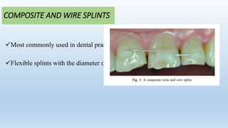
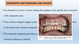
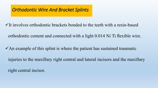
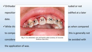
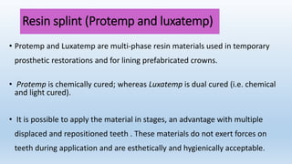
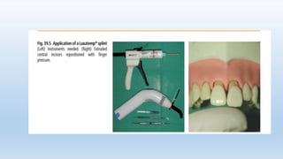
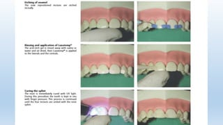
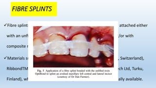
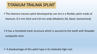
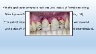
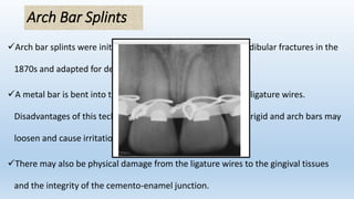
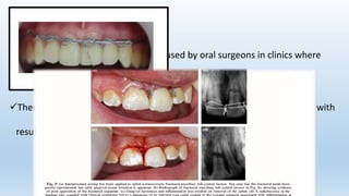
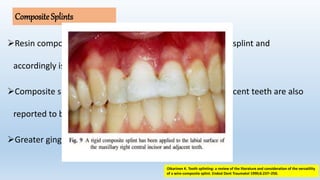
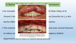
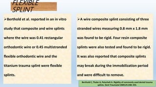
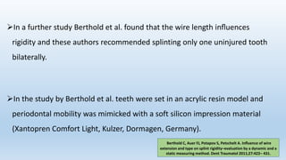
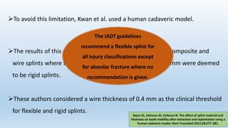
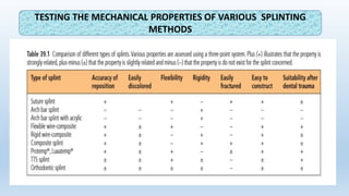
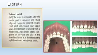
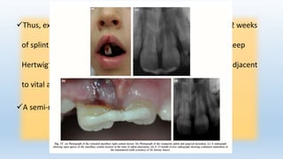
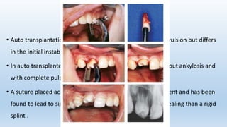
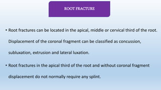
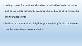
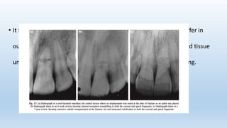
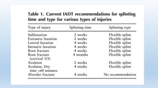
SPLINTING OF
TRAUMATIZED TEETH
PRESENTER- DR RUPALI
2nd year
Dept. of Pedodontics and Preventive dentistry
CONTENTS
INTRODUCTION
REQUISITES OF IDEAL SPLINTS
INFLUENCE OF SPLINTING ON DENTAL TISSUES
TYPES OF SPLINTS
TESTING THE MECHANICAL PROPERTIES OF VARIOUS SPLINTING
METHODS
INDICATIONS
CONTRAINDICATIONS
STEPS OF SPLINTING
RECOMMENDATIONS FOR SPLINTING TYPE AND DURATION
SPLINT REMOVAL
IADT GUIDELINES
CONCLUSION
REFERENCES
• A splint has been defined as ‘an apparatus used to support, protect or
immobilize teeth that have been loosened, replanted, fractured or subjected
to certain endodontic surgical procedures’.
American Association of Endodontists. Glossary
of Endodontic Terms. 8th ed. September 2015.
• The Glossary of Prosthodontic terms defines Splinting as: A rigid or
flexible device that maintains in position a displaced or movable part;
also used to keep in place and protect an injured part.
• According to Glossary of Periodontic terms it is: An appliance designed
to stabilize mobile teeth.
REQUIREMENTS FOR AN ACCEPTABLE
SPLINT
• Splint should preferably have slightly vertical and horizontal flexibility in
order to support healing.
• Rigidity of splints will be described as:
Flexible – more mobility than a non-injured tooth
Semi-rigid – equal to normal tooth mobility
Rigid – less than normal tooth mobility
An optimal splint should fulfil most or all of the following
requirements:
Direct intraoral application.
Easy to construct with materials available in dental practice.
Does not increase periodontal injuries or promote caries.
Does not irritate oral soft tissues.
Passive does not exert any orthodontic force on teeth.
Versatile in achieving rigid, semi-rigid or flexible splint.
Easy to remove and causes minimal or no permanent damage to the
dentition.
Allows pulp testing and endodontic treatment.
Hygienic and esthetic.
INFLUENCE OF SPLINTING ON DENTAL TISSUES
Influence upon gingiva
• The presence of wires placed in contact with the gingiva may lead to
invasion of bacteria through rupture or tears in the epithelial attachment.
• Wire‐loop splints (used in intermaxillary splinting) have been found to
lead to gingival changes which are, however, reversible after wire
removal.
Influence upon periodontal healing
• Experimental studies demonstrated that optimal periodontal
healing (i.e. with minimal ankylosis) after extraction and
replantation of teeth in animals was obtained in a non-splinted
situation compared to rigid splinting.
• It is assumed that slight mobility in the initial healing period
activates resorption of initially formed ankyloses sites.
Influence upon pulp healing
• Splinting could decrease pulp revascularization and increase the extent
of pulp necrosis and inflammatory root resorption compared to non-
splinting.
• In humans, splinting of auto transplanted teeth for only one week (with
a suture splint) has been found to improve pulpal healing as compared
to rigid splinting for four weeks.
Enamel changes after splinting
• The staining of the labial enamel takes place because of acid etching
and can be easily removed by careful polishing.
To stabilize moderate to advanced tooth mobility that cannot be reduced
by other means that has not responded to occlusal adjustment and
periodontal therapy.
Stabilize teeth in secondary occlusal trauma.
Stabilize teeth after orthodontic movement.
INDICATIONS
Stabilize teeth with increased tooth mobility, which interfere with
normal masticatory function.
Stabilize teeth after acute dental trauma i.e., subluxation, avulsion etc.
Facilitates scaling and surgical procedures.
Prevent tipping and drifting of teeth. Prevent extrusion of unopposed
teeth.
Lemmerman K. Rationale for stabilization. J Periodontol
1976;47:405-11.
Splinting teeth is not recommended if occlusal stability and optimal
periodontal conditions cannot be attained .
Any tooth mobility present before treatment must be reduced by means of
occlusal equilibration combined with periodontal therapy; otherwise if the
involved tooth does not respond, it must be extracted prior to continuing
from provisional restorations to definitive treatment.
CONTRAINDICATIONS
TYPES OF SPLINTING
Composite and wire splints
Composite and fishing line splints
Orthodontic wire and bracket
splints
Fibre splints
Titanium trauma
splints
Wire ligature splints
Arch bar splints
Composite splints
A splint for medical
emergency departments
A flexible splint
Resin splint
COMPOSITE AND WIRE SPLINTS
Most commonly used in dental practice.
Flexible splints with the diameter of 0.3 -0.4 mm
COMPOSITE AND FINISHING LINE SPLINTS
An alternative to wire is where fishing line replaces wire and the line is secured
with composite resin.
They utilizes whipper snipper nylon purchased from a hardware store attached
to the teeth with coloured composite resin (Ultradent; flowable purple.
The coloured composite provides a guide for the removal of the splint to
minimize damage to enamel.
Orthodontic Wire And Bracket Splints
It involves orthodontic brackets bonded to the teeth with a resin-based
orthodontic cement and connected with a light 0.014 Ni Ti flexible wire.
An example of this splint is where the patient has sustained traumatic
injuries to the maxillary right central and lateral incisors and the maxillary
right central incisor.
Orthodontic bracket splints allow teeth that have been intruded or not
repositioned correctly to have the occlusal relationships modified at a later
date.
While this type of splint was found to be irritating to the lips when compared
to composite and wire splints and titanium trauma splints, this is generally not
considered to be a clinical problem as any lip irritation can be avoided with
the application of wax.
Resin splint (Protemp and luxatemp)
• Protemp and Luxatemp are multi‐phase resin materials used in temporary
prosthetic restorations and for lining prefabricated crowns.
• Protemp is chemically cured; whereas Luxatemp is dual cured (i.e. chemical
and light cured).
• It is possible to apply the material in stages, an advantage with multiple
displaced and repositioned teeth . These materials do not exert forces on
teeth during application and are esthetically and hygienically acceptable.
FIBRE SPLINTS
Fibre splints use a polyethylene or Kevlar fibre mesh and are attached either
with an unfilled resin such as OptibondTM FL (Kerr, USA) and/or with
composite resin.
Materials such as Fiber-Splint (Polydentia SA Mezzovico-Vira, Switzerland),
RibbondTM (Ribbond Inc., Seattle, USA) or EverStick (Stick Tech Ltd, Turku,
Finland), which is a silinated E-type glass fibre, are commercially available.
TITANIUM TRAUMA SPLINT
The titanium trauma splint developed by von Arx is a flexible splint made of
titanium, 0.2 mm thick and 2.8 mm wide (Medartis AG, Basel, Switzerland).
It has a rhomboid mesh structure which is secured to the tooth with flowable
composite resin.
 A disadvantage of this splint type is its relatively high cost.
In this application composite resin was used instead of flowable resin (e.g.
Filtek Supreme Plus flowable restorative; 3M ESPE, St Paul, MN, USA).
The patient initially presented with an arch bar splint which was replaced
with a titanium trauma splint because of gross irritation to the gingival tissues.
Arch Bar Splints
Arch bar splints were initially adopted for maxillary and mandibular fractures in the
1870s and adapted for dento-alveolar trauma.
A metal bar is bent into the shape of the arch and fixed with ligature wires.
Disadvantages of this technique are that this type of splint is rigid and arch bars may
loosen and cause irritation.
There may also be physical damage from the ligature wires to the gingival tissues
and the integrity of the cemento-enamel junction.
Wire Ligature Splints
Wire ligature splints are sometimes used by oral surgeons in clinics where
dental splinting materials may not be available.
These splint types are generally rigid and impinge on the gingival tissues with
resulting inflammation.
CompositeSplints
Resin composite applied to the surfaces of teeth is a rigid splint and
accordingly is not recommended in the IADT guidelines.
Composite splints that are bonded interproximally to adjacent teeth are also
reported to be prone to fracture.
Greater gingival irritation.
Oikarinen K. Tooth splinting: a review of the literature and consideration of the versatility
of a wire-composite splint. Endod Dent Traumatol 1990;6:237–250.
A Splint For Medical Emergency Department.
An innovative approach in splinting developed by Dr Peter Foltyn at St
Vincent’s Hospital, Sydney, is the use of Stomahesive (ConvaTec Inc.), a skin
barrier adhesive material used for superficial skin trauma.
This material can crudely hold teeth in the socket and cover coronal fractures
to reduce sensitivity when patients are seen in emergency medical
departments out of hours of routine dental practice.
Though this, approach could require further repositioning with subsequent
splinting in a dental office.
Other temporary splinting may be provided by the use of the patient’s
mouthguards, orthodontic retainers, or aluminium foil adapted to the dental
arch and covered with thermoplastic acrylic.
DeAngelis AF, Barrowman RA, Harrod R, Nastri AL. Review article: Maxillofacial emergencies:
dentoalveolar and temporomandibular joint trauma. Emerg Med Australas 2014;26:439– 445.
FLEXIBLE
SPLINT
Berthold C, Thaler A, Petschelt A. Rigidity of commonly used dental trauma
splints. Dent Traumatol 2009;25:248–255.
Berthold et al. reported in an in vitro
study that composite and wire splints
where the wire was 0.41 rectangular
orthodontic wire or 0.45 multistranded
flexible orthodontic wire and the
titanium trauma splint were flexible
splints.
A wire composite splint consisting of three
stranded wires measuring 0.8 mm x 1.8 mm
was found to be rigid. Four resin composite
splints were also tested and found to be rigid.
It was also reported that composite splints
may break during the immobilization period
and were difficult to remove.
In a further study Berthold et al. found that the wire length influences
rigidity and these authors recommended splinting only one uninjured tooth
bilaterally.
In the study by Berthold et al. teeth were set in an acrylic resin model and
periodontal mobility was mimicked with a soft silicon impression material
(Xantopren Comfort Light, Kulzer, Dormagen, Germany).
Berthold C, Auer FJ, Potapov S, Petschelt A. Influence of wire
extension and type on splint rigidity–evaluation by a dynamic and a
static measuring method. Dent Traumatol 2011;27:422– 431.
To avoid this limitation, Kwan et al. used a human cadaveric model.
The results of this study also found composite splints and composite and
wire splints where the wire diameter was greater than 0.4 mm were deemed
to be rigid splints.
These authors considered a wire thickness of 0.4 mm as the clinical threshold
for flexible and rigid splints.
Kwan SC, Johnson JD, Cohenca N. The effect of splint material and
thickness on tooth mobility after extraction and replantation using a
human cadaveric model. Dent Traumatol 2012;28:277–281.
The IADT guidelines
recommend a flexible splint for
all injury classifications except
for alveolar fracture where no
recommendation is given.
TESTING THE MECHANICAL PROPERTIES OF VARIOUS SPLINTING
METHODS
 STEP 1
 STEP-2
 STEP 3
 STEP 4
RECOMMENDATIONS
FOR SPLINTING TYPE
AND DURATION
• Length and rigidity of splinting is primarily determined by the trauma
scenario.
 Extrusive luxation implies rupture of the periodontal ligament fibers. It has
been shown that the periodontal ligament achieves approximately 70% of its
original strength 2–3 weeks after injury.
EXTRUSION
Thus, extrusion represents a simple healing scenario and only 2 weeks
of splinting is needed to allow healing of the PDL as well as to keep
Hertwig’s epithelial root sheath in immature teeth in position adjacent
to vital apical tissues to ensure continued root development.
A semi‐rigid splint is recommended.
Traumatic dislocation in a lateral direction often causes damage to the PDL
and bone.
Hence the splinting time must be longer than for extrusively‐luxated teeth
in which the alveolar socket is intact. In these cases, a semi‐rigid splint
should be used for 4 weeks.
LATERAL LUXATION
Before removal of a splint a radiograph should be taken. If there are
radiographic signs of periodontal breakdown, additional splinting
time is necessary (usually 3–4 weeks)
• Intrusive luxation causes serious damage to the alveolar socket.
• If the tooth is surgically repositioned, the splinting time must be long enough
to support the tooth during remodeling of the bony socket, a process which
normally takes 6–8 weeks.
• To prevent permanent ankylosis a semi‐rigid splint should be used.
INTRUSION
• The duration of splinting is dependent upon the extent and nature of additional
damage to the socket (fracture).
• Replantation into an intact alveolar socket requires a splinting period of no
longer than 7–10 days to prevent ankylosis.
• In cases with incomplete eruption a suture placed over the incisal edge can
provide adequate support for an avulsed replanted tooth.
AVULSION
• Auto transplantation can be considered a well‐controlled avulsion but differs
in the initial instability due to the expanded socket.
• In auto transplanted teeth, healing usually takes place without ankylosis and
with complete pulp revascularization.
• A suture placed across the occlusal surface is usually sufficient and has been
found to lead to significantly better pulp and periodontal healing than a rigid
splint .
AUTOTRANSPLANTATION
• Root fractures can be located in the apical, middle or cervical third of the root.
Displacement of the coronal fragment can be classified as concussion,
subluxation, extrusion and lateral luxation.
• Root fractures in the apical third of the root and without coronal fragment
displacement do not normally require any splint.
ROOT FRACTURE
• In the past, root fractured teeth have been stabilized by a variety of splints,
such as cap splints, orthodontic appliances, bonded metal wires, composites
and fiber‐glass splints.
• Previous recommendations of rigid, long‐term splinting for all root fractures
have been questioned in recent studies.
• It has been shown that splinted and non‐splinted teeth did not differ in
outcome of treatment, i.e. whether the healing takes place by hard tissue
union, inter‐position of connective tissue and/or bone or no healing.
• The type of splinting – cap splint or wire‐composite splint – does not
influence outcome either in transverse or in oblique cervical root fractures.
Based on controversial results in recent studies, it has been recommended
to splint root fractures located at mid‐root for 4 weeks.
• In cervical root fractures, a longer splinting time might be indicated.
• The principles of treatment of alveolar fractures are related to the
treatment of both jaw fractures and tooth luxations.
• After proper repositioning, splinting is assumed to support both PDL and
bone healing.
• Fixation should be semi‐rigid and the splinting period 4 weeks.
ALVEOLAR BONE FRACTURES
• In most cases, splinting of luxated primary teeth is not possible due to lack of
patient cooperation.
• In the case of alveolar or mandibular fractures, a resin or a cap splint is
indicated.
• The cap splint should be cemented only on non‐traumatized teeth, leaving the
injured teeth free within the splint in order not to damage them when the
splint is removed.
SPLINTING IN PRIMARY
DENTITION
• Gingival fibers heal within one week, which is enough to provide some
stability.
• Where ankylosis may be a problem (avulsions), a short fixation period may
in some cases prevent permanent ankyloses.
GENERAL COMMENTS
• In cases where ankyloses is not a significant risk, a fixation period of 2–3
weeks is indicated.
• During the splinting period, it is essential that good oral hygiene be
maintained.
• Careful tooth brushing and rinsing with chlorhexidine are recommended.
When there is an associated injury to the bone, an additional 1–2 weeks is
indicated.
• If remodeling of the socket takes place (e.g. following crushing injuries,
as after intrusions and lateral luxation), 6–8 weeks of splinting may be
required.
• Concerning length of the splint (number of adjacent teeth), mobility
tests have shown that there is no need to extend the splint to more
than one non‐injured adjacent tooth to the injured tooth.
TYPE OF INJURY EXTRUSIVE
LUXATION
LATERAL
LUXATION
INTRUSIVE
LUXATION
AVULSION ROOT
FRACTURE
IN THE
CERVICAL
THIRD
ROOT
FRACTURE IN
THE MIDDLE OR
APICAL THIRD
ALVEOLAR
FRACTURE
FIXATION
PERIOD
TYPE OF
FIXATION
2 WEEKS
FLEXIBLE
4 WEEKS
FLEXIBLE
6-8 WEEKS
FLEXIBLE
1-2 WEEKS
FLEXIBLE
4 MONTHS
RIGID
4 WEEKS
FLEXIBLE
4 WEEKS
FLEXIBLE
FIXATION PERIOD AFTER VARIOUS DENTAL INJURIES
Splinting of Traumatized Teeth
J. O. Andreasen & K. S. Oikarinen ,5th edition
Splint Removal
• Removal of rigid arch bar splints or interdental wiring is often a difficult
process involving unwiring and cutting of wires close to the gingival
margins with potential damage to soft tissues.
• Removing a splint in which composite resin has been used is not only
time consuming, but iatrogenic injury to the enamel is an inevitable
outcome.
• Techniques of composite removal may involve debonding pliers,
handscalers, ultrasonic scalers, tungsten carbide burs, diamond burs,
Soflex disks (3M ESPE, St Paul, MN, USA), rubber wheels and cups.
• It has been shown experimentally that debonding pliers generate shearing
forces that result in irreversible damage to the enamel. Additionally, the
forces exerted may disturb the periodontal healing of the injured tooth.
• Both hand and ultrasonic scalers caused distinctive patterns of enamel
detachment and therefore it was concluded that they should not be used
for composite removal.
• It was found that tungsten carbide burs and Soflex discs resulted in
the least damage to the enamel.
Cehreli ZC, Lakshmipathy M, Yazici R. Effect of different splint removal techniques on
the surface roughness of human enamel: a three-dimensional optical profilometry
analysis. Dent Traumatol 2008;24:177–182.
CONCLUSIONS
While the type of splint and the splinting duration have not been generally
shown to affect healing outcomes, the IADT guidelines support the use of
flexible splints whenever possible.
This has often been achieved with the use of composite resin or orthodontic
brackets and light wire. Both of these techniques have been shown to cause
iatrogenic damage to the enamel.
A new protocol using a resin activated glass ionomer cement has been
proposed that offers ease of application and removal with minimal or no
iatrogenic damage to enamel.
REFERENCES
• American Association of Endodontists. Glossary of Endodontic Terms. 8th ed.
September 2015.
• Lemmerman K. Rationale for stabilization. J Periodontol 1976;47:405-11.
• Berthold C, Thaler A, Petschelt A. Rigidity of commonly used dental trauma
splints. Dent Traumatol 2009;25:248–255.
• Siegel SC, Driscoll CF, Feldman S. Tooth stabilization and splinting before and after
periodontal therapy with fixed partial dentures. Dent Clin North Am 1999;43:45-76.
• Splinting of Traumatized Teeth J. O. Andreasen & K. S. Oikarinen ,5th edition
Oikarinen K. Tooth splinting: a review of the literature and consideration of the
versatility of a wire-composite splint. Endod Dent Traumatol 1990;6:237–250.
Splinting of traumatized teeth

Splinting of traumatized teeth

  • 1.
    SPLINTING OF TRAUMATIZED TEETH PRESENTER-DR RUPALI 2nd year Dept. of Pedodontics and Preventive dentistry
  • 2.
    CONTENTS INTRODUCTION REQUISITES OF IDEALSPLINTS INFLUENCE OF SPLINTING ON DENTAL TISSUES TYPES OF SPLINTS TESTING THE MECHANICAL PROPERTIES OF VARIOUS SPLINTING METHODS INDICATIONS CONTRAINDICATIONS
  • 3.
    STEPS OF SPLINTING RECOMMENDATIONSFOR SPLINTING TYPE AND DURATION SPLINT REMOVAL IADT GUIDELINES CONCLUSION REFERENCES
  • 5.
    • A splinthas been defined as ‘an apparatus used to support, protect or immobilize teeth that have been loosened, replanted, fractured or subjected to certain endodontic surgical procedures’. American Association of Endodontists. Glossary of Endodontic Terms. 8th ed. September 2015.
  • 6.
    • The Glossaryof Prosthodontic terms defines Splinting as: A rigid or flexible device that maintains in position a displaced or movable part; also used to keep in place and protect an injured part. • According to Glossary of Periodontic terms it is: An appliance designed to stabilize mobile teeth.
  • 7.
    REQUIREMENTS FOR ANACCEPTABLE SPLINT • Splint should preferably have slightly vertical and horizontal flexibility in order to support healing. • Rigidity of splints will be described as: Flexible – more mobility than a non-injured tooth Semi-rigid – equal to normal tooth mobility Rigid – less than normal tooth mobility
  • 8.
    An optimal splintshould fulfil most or all of the following requirements: Direct intraoral application. Easy to construct with materials available in dental practice. Does not increase periodontal injuries or promote caries. Does not irritate oral soft tissues.
  • 9.
    Passive does notexert any orthodontic force on teeth. Versatile in achieving rigid, semi-rigid or flexible splint. Easy to remove and causes minimal or no permanent damage to the dentition. Allows pulp testing and endodontic treatment. Hygienic and esthetic.
  • 10.
    INFLUENCE OF SPLINTINGON DENTAL TISSUES Influence upon gingiva • The presence of wires placed in contact with the gingiva may lead to invasion of bacteria through rupture or tears in the epithelial attachment. • Wire‐loop splints (used in intermaxillary splinting) have been found to lead to gingival changes which are, however, reversible after wire removal.
  • 11.
    Influence upon periodontalhealing • Experimental studies demonstrated that optimal periodontal healing (i.e. with minimal ankylosis) after extraction and replantation of teeth in animals was obtained in a non-splinted situation compared to rigid splinting. • It is assumed that slight mobility in the initial healing period activates resorption of initially formed ankyloses sites.
  • 12.
    Influence upon pulphealing • Splinting could decrease pulp revascularization and increase the extent of pulp necrosis and inflammatory root resorption compared to non- splinting. • In humans, splinting of auto transplanted teeth for only one week (with a suture splint) has been found to improve pulpal healing as compared to rigid splinting for four weeks.
  • 13.
    Enamel changes aftersplinting • The staining of the labial enamel takes place because of acid etching and can be easily removed by careful polishing.
  • 14.
    To stabilize moderateto advanced tooth mobility that cannot be reduced by other means that has not responded to occlusal adjustment and periodontal therapy. Stabilize teeth in secondary occlusal trauma. Stabilize teeth after orthodontic movement. INDICATIONS
  • 15.
    Stabilize teeth withincreased tooth mobility, which interfere with normal masticatory function. Stabilize teeth after acute dental trauma i.e., subluxation, avulsion etc. Facilitates scaling and surgical procedures. Prevent tipping and drifting of teeth. Prevent extrusion of unopposed teeth. Lemmerman K. Rationale for stabilization. J Periodontol 1976;47:405-11.
  • 16.
    Splinting teeth isnot recommended if occlusal stability and optimal periodontal conditions cannot be attained . Any tooth mobility present before treatment must be reduced by means of occlusal equilibration combined with periodontal therapy; otherwise if the involved tooth does not respond, it must be extracted prior to continuing from provisional restorations to definitive treatment. CONTRAINDICATIONS
  • 17.
  • 18.
    Composite and wiresplints Composite and fishing line splints Orthodontic wire and bracket splints Fibre splints Titanium trauma splints Wire ligature splints Arch bar splints Composite splints A splint for medical emergency departments A flexible splint Resin splint
  • 19.
    COMPOSITE AND WIRESPLINTS Most commonly used in dental practice. Flexible splints with the diameter of 0.3 -0.4 mm
  • 20.
    COMPOSITE AND FINISHINGLINE SPLINTS An alternative to wire is where fishing line replaces wire and the line is secured with composite resin. They utilizes whipper snipper nylon purchased from a hardware store attached to the teeth with coloured composite resin (Ultradent; flowable purple. The coloured composite provides a guide for the removal of the splint to minimize damage to enamel.
  • 21.
    Orthodontic Wire AndBracket Splints It involves orthodontic brackets bonded to the teeth with a resin-based orthodontic cement and connected with a light 0.014 Ni Ti flexible wire. An example of this splint is where the patient has sustained traumatic injuries to the maxillary right central and lateral incisors and the maxillary right central incisor.
  • 22.
    Orthodontic bracket splintsallow teeth that have been intruded or not repositioned correctly to have the occlusal relationships modified at a later date. While this type of splint was found to be irritating to the lips when compared to composite and wire splints and titanium trauma splints, this is generally not considered to be a clinical problem as any lip irritation can be avoided with the application of wax.
  • 23.
    Resin splint (Protempand luxatemp) • Protemp and Luxatemp are multi‐phase resin materials used in temporary prosthetic restorations and for lining prefabricated crowns. • Protemp is chemically cured; whereas Luxatemp is dual cured (i.e. chemical and light cured). • It is possible to apply the material in stages, an advantage with multiple displaced and repositioned teeth . These materials do not exert forces on teeth during application and are esthetically and hygienically acceptable.
  • 26.
    FIBRE SPLINTS Fibre splintsuse a polyethylene or Kevlar fibre mesh and are attached either with an unfilled resin such as OptibondTM FL (Kerr, USA) and/or with composite resin. Materials such as Fiber-Splint (Polydentia SA Mezzovico-Vira, Switzerland), RibbondTM (Ribbond Inc., Seattle, USA) or EverStick (Stick Tech Ltd, Turku, Finland), which is a silinated E-type glass fibre, are commercially available.
  • 27.
    TITANIUM TRAUMA SPLINT Thetitanium trauma splint developed by von Arx is a flexible splint made of titanium, 0.2 mm thick and 2.8 mm wide (Medartis AG, Basel, Switzerland). It has a rhomboid mesh structure which is secured to the tooth with flowable composite resin.  A disadvantage of this splint type is its relatively high cost.
  • 28.
    In this applicationcomposite resin was used instead of flowable resin (e.g. Filtek Supreme Plus flowable restorative; 3M ESPE, St Paul, MN, USA). The patient initially presented with an arch bar splint which was replaced with a titanium trauma splint because of gross irritation to the gingival tissues.
  • 29.
    Arch Bar Splints Archbar splints were initially adopted for maxillary and mandibular fractures in the 1870s and adapted for dento-alveolar trauma. A metal bar is bent into the shape of the arch and fixed with ligature wires. Disadvantages of this technique are that this type of splint is rigid and arch bars may loosen and cause irritation. There may also be physical damage from the ligature wires to the gingival tissues and the integrity of the cemento-enamel junction.
  • 30.
    Wire Ligature Splints Wireligature splints are sometimes used by oral surgeons in clinics where dental splinting materials may not be available. These splint types are generally rigid and impinge on the gingival tissues with resulting inflammation.
  • 31.
    CompositeSplints Resin composite appliedto the surfaces of teeth is a rigid splint and accordingly is not recommended in the IADT guidelines. Composite splints that are bonded interproximally to adjacent teeth are also reported to be prone to fracture. Greater gingival irritation. Oikarinen K. Tooth splinting: a review of the literature and consideration of the versatility of a wire-composite splint. Endod Dent Traumatol 1990;6:237–250.
  • 32.
    A Splint ForMedical Emergency Department. An innovative approach in splinting developed by Dr Peter Foltyn at St Vincent’s Hospital, Sydney, is the use of Stomahesive (ConvaTec Inc.), a skin barrier adhesive material used for superficial skin trauma. This material can crudely hold teeth in the socket and cover coronal fractures to reduce sensitivity when patients are seen in emergency medical departments out of hours of routine dental practice.
  • 33.
    Though this, approachcould require further repositioning with subsequent splinting in a dental office. Other temporary splinting may be provided by the use of the patient’s mouthguards, orthodontic retainers, or aluminium foil adapted to the dental arch and covered with thermoplastic acrylic. DeAngelis AF, Barrowman RA, Harrod R, Nastri AL. Review article: Maxillofacial emergencies: dentoalveolar and temporomandibular joint trauma. Emerg Med Australas 2014;26:439– 445.
  • 34.
    FLEXIBLE SPLINT Berthold C, ThalerA, Petschelt A. Rigidity of commonly used dental trauma splints. Dent Traumatol 2009;25:248–255. Berthold et al. reported in an in vitro study that composite and wire splints where the wire was 0.41 rectangular orthodontic wire or 0.45 multistranded flexible orthodontic wire and the titanium trauma splint were flexible splints. A wire composite splint consisting of three stranded wires measuring 0.8 mm x 1.8 mm was found to be rigid. Four resin composite splints were also tested and found to be rigid. It was also reported that composite splints may break during the immobilization period and were difficult to remove.
  • 35.
    In a furtherstudy Berthold et al. found that the wire length influences rigidity and these authors recommended splinting only one uninjured tooth bilaterally. In the study by Berthold et al. teeth were set in an acrylic resin model and periodontal mobility was mimicked with a soft silicon impression material (Xantopren Comfort Light, Kulzer, Dormagen, Germany). Berthold C, Auer FJ, Potapov S, Petschelt A. Influence of wire extension and type on splint rigidity–evaluation by a dynamic and a static measuring method. Dent Traumatol 2011;27:422– 431.
  • 36.
    To avoid thislimitation, Kwan et al. used a human cadaveric model. The results of this study also found composite splints and composite and wire splints where the wire diameter was greater than 0.4 mm were deemed to be rigid splints. These authors considered a wire thickness of 0.4 mm as the clinical threshold for flexible and rigid splints. Kwan SC, Johnson JD, Cohenca N. The effect of splint material and thickness on tooth mobility after extraction and replantation using a human cadaveric model. Dent Traumatol 2012;28:277–281. The IADT guidelines recommend a flexible splint for all injury classifications except for alveolar fracture where no recommendation is given.
  • 37.
    TESTING THE MECHANICALPROPERTIES OF VARIOUS SPLINTING METHODS
  • 39.
  • 40.
  • 41.
  • 42.
  • 43.
  • 44.
    • Length andrigidity of splinting is primarily determined by the trauma scenario.  Extrusive luxation implies rupture of the periodontal ligament fibers. It has been shown that the periodontal ligament achieves approximately 70% of its original strength 2–3 weeks after injury. EXTRUSION
  • 45.
    Thus, extrusion representsa simple healing scenario and only 2 weeks of splinting is needed to allow healing of the PDL as well as to keep Hertwig’s epithelial root sheath in immature teeth in position adjacent to vital apical tissues to ensure continued root development. A semi‐rigid splint is recommended.
  • 46.
    Traumatic dislocation ina lateral direction often causes damage to the PDL and bone. Hence the splinting time must be longer than for extrusively‐luxated teeth in which the alveolar socket is intact. In these cases, a semi‐rigid splint should be used for 4 weeks. LATERAL LUXATION
  • 47.
    Before removal ofa splint a radiograph should be taken. If there are radiographic signs of periodontal breakdown, additional splinting time is necessary (usually 3–4 weeks)
  • 48.
    • Intrusive luxationcauses serious damage to the alveolar socket. • If the tooth is surgically repositioned, the splinting time must be long enough to support the tooth during remodeling of the bony socket, a process which normally takes 6–8 weeks. • To prevent permanent ankylosis a semi‐rigid splint should be used. INTRUSION
  • 49.
    • The durationof splinting is dependent upon the extent and nature of additional damage to the socket (fracture). • Replantation into an intact alveolar socket requires a splinting period of no longer than 7–10 days to prevent ankylosis. • In cases with incomplete eruption a suture placed over the incisal edge can provide adequate support for an avulsed replanted tooth. AVULSION
  • 50.
    • Auto transplantationcan be considered a well‐controlled avulsion but differs in the initial instability due to the expanded socket. • In auto transplanted teeth, healing usually takes place without ankylosis and with complete pulp revascularization. • A suture placed across the occlusal surface is usually sufficient and has been found to lead to significantly better pulp and periodontal healing than a rigid splint . AUTOTRANSPLANTATION
  • 51.
    • Root fracturescan be located in the apical, middle or cervical third of the root. Displacement of the coronal fragment can be classified as concussion, subluxation, extrusion and lateral luxation. • Root fractures in the apical third of the root and without coronal fragment displacement do not normally require any splint. ROOT FRACTURE
  • 52.
    • In thepast, root fractured teeth have been stabilized by a variety of splints, such as cap splints, orthodontic appliances, bonded metal wires, composites and fiber‐glass splints. • Previous recommendations of rigid, long‐term splinting for all root fractures have been questioned in recent studies.
  • 53.
    • It hasbeen shown that splinted and non‐splinted teeth did not differ in outcome of treatment, i.e. whether the healing takes place by hard tissue union, inter‐position of connective tissue and/or bone or no healing.
  • 54.
    • The typeof splinting – cap splint or wire‐composite splint – does not influence outcome either in transverse or in oblique cervical root fractures. Based on controversial results in recent studies, it has been recommended to splint root fractures located at mid‐root for 4 weeks. • In cervical root fractures, a longer splinting time might be indicated.
  • 56.
    • The principlesof treatment of alveolar fractures are related to the treatment of both jaw fractures and tooth luxations. • After proper repositioning, splinting is assumed to support both PDL and bone healing. • Fixation should be semi‐rigid and the splinting period 4 weeks. ALVEOLAR BONE FRACTURES
  • 57.
    • In mostcases, splinting of luxated primary teeth is not possible due to lack of patient cooperation. • In the case of alveolar or mandibular fractures, a resin or a cap splint is indicated. • The cap splint should be cemented only on non‐traumatized teeth, leaving the injured teeth free within the splint in order not to damage them when the splint is removed. SPLINTING IN PRIMARY DENTITION
  • 58.
    • Gingival fibersheal within one week, which is enough to provide some stability. • Where ankylosis may be a problem (avulsions), a short fixation period may in some cases prevent permanent ankyloses. GENERAL COMMENTS
  • 59.
    • In caseswhere ankyloses is not a significant risk, a fixation period of 2–3 weeks is indicated. • During the splinting period, it is essential that good oral hygiene be maintained. • Careful tooth brushing and rinsing with chlorhexidine are recommended. When there is an associated injury to the bone, an additional 1–2 weeks is indicated.
  • 60.
    • If remodelingof the socket takes place (e.g. following crushing injuries, as after intrusions and lateral luxation), 6–8 weeks of splinting may be required. • Concerning length of the splint (number of adjacent teeth), mobility tests have shown that there is no need to extend the splint to more than one non‐injured adjacent tooth to the injured tooth.
  • 61.
    TYPE OF INJURYEXTRUSIVE LUXATION LATERAL LUXATION INTRUSIVE LUXATION AVULSION ROOT FRACTURE IN THE CERVICAL THIRD ROOT FRACTURE IN THE MIDDLE OR APICAL THIRD ALVEOLAR FRACTURE FIXATION PERIOD TYPE OF FIXATION 2 WEEKS FLEXIBLE 4 WEEKS FLEXIBLE 6-8 WEEKS FLEXIBLE 1-2 WEEKS FLEXIBLE 4 MONTHS RIGID 4 WEEKS FLEXIBLE 4 WEEKS FLEXIBLE FIXATION PERIOD AFTER VARIOUS DENTAL INJURIES Splinting of Traumatized Teeth J. O. Andreasen & K. S. Oikarinen ,5th edition
  • 63.
    Splint Removal • Removalof rigid arch bar splints or interdental wiring is often a difficult process involving unwiring and cutting of wires close to the gingival margins with potential damage to soft tissues. • Removing a splint in which composite resin has been used is not only time consuming, but iatrogenic injury to the enamel is an inevitable outcome.
  • 64.
    • Techniques ofcomposite removal may involve debonding pliers, handscalers, ultrasonic scalers, tungsten carbide burs, diamond burs, Soflex disks (3M ESPE, St Paul, MN, USA), rubber wheels and cups.
  • 65.
    • It hasbeen shown experimentally that debonding pliers generate shearing forces that result in irreversible damage to the enamel. Additionally, the forces exerted may disturb the periodontal healing of the injured tooth. • Both hand and ultrasonic scalers caused distinctive patterns of enamel detachment and therefore it was concluded that they should not be used for composite removal.
  • 66.
    • It wasfound that tungsten carbide burs and Soflex discs resulted in the least damage to the enamel. Cehreli ZC, Lakshmipathy M, Yazici R. Effect of different splint removal techniques on the surface roughness of human enamel: a three-dimensional optical profilometry analysis. Dent Traumatol 2008;24:177–182.
  • 67.
    CONCLUSIONS While the typeof splint and the splinting duration have not been generally shown to affect healing outcomes, the IADT guidelines support the use of flexible splints whenever possible. This has often been achieved with the use of composite resin or orthodontic brackets and light wire. Both of these techniques have been shown to cause iatrogenic damage to the enamel. A new protocol using a resin activated glass ionomer cement has been proposed that offers ease of application and removal with minimal or no iatrogenic damage to enamel.
  • 68.
    REFERENCES • American Associationof Endodontists. Glossary of Endodontic Terms. 8th ed. September 2015. • Lemmerman K. Rationale for stabilization. J Periodontol 1976;47:405-11. • Berthold C, Thaler A, Petschelt A. Rigidity of commonly used dental trauma splints. Dent Traumatol 2009;25:248–255. • Siegel SC, Driscoll CF, Feldman S. Tooth stabilization and splinting before and after periodontal therapy with fixed partial dentures. Dent Clin North Am 1999;43:45-76. • Splinting of Traumatized Teeth J. O. Andreasen & K. S. Oikarinen ,5th edition Oikarinen K. Tooth splinting: a review of the literature and consideration of the versatility of a wire-composite splint. Endod Dent Traumatol 1990;6:237–250.