Spinal Nerve
•Download as PPT, PDF•
211 likes•106,120 views

Spinal nerves emerge from the spinal cord and carry sensory and motor information between the spinal cord and specific body regions. There are 31 pairs of spinal nerves that are categorized based on the region of the spinal cord they emerge from. The anterior rami of spinal nerves form plexuses that further distribute nerves to various body structures, while the thoracic spinal nerves directly innervate the intercostal muscles and skin as intercostal nerves.

Recommended
More Related Content
What's hot
What's hot (20)
Similar to Spinal Nerve
Similar to Spinal Nerve (20)
More from Sado Anatomist
More from Sado Anatomist (20)
Recently uploaded
Recently uploaded (20)
Spinal Nerve
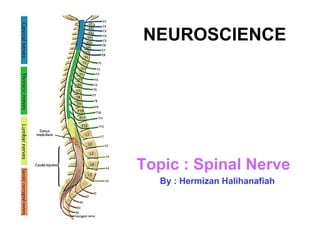
- 1. NEUROSCIENCE Topic : Spinal Nerve By : Hermizan Halihanafiah
- 2. Spinal Nerve • Spinal nerve are the path of communication between the spinal cord and the specific region of the body neuroscienceMizan
- 3. Spinal Nerve • 31 pairs • Spinal nerve follows the name of corresponding vertebra column. • Consists cervical spinal nerve, thoracic spinal nerve, lumbar spinal nerve, sacral spinal nerve and coccyx spinal nerve. • Emerge from spinal cord and through the intervertebral foramina of vertebra. neuroscienceMizan
- 4. Spinal Nerve • Spinal nerves: 1. 8 pairs of cervical spinal nerves 2. 12 pairs of thoracic spinal nerves 3. 5 pairs of lumbar spinal nerves. 4. 5 pairs of sacral spinal nerves 5. 1 pairs of coccyx spinal nerves. neuroscienceMizan
- 5. Spinal Nerve • Cervical and thoracic spinal nerves arise and leave at corresponding vertebra . • Because the spinal cord are shorter than vertebra column, nerve that arise from lumbar, sacral and coccyx region of spinal cord do not leave the vertebra column at the same level where they exit the cord. • The root of these spinal nerves angle inferiorly in the vertebral canal from the end of spinal cord like wisps of hair. neuroscienceMizan
- 6. Spinal Nerves neuroscienceMizan
- 7. • These root of this nerve, collectively called cauda equina. • Typical spinal nerve has 2 connection to spinal cord; posterior / dorsal and anterior/ ventral root. • Posterior and anterior root unite to form spinal nerve at intervertebral foramina. • Since posterior root contain sensory axons and anterior root contain motor axons, spinal nerves is classified as a mixed nerve. • Posterior root contain posterior root ganglion which cell bodies of sensory neuron is located. neuroscienceMizan
- 8. Spinal Nerves neuroscienceMizan
- 11. Spinal Nerves Cervical spinal nerves • 8 Pairs • 1st pair emerge between atlas and occipital bone • The remaining emerge from the vertebral column through intervertebral foramina. • Spinal nerves C1 – C7 exits the vertebral canal above their corresponding vertebra. • Spinal nerve C8 exits vertebral canal between C7 and T1 neuroscienceMizan
- 12. Cervical Spinal Nerves neuroscienceMizan
- 13. Spinal Nerves Thoracic spinal nerve • 12 pairs • Exits the vertebral canal below their corresponding vertebra. • Emerge from thoracic vertebra • Continuous to form intercostals nerves. neuroscienceMizan
- 14. Spinal Nerves Lumbar Spinal Nerve • 5 pairs • Emerge from lumbar vertebra. • Exits the vertebral canal below their corresponding vertebra. neuroscienceMizan
- 15. Spinal Nerves Sacral and Coccyx Spinal Nerve • 5 pairs • From the spinal cord, the root of the sacral spinal nerve enter the sacral canal (part of the vertebral canal). • Sacral nerves (S1-S4) exits the vertebral canal via 4 pairs of anterior and posterior sacral foramen. • Spinal nerves S5 and Co1 exits from sacral hiatus. neuroscienceMizan
- 16. Distribution of Spinal Nerve Branches • From the root, after passing the intervetebral foramen, a spinal nerve divide into several branches. • Theses branches are call rami (ramus) ; posterior (dorsal) ramus and anterior (ventral) ramus. • Posterior (dorsal) ramus serve the deep muscles and skin of the posterior surface of the trunk. • Anterior (ventral) ramus serve muscles and structure of the upper and lower limbs and the skin of the lateral and anterior surface of the trunk. neuroscienceMizan
- 17. Distribution of Spinal Nerve Branches • Spinal nerve also give off a meningeal branch, where this branch reenter the vertebral cavity through intervertebral foramen and supply the vertebra, vertebra ligament and blood vessels of spinal cord and meninges. • Other branches from spinal nerve are the rami communicantes ( components of ANS) neuroscienceMizan
- 19. Distribution of Spinal Nerve Plexuses • Axons from the anterior (ventral) rami of spinal nerves, except for the thoracic nerves T2-T12, do not go directly to the body structures they supply. • They form network on both the left and right side of the body by joining with various numbers of axons from anterior rami of adjacent nerves. neuroscienceMizan
- 20. Distribution of Spinal Nerve Plexuses • Network of axons is call plexuses. • The principle plexus: 1. Cervical plexus 2. Brachial plexus 3. Lumbar plexus 4. Sacral plexus 5. Coccygeal plexus neuroscienceMizan
- 22. Distribution of Spinal Nerve Plexuses • Emerging from the plexus are nerves bearing names that are often descriptive of the general regions they serve or the course they take. • Each of the nerves in turn may have several branches named for the specific structures they innervate. neuroscienceMizan
- 23. Plexuses Cervical Plexus • Formed by the roots of anterior rami of the 1st four cervical nerve (C1-C4), with contribution from C5. • There is one on each side of the neck alongside the first 4 cervical vertebrae. • Supply the skin (sensory / cutaneous innervations) and muscles (motor innervations) of the: head, neck superior part of the shoulder and chest. neuroscienceMizan
- 25. Plexuses Brachial Plexus • Form by roots of anterior rami of spinal nerves of C5-C8 and T1. • Extends inferiorly and laterally on either side of the last 4 and 5 cervical vertebrae. • Divides into roots, trunks, divisions, cords and terminal branches (nerves). • 5 important nerves arise from brachial plexus are musculocutaneous, axillary, radial, ulnar and median nerves. • Give skin (sensory) innervations and muscles (motor) innervations for the most of the shoulder and upper limbs region. neuroscienceMizan
- 27. Plexuses Lumbar Plexus • The roots of anterior rami of spinal nerves L1-L4 form lumbar plexus. • Minimal intermingling axons compare to brachial plexus. • The main nerve is a femoral (the largest) and obturator nerves. • Supply the skin and muscles of the: – anterolateral abdominal wall – external genitals (cremaster muscles, skin on scrotum, labia majora etc) – part of the lower limbs (adductor muscles of tight, flexor muscles of hip and extensor muscles of knee joint) neuroscienceMizan
- 29. Plexuses Sacral Plexus • Form by the roots of anterior rami of spinal nerves L4-L5 and S1-S4. • This plexus is situated largely anterior to the sacrum • The largest nerve in the body, sciatic nerve arise from the sacral plexus. • Also pudendal nerve, superior gluteal, inferior gluteal etc. • Supply the skin (sensory) and muscles (motor) of the: – Buttock – Perineum – Lower limbs neuroscienceMizan
- 31. Plexuses Coccygeal Plexus • Form from roots of anterior rami of spinal nerves S4 and S5 and the coccygeal nerves. • Supplies a small area of skin in the coccygeal region. neuroscienceMizan
- 33. Intercostal Nerves • The anterior rami of spinal nerves T2-T12 do not enter into the formation of plexuses and are known as intercostal or thoracic nerves. • These nerves directly connect to the structure they supply in the intercostal spaces. • After leaving its intervertebral foramen, anterior ramus of nerve T2 innervates the intercostal muscles of the second intercostal space and supplies the skin of the axilla and posteromedial aspect of the arm. neuroscienceMizan
- 34. Intercostal Nerves • Nerves T3-T6 extend along the costal groove of the ribs and then to the intercostal muscles and skin of the anterior and lateral chest wall. • Nerves T7-T12 supply the intercostal muscles and abdominals muscles , along with the overlying skin. • The posterior rami of the intercostal nerves supply the deep back muscles and skin of the posterior aspect of the thorax. neuroscienceMizan