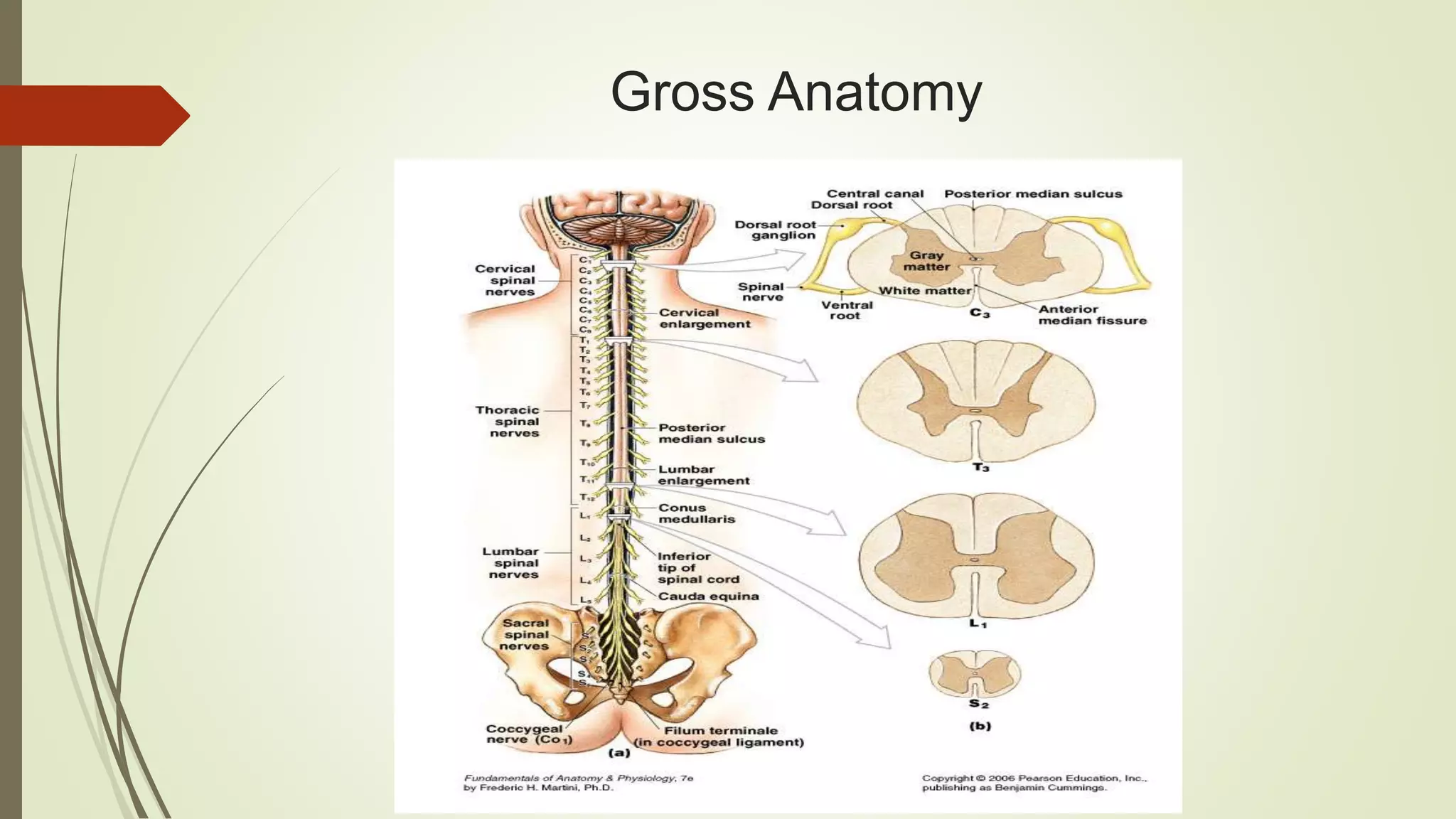
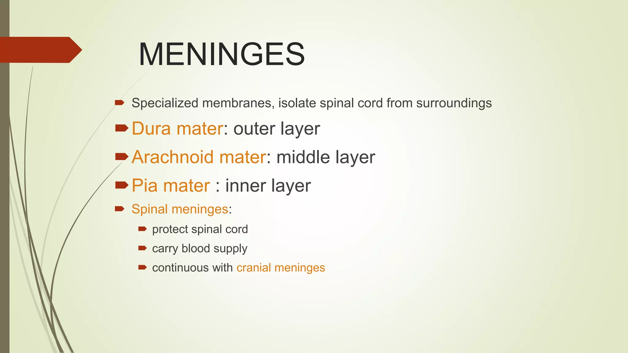
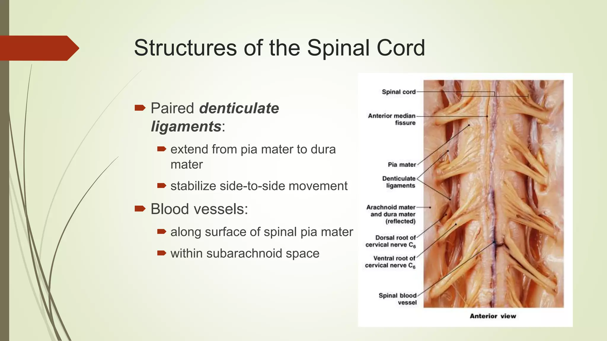
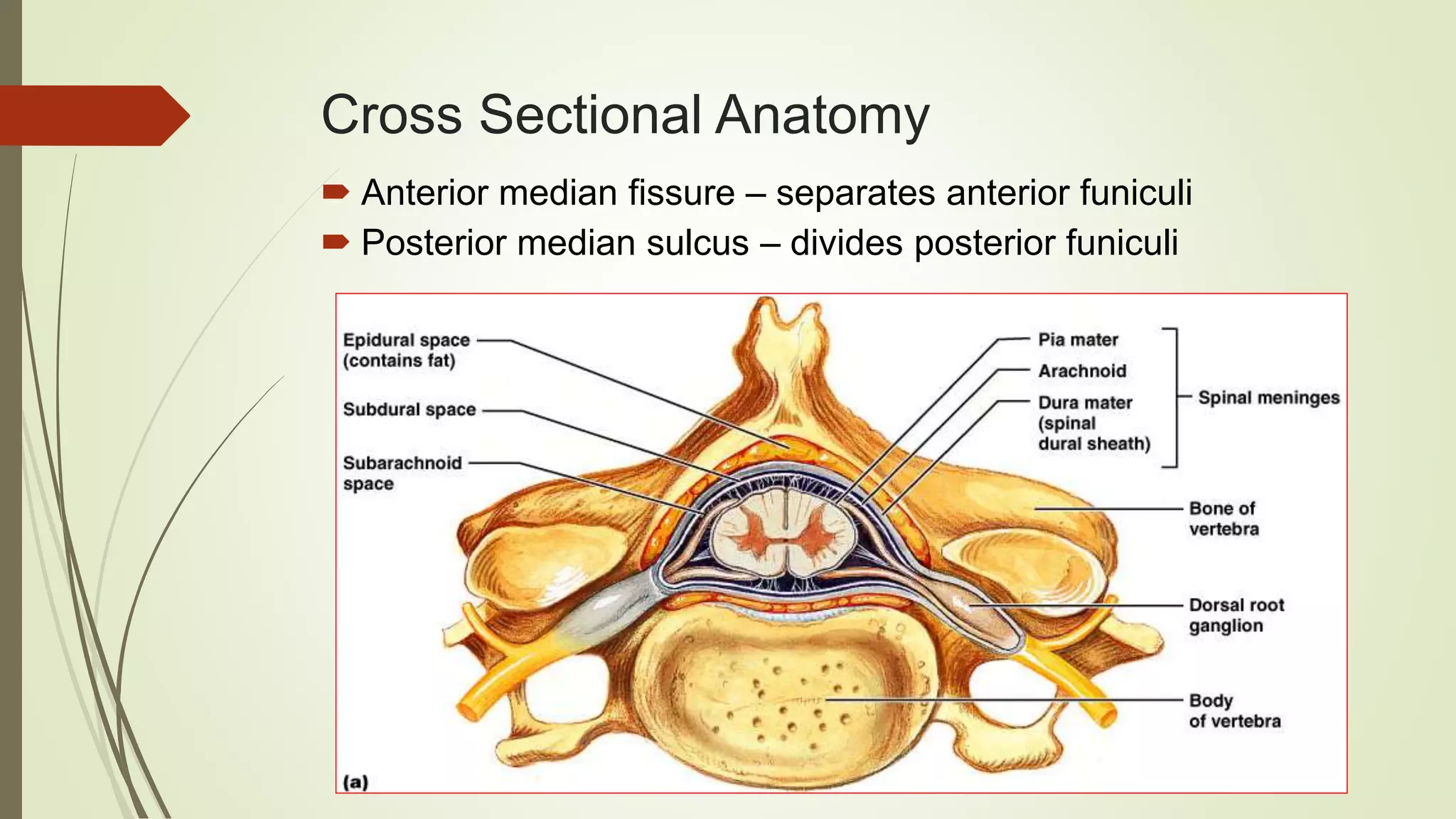
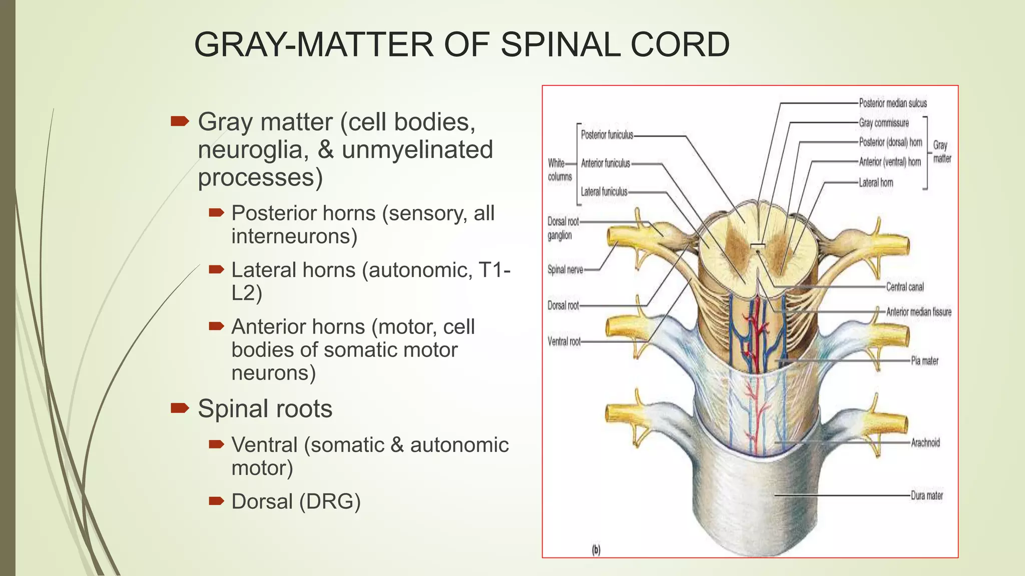
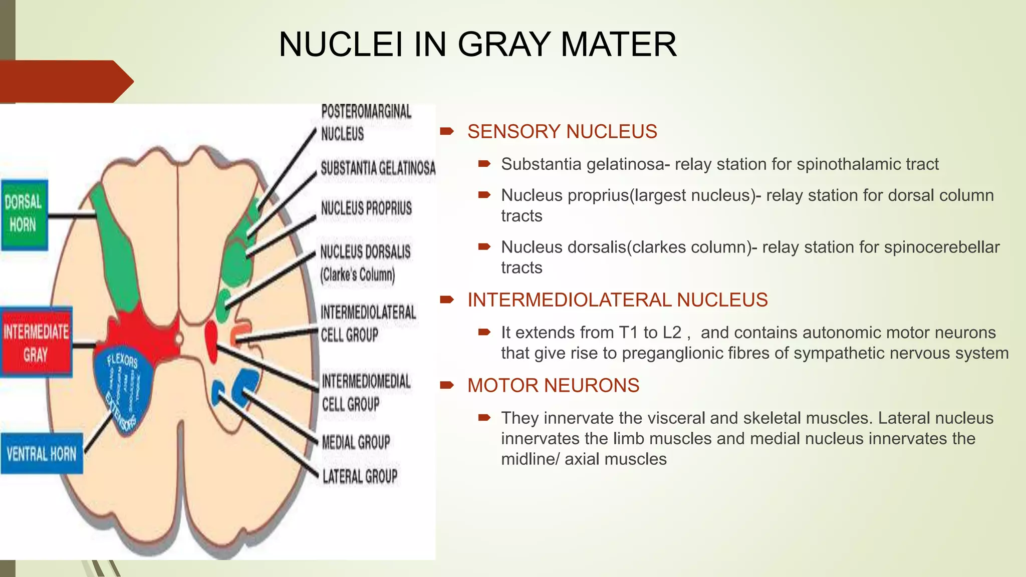
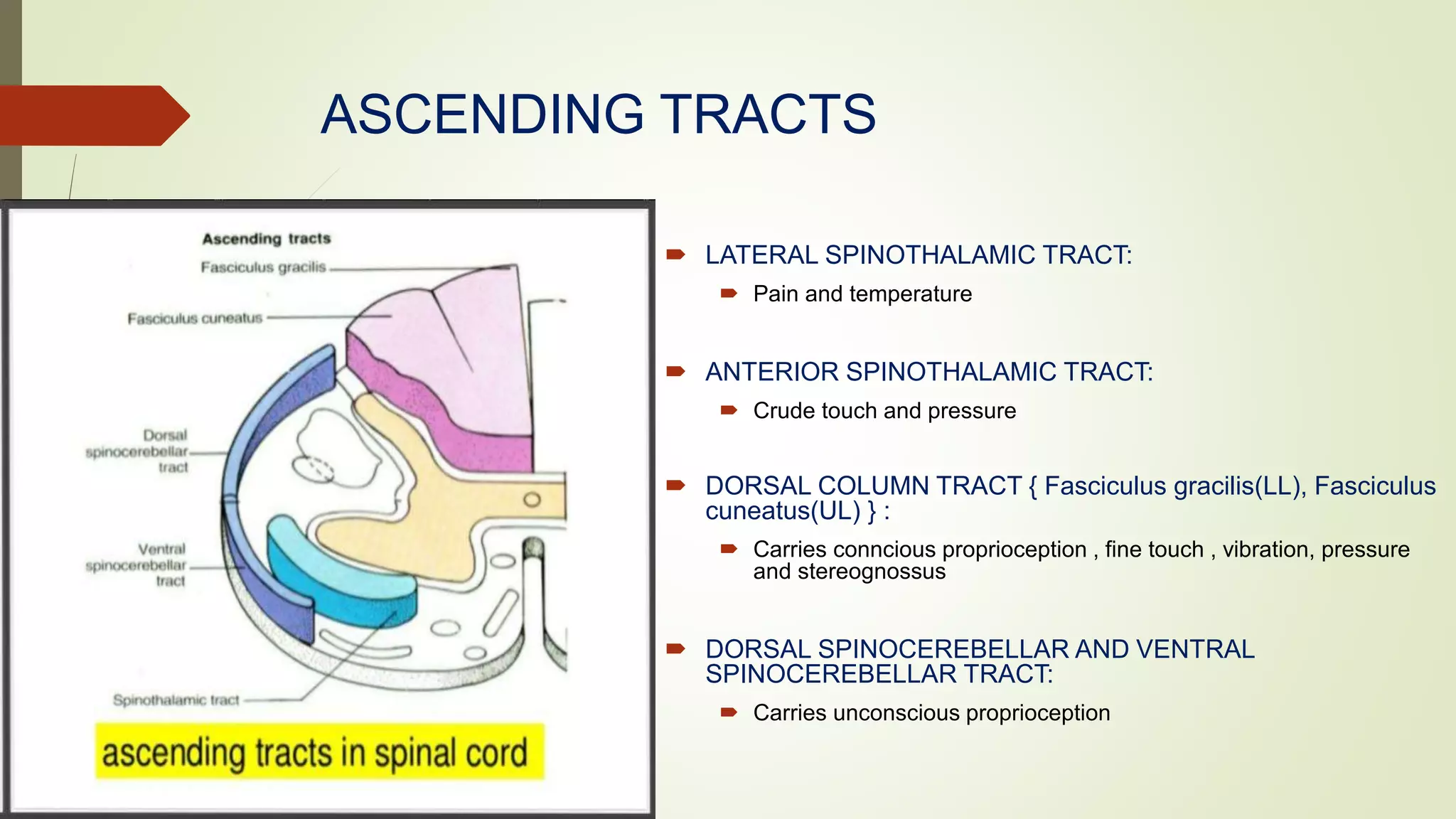
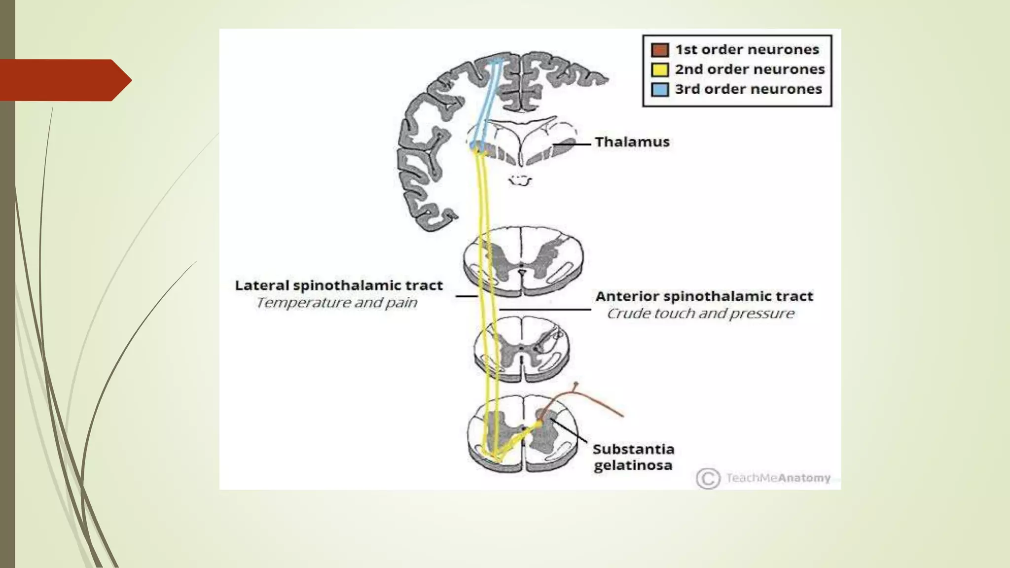
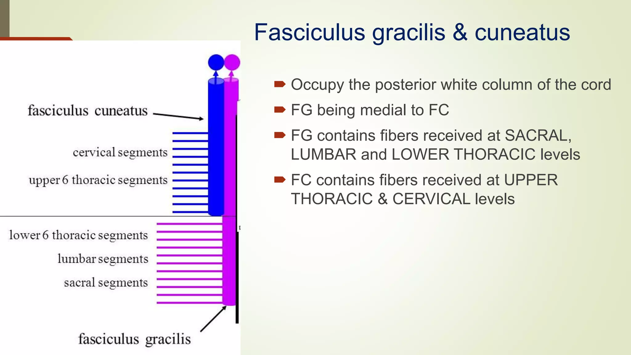
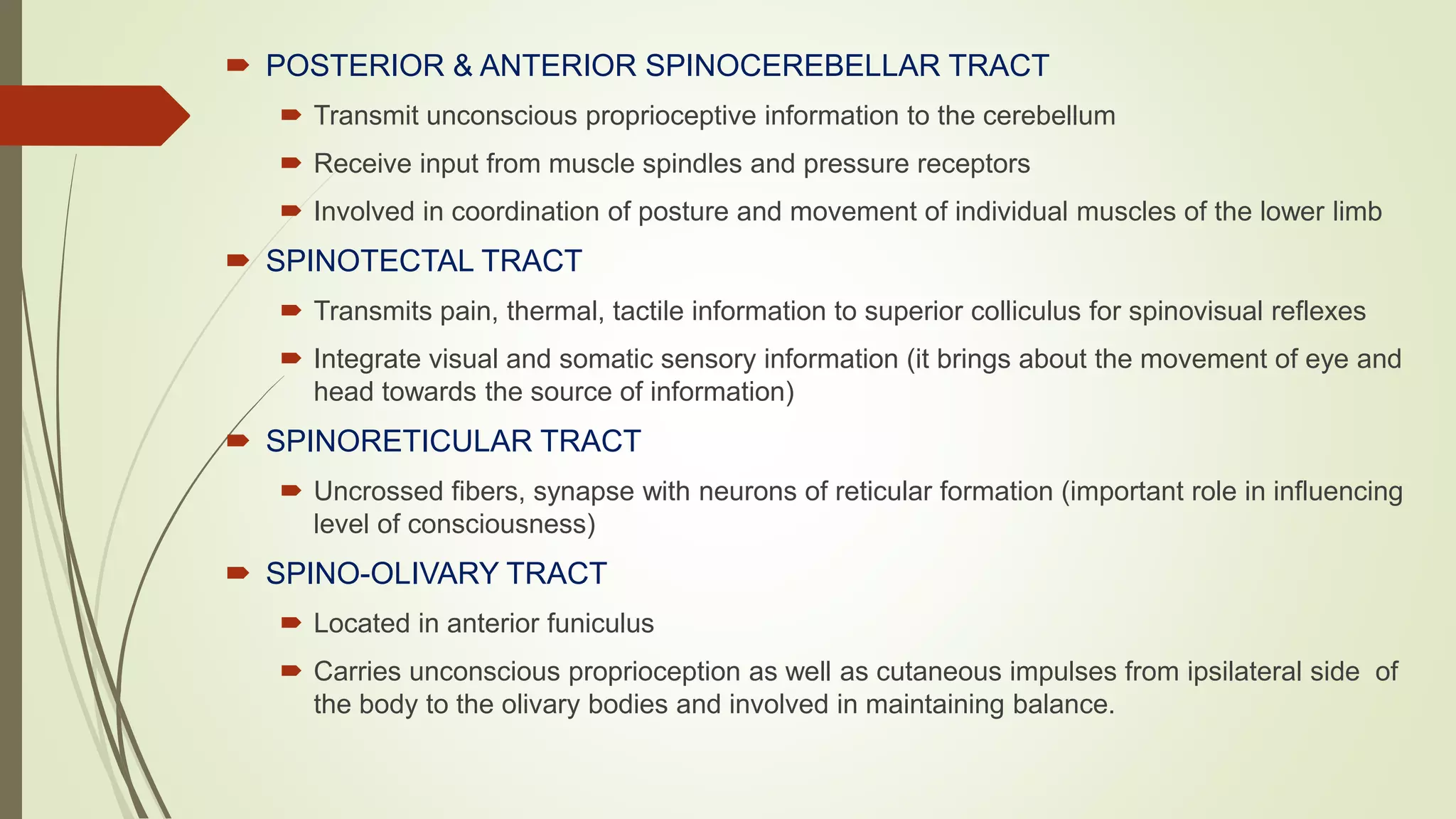
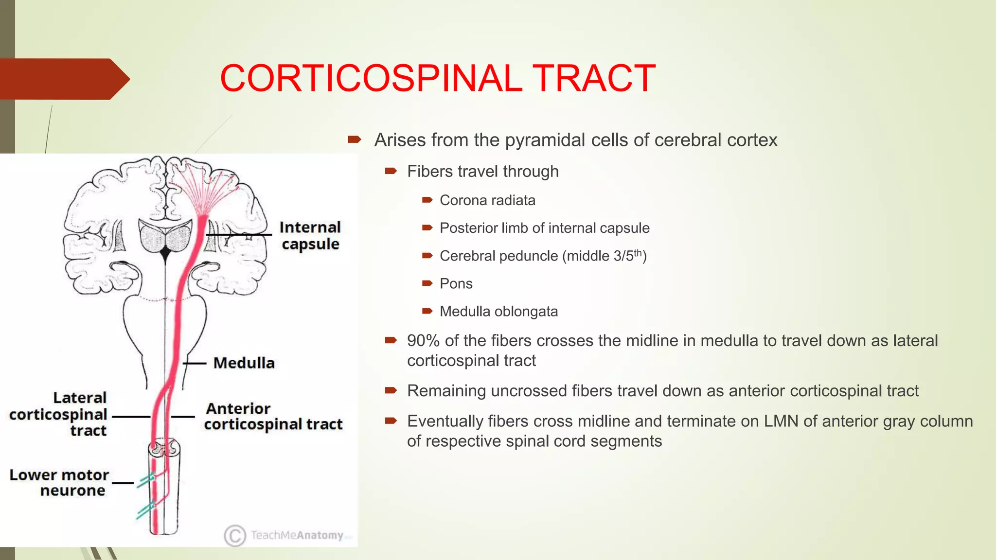
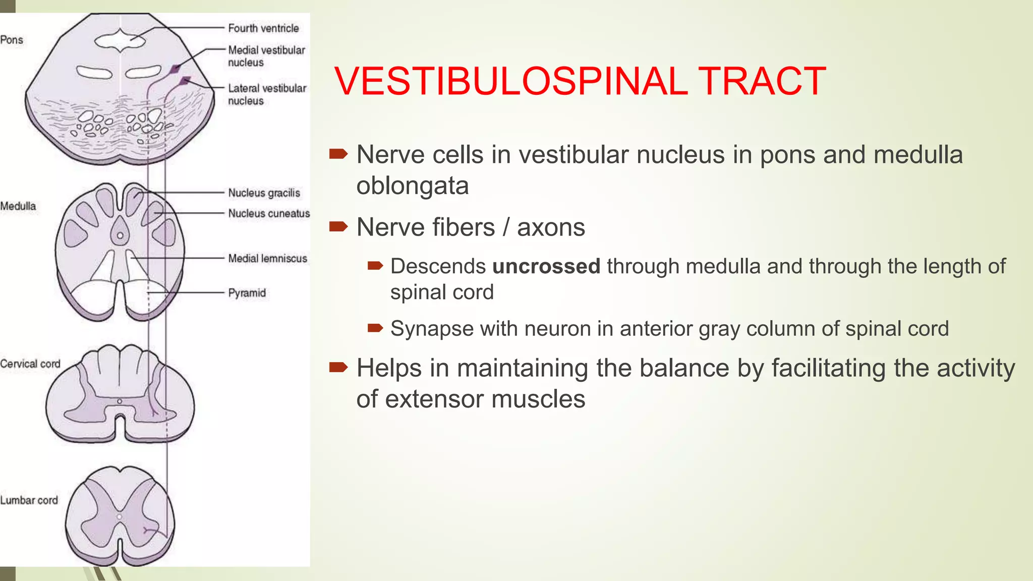
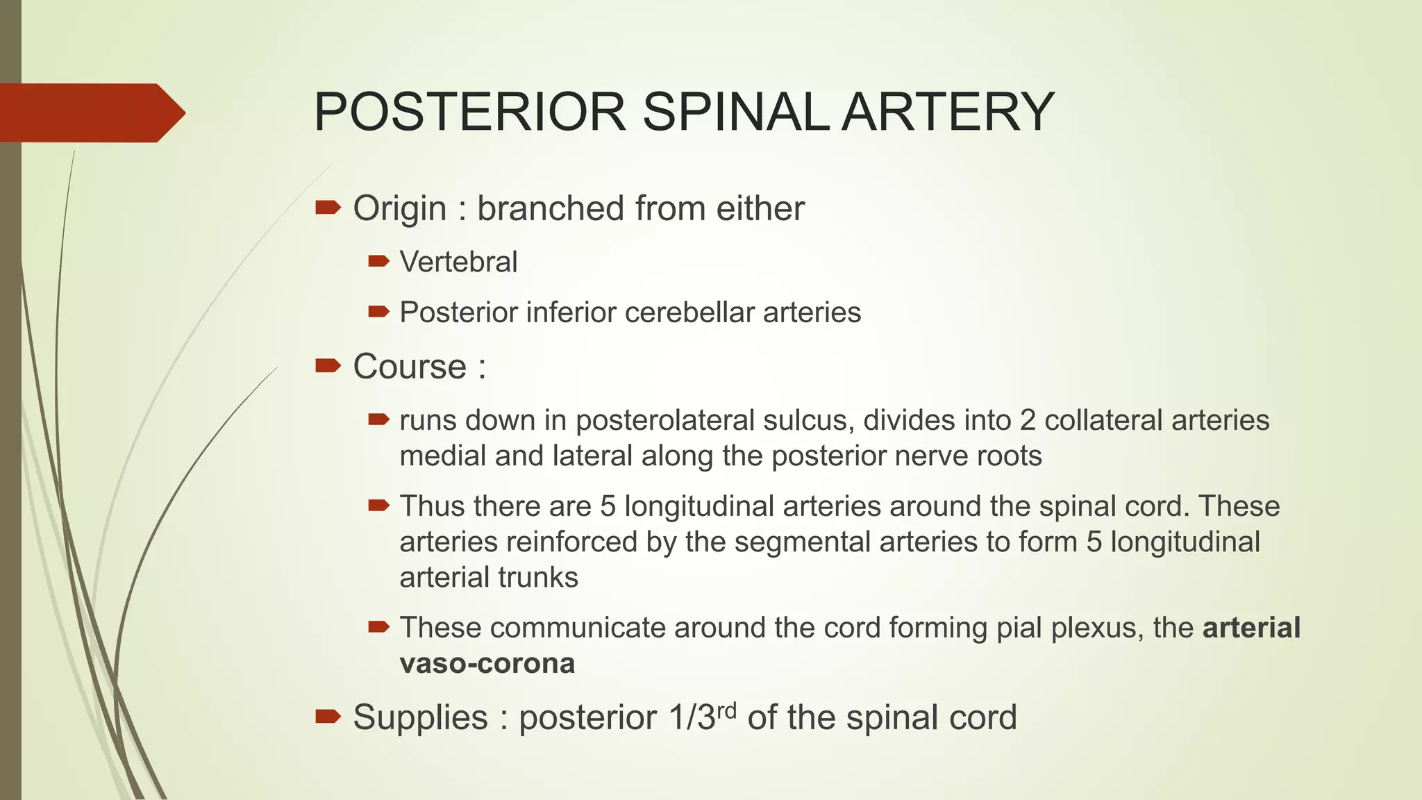
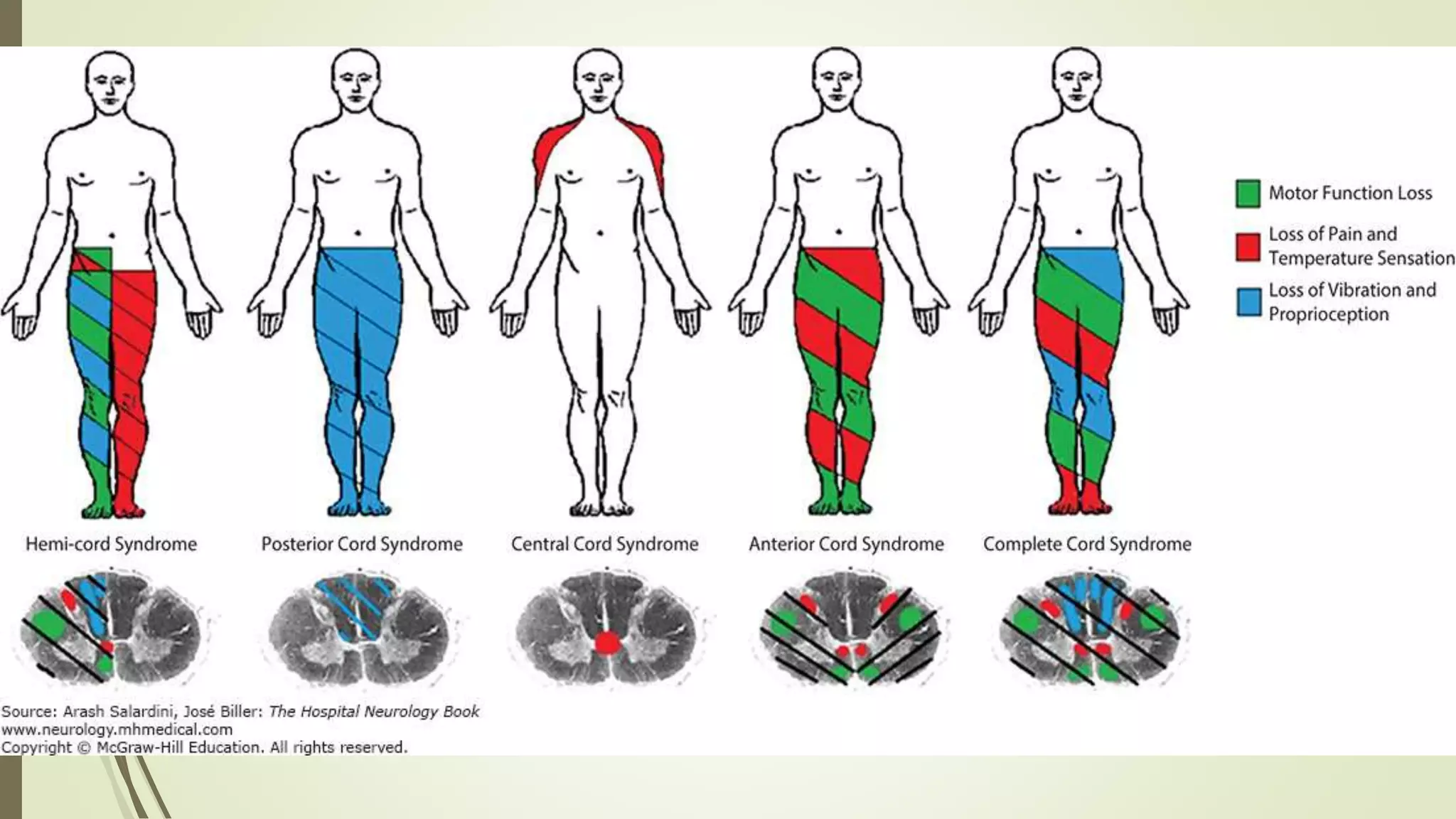
1. Neuroanatomy of Spinal cord
Presentor: Dr.Deepika.T
Post graduate student
Moderator: Dr.Byrareddy
Professor
Department of General Medicine,
RRMCH, Bengaluru
3. Gross Anatomy
ī´ Spinal cord lies in vertebral canal
ī´ Extends from level of cranial border of atlas to lower border of L1
or upper border of L2 vertebrae in adults
ī´ About 18 inches (45 cm) long
ī´ 1/2 inch (14 mm) wide
ī´ Approx weight = 30 gms
ī´ Corresponding average length of spinal column is 70cm
ī´ Anchored to duramater by dentate ligament
ī´ Cylindrical in shape & flattened dorso ventrally
4. Gross Anatomy
ī´ Has cervical (C5 to T1) and lumbar (L3 to S2) enlargements
ī´ Below Lumbar enlargement, spinal cord narrows ending as conus
medullaris
ī´ CNS tissue ends between vertebrae L1 and L2 whereas in neonates
ends at upper border of L3
ī´ At birth, cord and vertebrae are about the same size but cord stops
elongating at around age 4
ī´ Although it is a continuous & non segmental structure, 31 pair of
originating nerves give it a segmental appearance
ī´ 31 pairs of spinal nerves: 8C, 12T, 5L, 5S & 1Co
ī´ Each pair of nerves exits the vertebral column at the level it initially
lined up with at birth
5. Gross Anatomy
ī´Conus medullaris:
ī´thin, conical end of the spinal cord
ī´Cauda equina:
ī´nerve roots extending below conus medullaris
ī´Filum terminale:
ī´thin thread of fibrous tissue at end of conus medullaris
ī´attaches to coccygeal ligament
8. ī´ Epidural space :
between spinal duramater and walls of spinal column,
contains loose connective and areolar tissue
ī´ Subdural space
ī´ between arachnoid mater and dura mater
ī´ Subarachnoid space:
ī´ between arachnoid mater and pia mater
ī´ filled with cerebrospinal fluid (CSF)
SPACES
9. Structures of the Spinal Cord
ī´ Paired denticulate
ligaments:
ī´ extend from pia mater to dura
mater
ī´ stabilize side-to-side movement
ī´ Blood vessels:
ī´ along surface of spinal pia mater
ī´ within subarachnoid space
10.
11. Cross Sectional Anatomy
ī´ Anterior median fissure â separates anterior funiculi
ī´ Posterior median sulcus â divides posterior funiculi
12. Cross Sectional Anatomy
ī´Spinal cord has a narrow, fluid filled central canal
ī´Central canal is surrounded by butterfly or H-shaped
gray matter containing sensory and motor nuclei
(soma), unmyelinated processes, and neuroglia
ī´White matter is on the outside of the gray matter
(opposite of the brain) and contains myelinated and
unmyelinated fibers
14. Gray Matter: Organization
ī´ Dorsal half â sensory roots and ganglia
ī´ Ventral half â motor roots
ī´ Dorsal and ventral roots fuse laterally to form spinal nerves
ī´ Four zones are evident within the gray matter â somatic sensory
(SS), visceral sensory (VS), visceral motor (VM), and somatic
motor (SM)
15. ī´ SENSORY NUCLEUS
ī´ Substantia gelatinosa- relay station for spinothalamic tract
ī´ Nucleus proprius(largest nucleus)- relay station for dorsal column
tracts
ī´ Nucleus dorsalis(clarkes column)- relay station for spinocerebellar
tracts
ī´ INTERMEDIOLATERAL NUCLEUS
ī´ It extends from T1 to L2 , and contains autonomic motor neurons
that give rise to preganglionic fibres of sympathetic nervous system
ī´ MOTOR NEURONS
ī´ They innervate the visceral and skeletal muscles. Lateral nucleus
innervates the limb muscles and medial nucleus innervates the
midline/ axial muscles
NUCLEI IN GRAY MATER
16. White matter
ī´ Tracts (or fasciculi):
ī´ bundles of axons in the white columns
ī´ relay certain type of information in same direction
ī´ Ascending tracts:
ī´ carry information to brain
ī´ Descending tracts:
ī´ carry motor commands to spinal cord
17. Cross-sectional anatomy â white mater
ī´ White matter
ī´ 3 funiculi (posterior, lateral,
anterior)
ī´Ascending, descending,
transverse
ī´ Consist of âtractsâ containing
similarly functional axons
ī´All tracts are paired
ī´Most cross over (decussate) at
some point
ī´Most consist of a chain of 2 or 3
successive neurons
19. ASCENDING TRACTS
ī´ LATERAL SPINOTHALAMIC TRACT:
ī´ Pain and temperature
ī´ ANTERIOR SPINOTHALAMIC TRACT:
ī´ Crude touch and pressure
ī´ DORSAL COLUMN TRACT { Fasciculus gracilis(LL), Fasciculus
cuneatus(UL) } :
ī´ Carries conncious proprioception , fine touch , vibration, pressure
and stereognossus
ī´ DORSAL SPINOCEREBELLAR AND VENTRAL
SPINOCEREBELLAR TRACT:
ī´ Carries unconscious proprioception
20. Spinothalamic tract
First order neuron
impulses from free nerve endings transmitted to spinal cord
Central processes enters the spinal cord through posterior nerve root, proceed to the tip
of dorsal gray column
Second order neuron
In the dorsal horn cross to the opposite side (decussates)
Ascends in the contralateral ventral & lateral column
Ends in VPL nucleus of thalamus
Third order neuron
From the VPL nucleus of thalamus projects to cerebral cortex (area 3,1,2)
21.
22. Lateral spinothalamic tract
ī´ Clinical application
ī´ Destruction of LSTT
ī´ Loss of pain & thermal sensation
- Below the level of lesion
- On the contralateral side of the body
ī´ Patient will not respond to pinprick & cannot recognise
hot & cold
23. Anterior spinothalamic tract
ī´ Clinical application
ī´ Destruction of ASTT
ī´ Loss of light touch & pressure sense
- Below the level of lesion
- On the contralateral side of the body
ī´ (Discriminative touch will still be present, as this infprmation is carried by posterior
column)
24. Fasciculus gracilis & cuneatus
ī´ Occupy the posterior white column of the cord
ī´ FG being medial to FC
ī´ FG contains fibers received at SACRAL,
LUMBAR and LOWER THORACIC levels
ī´ FC contains fibers received at UPPER
THORACIC & CERVICAL levels
25. Fasciculus gracilis & cuneatus
ī´ First order neuron
Ascend without interruption and terminate upon 2nd
order neurons in NUCLEUS GRACILIS & NUCLEUS
CUNEATUS
ī´ Second order neuron
Axons of 2nd order neurons decussate in the
medulla as internal arcuate fibers and ascend through
the brain as medial lemniscus
ī´ Third order neuron
Medial lemniscus terminates in the VENTRAL
POSTERIOR nucleus of THALAMUS,3rd order neurons
project to somatosensory cortex
26. Fasciculus gracilis & cuneatus
ī´ Clinical application
ī´ Destruction of Fasciculus gracilia & cuneatus
ī´ Loss of muscle joint sense, position sense, vibration sense
& tactile discrimination light touch & pressure sense
- Below the level of lesion
- On the same side of the body
27. ī´ POSTERIOR & ANTERIOR SPINOCEREBELLAR TRACT
ī´ Transmit unconscious proprioceptive information to the cerebellum
ī´ Receive input from muscle spindles and pressure receptors
ī´ Involved in coordination of posture and movement of individual muscles of the lower limb
ī´ SPINOTECTAL TRACT
ī´ Transmits pain, thermal, tactile information to superior colliculus for spinovisual reflexes
ī´ Integrate visual and somatic sensory information (it brings about the movement of eye and
head towards the source of information)
ī´ SPINORETICULAR TRACT
ī´ Uncrossed fibers, synapse with neurons of reticular formation (important role in influencing
level of consciousness)
ī´ SPINO-OLIVARY TRACT
ī´ Located in anterior funiculus
ī´ Carries unconscious proprioception as well as cutaneous impulses from ipsilateral side of
the body to the olivary bodies and involved in maintaining balance.
29. ī´ PYRAMIDAL TRACT ( ANTERIOR AND LATERAL
CORTICOSPINAL TRACT):
ī´ Skilled voluntary movements
ī´ RUBROSPINAL TRACT:
ī´ Facilitates flexors
ī´ Inhibits extensors
ī´ TECTOSPINAL TRACT:
ī´ Responsible for coordinated movements of head and neck,
eyeball and limbs based on visual stimulus
ī´ VESTIBULOSPINAL TRACT
ī´ Also called as postural tract
ī´ Helps in maintaining posture
DESCENDING TRACTS
30. CORTICOSPINAL TRACT
ī´ Arises from the pyramidal cells of cerebral cortex
ī´ Fibers travel through
ī´ Corona radiata
ī´ Posterior limb of internal capsule
ī´ Cerebral peduncle (middle 3/5th)
ī´ Pons
ī´ Medulla oblongata
ī´ 90% of the fibers crosses the midline in medulla to travel down as lateral
corticospinal tract
ī´ Remaining uncrossed fibers travel down as anterior corticospinal tract
ī´ Eventually fibers cross midline and terminate on LMN of anterior gray column
of respective spinal cord segments
31. RUBROSPINAL TRACT
ī´ Nerve cells in Red nucleus
ī´ Nerve fibers / axons
ī´ Cross the midline
ī´ Descend as rubrospinal tract
ī´ Terminate in anterior gray column of spinal cord
ī´ Facilitate the activity of flexor muscles
32. TECTOSPINAL TRACT
ī´ Nerve cells in superior colliculus of midbrain
ī´ Nerve fibers / axons
ī´ Cross the midline
ī´ Descend close to medial longitudinal fasciculus
ī´ Terminate in the anterior gray column of upper cervical segments of spinal
cord
ī´ Responsible for reflex movement of head & neck in response to visual
stimuli
33. VESTIBULOSPINAL TRACT
ī´ Nerve cells in vestibular nucleus in pons and medulla
oblongata
ī´ Nerve fibers / axons
ī´ Descends uncrossed through medulla and through the length of
spinal cord
ī´ Synapse with neuron in anterior gray column of spinal cord
ī´ Helps in maintaining the balance by facilitating the activity
of extensor muscles
34. RETICULOSPINAL TRACT
ī´ Nerve cells in reticular formation of caudal pons & rostral
medulla
ī´ Nerve fibers / axons
ī´ Consists of 2 separate pathways (Pontine reticulospinal tract &
medullary reticulospinal tract)
ī´ Pass through pons & medulla
ī´ Ends at anterior gray column (mainly uncrossed)
ī´ Facilitates voluntary and reflex activity
ī´ They influence the action of motor neurons of phrenic and
intercostal nerves, thus control RESPIRATION.
38. ANTERIOR SPINAL ARTERY
ī´Origin : formed by union of 2 small spinal branches
of right & left vertebral arteries in the upper cervical
canal
ī´Course : runs caudally in the anterior median fissure
ī´Termination : filum terminale
ī´Supplies : anterior 2/3rd of the cord
39. ANTERIOR SPINAL ARTERY
ī´Due to occlusion (thrombosis or compression) of
anterior spinal artery
ī´Results in
ī´Motor symptoms: coz of involvement of corticospinal tracts
and anterior gray columns
ī´Bilateral loss of pain & temperature sensation due to
ischemia of spinothalamic tracts
40. POSTERIOR SPINAL ARTERY
ī´ Origin : branched from either
ī´ Vertebral
ī´ Posterior inferior cerebellar arteries
ī´ Course :
ī´ runs down in posterolateral sulcus, divides into 2 collateral arteries
medial and lateral along the posterior nerve roots
ī´ Thus there are 5 longitudinal arteries around the spinal cord. These
arteries reinforced by the segmental arteries to form 5 longitudinal
arterial trunks
ī´ These communicate around the cord forming pial plexus, the arterial
vaso-corona
ī´ Supplies : posterior 1/3rd of the spinal cord
41.
42. SEGMENTAL ARTERIES
ī´ They reach the spinal cord as anterior and posterior radicular
arteries along the corresponding roots of the spinal nerves
respectively and nourish the nerve roots
ī´ Anterior radicular arteries: larger and less in number
ī´ Posterior radicular arteries: smaller and more in number
ī´ Importance: end arteries, if anyone of them is blocked, there
will not be any collateral circulation
ī´ Branches of
ī´ Deep cervical
ī´ Ascending cervical
ī´ Intercostal &
ī´ Lumbar
43. CLINICAL CORRELATION
ī´ Artery of T11/ T12 spinal segment (arteria radicularis magna)
is remarkably large, arises directly from aorta on its left side
ī´ A fracture of vertebra involving this artery leads to ischemia of
several segments of the cord
ī´ T1, T4 and L1 segments â the meeting places of different
major arteries are vulnerable to ischemic necrosis
44. VENOUS DRAINAGE
ī´Two median longitudinal â one in the anterior median
fissure another in posteromedian sulcus
ī´Two anterolateral - one on either side posterior to
the anterior nerve roots
ī´Two posterlateral â one on either side posterior to
posterior nerve roots
45. VENOUS DRAINAGE
ī´ Drain below through internal vertebral venous
plexus into the vertebral posterior intercostal, lumbar
and lateral scral veins
ī´ Internal vertebral venous plexus communicates
above with the basilar venous plexus.