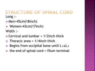
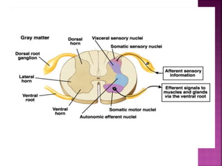
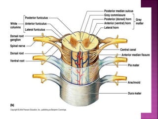
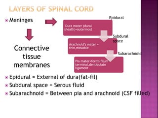
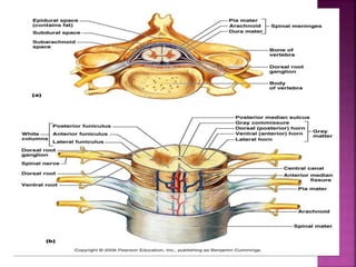
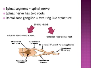
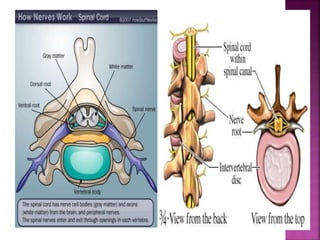
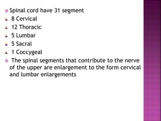
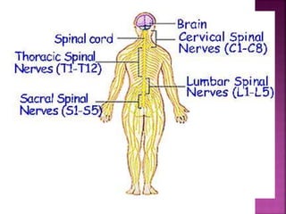
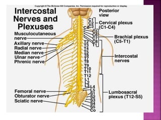
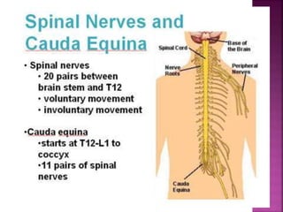
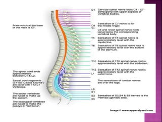
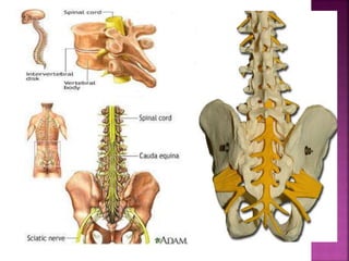
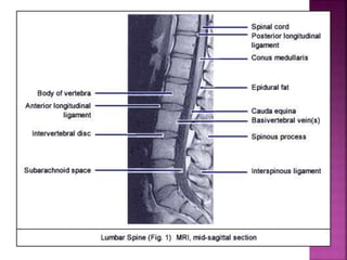
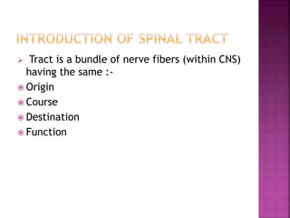
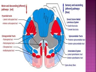
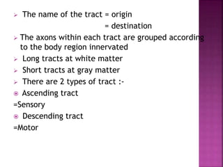
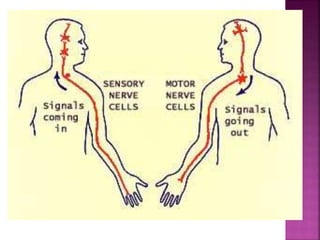
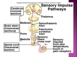
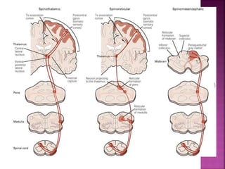
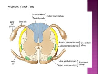
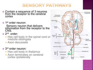
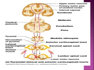
 Structure
 Function
 Tracts
Long :-
 Men=45cm(18inch)
 Women=43cm(17inch)
Width :-
 Cervical and lumbar = 1/2inch thick
 Thoracic area = 1/4inch thick
 Begins from occipital bone until L1&L2
 the end of spinal cord = filum terminal
 Meninges
 Epidural = External of dura(fat-fil)
 Subdural space = Serous fluid
 Subarachnoid = Between pia and arachnoid (CSF filled)
Dura mater (dural
sheath)=outermost
Arachnoid’s mater =
thin,movable
Pia mater=forms filum
terminal,denticulate
ligament
Connective
tissue
membranes
Subdural
space
Subarachnoid
Epidural
 Spinal segment = spinal nerve
 Spinal nerve has two roots
 Dorsal root ganglion = swelling like structure
Anterior root= ventral root
Posterior root=dorsal root
SPINAL NERVE
 Spinal cord have 31 segment
8 Cervical
12 Thoracic
5 Lumbar
5 Sacral
1 Coccygeal
 The spinal segments that contribute to the nerve
of the upper are enlargement to the form cervical
and lumbar enlargements
vertebrae spinal segments
C1 to C4 (upper cervical) same
C4 To C7 (lower cervical) +1
T1 to T7 +2
T7 to T9 +3
T10 L1,L2
T11 L3,L4
T12 L5,S1
L1 sacral and coccygeal nerve
 Spinal Arteries :-
 Anterior (1) & Posterior (2) Spinal Artery from
Vertebral artery
 Radicular Arteries :-
 Segmental arteries from Vertebral,Ascending
Cervical,Intercostal and Lumbar Artery
 Venous Drainage veins :-
 Longitudinal & Radicular Veins Intervertebral
veins Internal Vertebral Venous Plexus
external vertebral venous plexus segmental
veins
 Tract is a bundle of nerve fibers (within CNS)
having the same :-
 Origin
 Course
 Destination
 Function
 The name of the tract = origin
= destination
 The axons within each tract are grouped according
to the body region innervated
 Long tracts at white matter
 Short tracts at gray matter
 There are 2 types of tract :-
 Ascending tract
=Sensory
 Descending tract
=Motor
 Decussation is the cross-over of the tract
from one side to the other.
 Therefore,there are instances where the left
side of the body is controlled by the right
brain hemisphere.
 Decussation occurs at different locations for
each tracts.
Ascending Spinal Tracts
 Contain a sequence of 3 neurons
from the receptor to the cerebral
cortex
 1st order neuron:
Sensory neuron that delivers
information from the receptor to the
CNS.
 2nd order:
 Has cell body in the spinal cord or
medulla oblongata
 Axon decussate
 3rd order neuron:
 Has cell body in thalamus
 Axon terminates on cerebral
cortex ipsilaterally
1
2
3
Modality: Discriminative Touch
Sensation Conscious
Proprioception
Receptor: Most receptors except
free nerve endings
Ist Neuron: Dorsal Root Ganglion
(Spinal Ganglion)
Dorsal Column Nuclei (Nucleus
Gracilis
Thalamus (VPLc) ,Corona Radiata
Termination: Primary Somesthetic
Area (S I)
Posterior White Column-Medial
Lemniscal Pathway
 Modality:pain and thermal
sensations.
Ist Neuron : dorsal horn
 2nd Neuron: mostly in the
nucleus proprius), decussate
within one segment by passing
through the ventral white
commissure
 3rd Neuron: ventral posterior
nucleus of the thalamus
 Thalamic neurons project to the
somatosensory cortex
 Modality:non- discriminative
touch and pressure
 Ist Neuron : dorsal horn
 2nd Neuron: nucleus
proprius,crossing to opposite
side by passing through the
ventral white commissure
 3rd Neuron: in ventral
posterior nucleus of the
thalamus
 Thalamic neurons project to the
somatosensory cortex
 Ist Neuron neuron lie in base of
the dorsal horn of the lumbosacral
segments
 cross to opposite side, ascend as
far as the midbrain
 2nd Neuron: terminating in the
cerebellar cortex
Both spinocerebellar tracts convey
sensory information to the same
side of the cerebellum
 Info:Present only above
level L3
 Ist Neuron in Clark’s
column
 2nd Neuron: terminate
ipsilaterally in the
cerebellar cortex
 2 Sequence of neuron(from cerebral
cortex/brain stem – muscle)
 Upper Motor Neuron(UMN)
o Spinal cord -> brain
o Cell body cross over before terminate on lower
motor neuron
 Lower Motor Neuron(LMN)
o Spinal cord -> muscle
o Cell body pass through posterior root of spinal
nerve
DESCENDINIG
TRACT
PYRAMIDAL TRACT
(DIRECT)
ANTERIOR
CORTICOSPINAL
TRACT
LATERAL
CORTICOSPINAL
TRACT
EXTRAPYRAMIDAL
TRACT
(INDIRECT)
RETICULOSPINAL
TRACT
VESTIBULOSPINAL
TRACT
TECTOSPINAL
TRACT
RUBROSPINAL
TRACT
Function:voluntary movement
: fine skill movement
 Anterior corticospinal tract
o Origin:motor cortex (4,6)
:somatosensory cortex (1,2,3)
o Course:pass through midbrain(cerebral peduncle)
pyramid of medulla oblongata
o Termination: anterior white column
 Lateral corticospinal tract
o Origin: motor cortex (4,6)
:somatosensory cortex (1,2,3)
o Course: pass through midbrain(cerebral peduncle)
(cross over)
pyramid of medulla oblongata
o Termination: lateral white column
o Corticobulbar  innervate cranial nerves
 motor nuclei of CNS
 Function: coordination of voluntary and reflex
movement
: control of muscle tone
: control of respiration and diameter of
blood vessel
 Origin: Reticular formation (pontine & medulla)
 Course :pass through medial pontine
lateral medulla
 Termination: ventral funniculus
 Function: coordination of head and neck to
the visual & audio stimuli
 Origin : superior colliculus
 Course : pass through periaquductal
(cross)
Dorsal tegmental
 Termination: Anterior median fissure
(cervical area)
 Function : controls flexor
muscle tone
 Origin : Red nucleus
 Course :cross to
(lower cervical segment)
 Termination: lateral
white column
 Lateral vestibulospinal tract
 Function: control extensor
muscle tone
: antigravity
maintenance
of posture
 Origin : lateral vestibular
(Derter’s) nucleus
 Termination :ventral
funniculus
• Medial vestibulospinal tract
Function: control
movement
of head
:maintain
equilibrium
Origin:medial vestibular
nucleus
Termination: ventral
funniculus
+
medial
funniculus

Spinal Cord

  • 2.
  • 3.
    Long :-  Men=45cm(18inch) Women=43cm(17inch) Width :-  Cervical and lumbar = 1/2inch thick  Thoracic area = 1/4inch thick  Begins from occipital bone until L1&L2  the end of spinal cord = filum terminal
  • 7.
     Meninges  Epidural= External of dura(fat-fil)  Subdural space = Serous fluid  Subarachnoid = Between pia and arachnoid (CSF filled) Dura mater (dural sheath)=outermost Arachnoid’s mater = thin,movable Pia mater=forms filum terminal,denticulate ligament Connective tissue membranes Subdural space Subarachnoid Epidural
  • 9.
     Spinal segment= spinal nerve  Spinal nerve has two roots  Dorsal root ganglion = swelling like structure Anterior root= ventral root Posterior root=dorsal root SPINAL NERVE
  • 12.
     Spinal cordhave 31 segment 8 Cervical 12 Thoracic 5 Lumbar 5 Sacral 1 Coccygeal  The spinal segments that contribute to the nerve of the upper are enlargement to the form cervical and lumbar enlargements
  • 13.
    vertebrae spinal segments C1to C4 (upper cervical) same C4 To C7 (lower cervical) +1 T1 to T7 +2 T7 to T9 +3 T10 L1,L2 T11 L3,L4 T12 L5,S1 L1 sacral and coccygeal nerve
  • 20.
     Spinal Arteries:-  Anterior (1) & Posterior (2) Spinal Artery from Vertebral artery  Radicular Arteries :-  Segmental arteries from Vertebral,Ascending Cervical,Intercostal and Lumbar Artery  Venous Drainage veins :-  Longitudinal & Radicular Veins Intervertebral veins Internal Vertebral Venous Plexus external vertebral venous plexus segmental veins
  • 21.
     Tract isa bundle of nerve fibers (within CNS) having the same :-  Origin  Course  Destination  Function
  • 23.
     The nameof the tract = origin = destination  The axons within each tract are grouped according to the body region innervated  Long tracts at white matter  Short tracts at gray matter  There are 2 types of tract :-  Ascending tract =Sensory  Descending tract =Motor
  • 25.
     Decussation isthe cross-over of the tract from one side to the other.  Therefore,there are instances where the left side of the body is controlled by the right brain hemisphere.  Decussation occurs at different locations for each tracts.
  • 30.
  • 31.
     Contain asequence of 3 neurons from the receptor to the cerebral cortex  1st order neuron: Sensory neuron that delivers information from the receptor to the CNS.  2nd order:  Has cell body in the spinal cord or medulla oblongata  Axon decussate  3rd order neuron:  Has cell body in thalamus  Axon terminates on cerebral cortex ipsilaterally 1 2 3
  • 32.
    Modality: Discriminative Touch SensationConscious Proprioception Receptor: Most receptors except free nerve endings Ist Neuron: Dorsal Root Ganglion (Spinal Ganglion) Dorsal Column Nuclei (Nucleus Gracilis Thalamus (VPLc) ,Corona Radiata Termination: Primary Somesthetic Area (S I) Posterior White Column-Medial Lemniscal Pathway
  • 33.
     Modality:pain andthermal sensations. Ist Neuron : dorsal horn  2nd Neuron: mostly in the nucleus proprius), decussate within one segment by passing through the ventral white commissure  3rd Neuron: ventral posterior nucleus of the thalamus  Thalamic neurons project to the somatosensory cortex
  • 34.
     Modality:non- discriminative touchand pressure  Ist Neuron : dorsal horn  2nd Neuron: nucleus proprius,crossing to opposite side by passing through the ventral white commissure  3rd Neuron: in ventral posterior nucleus of the thalamus  Thalamic neurons project to the somatosensory cortex
  • 35.
     Ist Neuronneuron lie in base of the dorsal horn of the lumbosacral segments  cross to opposite side, ascend as far as the midbrain  2nd Neuron: terminating in the cerebellar cortex Both spinocerebellar tracts convey sensory information to the same side of the cerebellum
  • 36.
     Info:Present onlyabove level L3  Ist Neuron in Clark’s column  2nd Neuron: terminate ipsilaterally in the cerebellar cortex
  • 38.
     2 Sequenceof neuron(from cerebral cortex/brain stem – muscle)  Upper Motor Neuron(UMN) o Spinal cord -> brain o Cell body cross over before terminate on lower motor neuron  Lower Motor Neuron(LMN) o Spinal cord -> muscle o Cell body pass through posterior root of spinal nerve
  • 39.
  • 41.
    Function:voluntary movement : fineskill movement  Anterior corticospinal tract o Origin:motor cortex (4,6) :somatosensory cortex (1,2,3) o Course:pass through midbrain(cerebral peduncle) pyramid of medulla oblongata o Termination: anterior white column
  • 42.
     Lateral corticospinaltract o Origin: motor cortex (4,6) :somatosensory cortex (1,2,3) o Course: pass through midbrain(cerebral peduncle) (cross over) pyramid of medulla oblongata o Termination: lateral white column o Corticobulbar  innervate cranial nerves  motor nuclei of CNS
  • 47.
     Function: coordinationof voluntary and reflex movement : control of muscle tone : control of respiration and diameter of blood vessel  Origin: Reticular formation (pontine & medulla)  Course :pass through medial pontine lateral medulla  Termination: ventral funniculus
  • 49.
     Function: coordinationof head and neck to the visual & audio stimuli  Origin : superior colliculus  Course : pass through periaquductal (cross) Dorsal tegmental  Termination: Anterior median fissure (cervical area)
  • 51.
     Function :controls flexor muscle tone  Origin : Red nucleus  Course :cross to (lower cervical segment)  Termination: lateral white column
  • 52.
     Lateral vestibulospinaltract  Function: control extensor muscle tone : antigravity maintenance of posture  Origin : lateral vestibular (Derter’s) nucleus  Termination :ventral funniculus
  • 53.
    • Medial vestibulospinaltract Function: control movement of head :maintain equilibrium Origin:medial vestibular nucleus Termination: ventral funniculus + medial funniculus