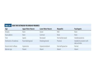
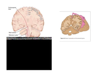
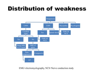
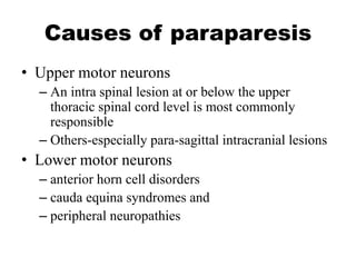
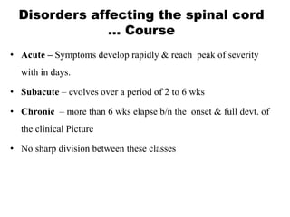
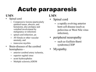
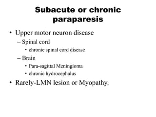
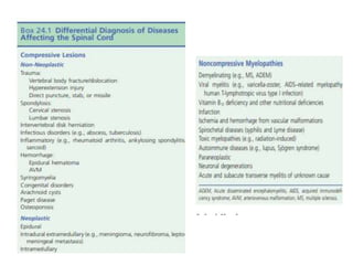
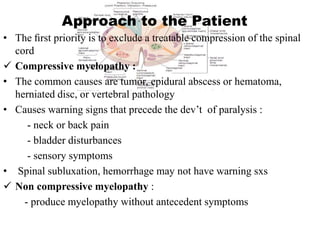
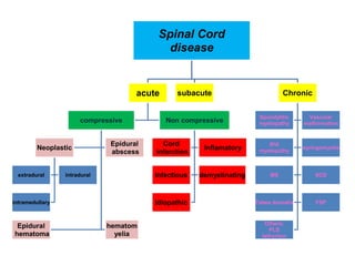
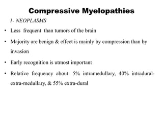
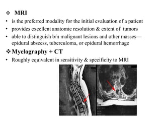
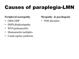
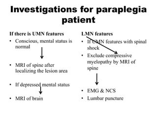
Paraparesis/paraplegia
Dr.Abiy F.
sep17,2015
Arsi University
Revision
• 1.which type of brain tumor is common?
• A.Astrocytoma
• B.Meningioma
• C.Glioblastoma multiforme
• D.Metastatic brain tumor
• 2.which one is focal(lateralie)sign of brain
tumor?
• A.Aphasia
• B.personality changes
• C.headache
• D.Gait abnormality
• 3.one of the statement is true about brain tumors.
• A.The most common source of metastatic brain
tumor is melanoma
• B.Brain metastsasis lesions are always multiple
B/c they disseminate hematogenously
• C.the commonest risk factor for primary brain
tumor is genetic factors
• D. Glioblastoma is the most common malignant
primary brain tumor in adults.
• 4.one of the following statement is false about
intracranial pressure.
• A. pathologic intracranial hypertension (ICH)
is present at pressures ≥20 mmHg
• B.Commonest cause of ICH is pseudotumor
cerebri
• C. Low ICP is caused by diminished CSF
volume
• D.Postural headache is typical for low ICP
Outline of presentation
• Definition
• Causes
• UMN lesion/LMN lesion
• Diseases of spinal cord
• DDx
• Work up
Definition
• Weakness is a reduction in the power that can
be exerted by one or more muscles.
• Paralysis or the suffix “-plegia” indicates
weakness so severe that a muscle cannot be
contracted at all, whereas paresis refers to less
severe weakness.
• The prefix “hemi-” refers to one-half of the
body, “para-” to both legs, “mono-” to one
limb and “quadri-” to all four limbs.
Spinal Shock
• When the corticospinal pathways are affected by a brain or
spinal cord lesion of sudden onset, there may also be a
period of flaccidity and areflexia accompanying the
paralysis below the level of the lesion.
• This is the period of “spinal shock,” which sooner or later
gives way, in most instances, to the corticospinal syndrome.
• During the neural shock phase, the plantar responses may be
mute and the superficial reflexes absent.
• The pyramidal, or upper motor neuron, syndrome gradually
emerges over hours to weeks with spasticity, hyperactive
tendon reflexes, extensor plantar responses, and continued
absence of superficial reflexes.
Pattern of weakness
• Distribution
• UMN/LMN or Others (myopathic,
psychogenic)
• Duration
– Acute, Subacute, or Chronic
Distribution of weakness
Paraparesis
UMN
Cerebral
signs
Yes
Brain MRI
or CT-scan
No
MRI of
spine
Non-
comprsesive
LP
Compresive
LMN
Yes
EMG &
NCS
Spinal shock
Myopathic
EMG &
NCS
Psychogenic
EMG=electromyelography, NCS=Nerve conduction study
Causes of paraparesis
• Upper motor neurons
– An intra spinal lesion at or below the upper
thoracic spinal cord level is most commonly
responsible
– Others-especially para-sagittal intracranial lesions
• Lower motor neurons
– anterior horn cell disorders
– cauda equina syndromes and
– peripheral neuropathies
Disorders affecting the spinal cord
… Course
• Acute – Symptoms develop rapidly & reach peak of severity
with in days.
• Subacute – evolves over a period of 2 to 6 wks
• Chronic – more than 6 wks elapse b/n the onset & full devt. of
the clinical Picture
• No sharp division between these classes
Acute paraparesis
UMN
• Spinal cord
– Compressive lesions (particularly
epidural tumor, abscess, and
hematoma ,disc prolapse and
vertebral involvement by
malignancy or infection)
– spinal cord infarction ,an
– AV-fistula or other vascular
anomaly, and
– transverse myelitis
• Brain-diseases of the cerebral
hemispheres –
– anterior cerebral artery ischemia,
– superior sagittal sinus
– acute hydrocephalus
– Multiple sclerosis,ADEM
LMN
• Spinal cord
– a rapidly evolving anterior
horn cell disease (such as
poliovirus or West Nile virus
infection),
• peripheral neuropathy
– such as Guillain-Barré
syndrome,CIDP
• Myopathy.
Subacute or chronic
paraparesis
• Upper motor neuron disease
– Spinal cord
• chronic spinal cord disease
– Brain
• Para-sagittal Meningioma
• chronic hydrocephalus
• Rarely-LMN lesion or Myopathy.
Approach to the Patient
• The first priority is to exclude a treatable compression of the spinal
cord
 Compressive myelopathy :
• The common causes are tumor, epidural abscess or hematoma,
herniated disc, or vertebral pathology
• Causes warning signs that precede the dev’t of paralysis :
- neck or back pain
- bladder disturbances
- sensory symptoms
• Spinal subluxation, hemorrhage may not have warning sxs
 Non compressive myelopathy :
- produce myelopathy without antecedent symptoms
Spinal Cord
disease
acute
compressive
Neoplastic
extradural intradural
intramedullary
Epidural
abscess
Epidural
hematoma
hematom
yelia
Non compressive
Cord
infarction
Inflamatory
Infectious demyelinating
Idiopathic
subacute Chronic
Spondylitic
myelopathy
Vascular
malformation
RVI
myelopathy
syringomyelia
MS SCD
Tabes dorsalis FSP
Others;
PLS
lathyrism
Compressive Myelopathies
1- NEOPLASMS
• Less frequent than tumors of the brain
• Majority are benign & effect is mainly by compression than by
invasion
• Early recognition is utmost important
• Relative frequency about: 5% intramedullary, 40% intradural-
extra-medullary, & 55% extra-dural
A- Epidural Neoplasms:
• Result from metastases to the adjacent spinal bones
• Any malignant tumor can metastasize to the spinal column
• Most frequent: breast, lung, prostate, kidney, lymphoma, and
plasma cell dyscrasia
• 60 % occur in the thoracic, 30 % in the lumbosacral, & 10 % in the
cervical spine
• Metastases from prostate & ovarian ca occur disproportionately in
the sacral and lumbar vertebrae
Epidural Neoplasm
• Presentation :
 Pain is usually the initial Sx in 83- 95 % of pts at the time of dx
– may be aching and localized or sharp and radiating
– Typically worsens with movement, coughing, or sneezing and
characteristically awakens pts at night.
– Pain is often worse with recumbency, a feature attributed to
distension of the epidural venous plexus.
– Over time, the pain may develop a radicular quality
Epidural Tumors
 Motor findings
• Weakness in 60 - 85 % of pts at the time of dx
• Weakness is from CST dysfunction when the lesion is at or
above the conus medullaris
• UMN lesion signs
Sensory findings
• Are less common than motor findings
• Ascending numbness and paresthesias
 Bladder and bowel dysfunction
• A late finding , may be present in as many as one-half of pts
• Most commonly presents as urinary retention and is rarely the sole
symptom of ESCC ( autonomic neuropathy + opiate use)
 Ataxia : Spinocerebellar tract dysfunction
Investigation:
 Plain radiographs and radionuclide bone scans :
- have only a limited role in dx
- they do not identify 15–20% of metastatic vertebral lesions
- fail to detect paravertebral masses that reach the epidural
space through the intervertebral foramina
 MRI
• is the preferred modality for the initial evaluation of a patient
• provides excellent anatomic resolution & extent of tumors
• able to distinguish b/n malignant lesions and other masses—
epidural abscess, tuberculoma, or epidural hemorrhage
Myelography + CT
• Roughly equivalent in sensitivity & specificity to MRI
 Treatment:
- Glucocorticoids to reduce cord edema
- Local RXT (initiated as early as possible) to the symptomatic lesion
- Specific therapy for the underlying tumor type
• Surgery
- decompression by laminectomy or vertebral body resection
- Indications:
B- Intradural mass lesions:
• Are slow-growing and benign
- Intramedullary = 10% (Glial tumors or 20 )
- Extramedullary = 90%
• Commonest : Meningiomas and neurofibromas
• Occasional cases : chordoma, lipoma, dermoid, or sarcoma
• Spinal cord gliomas are rare, probably because of the relative
paucity of glial tissue in the spinal cord
Intradural tumors
MRI of a thoracic
meningioma
MRI of an IM astrocytoma
Symptoms:
 EM: compression of nerve
roots, spinal cord or vessels
 IM:
- presents as central or hemicord
syndrome
- usually in cervical cord
- pain is poorly localized
&burning
- sacral sparing of sensation
Management: Surgery
(complete resection or debulking)
- The value of adjunctive RXT &
CXT is uncertain
Compressive Myelopathy… contd
2- Spinal Epidural Abscess:
•Abscesses that begin at one level extend to multiple levels
•Presents as a clinical triad of midline dorsal pain, fever, and
progressive limb weakness
•Aching pain is either over the spine or in a radicular pattern
•The duration of pain prior to presentation is generally 2 wks but
may on occasion be several months or longer
•Risk factors :
- an impaired immune status (DM, RF, alcoholism, HIV, Ca)
- intravenous drug abuse, Tatooing, acupancture
- infections of the skin or other tissues, trauma
• 2/3rd are due to hematogenous spread form the skin, soft tissue, or
deep viscera
• The other arise from direct extension of a local infection to the
subdural space… Vertebral OM, decubitus ulcers,
• Or direct inoculation… LP, epidural anesthesia, or spinal surgery
• Causative org : S.aureus( 63%) , GN-bacilli( 16%), Strept( 9%),
anaerobes(2%), and fungi ( 1%)
• TBC from an adjacent vertebral source, in the underdeveloped
world
• Mechanism of damage to the spinal cord:
• Direct compression
• Thrombosis and thrombophlebitis of nearby veins
• Interruption of the arterial blood supply
• Bacterial toxins and mediators of inflammation
 Invest- CBC, ESR , MRI, LP ( if signs of meningoencephalitis)
• MRI scans localize the abscess & exclude other causes of myelopathy

 Treatment :
• Decompressive laminectomy with debridement + long-term antibiotic Rx ( > 6 wks)
• Empiric Rx: Vancomycine + 3rd/4th cephalosporins + metronidazole
• Surgical evacuation prevents devt. of paralysis and may improve or reverse paralysis
in evolution.
• it is unlikely to improve deficits of more than several days duration.
Compressive Myelopathies… contd
3- Spinal Epidural Hematoma:
• Causes acute local/radicular pain ffed by variable loss of sensory,
motor, and bladder and bowel function
• The source of bleeding is usually venous and Sxs typically present
over days, although more abrupt presentations are also described
• Predisposing conditions:
-Therapeutic anticoagulation, trauma, tumor, or blood dyscrasia
• Rare cases complicate LP or epidural anesthesia, use of LMWH
 Investigation:
- MRI confirm the dx and delineate the extent of the bleeding
 Treatment :
• Prompt reversal of clotting disorder and surgical decompression
• Surgery may be followed by substantial recovery, in pts with some
preservation of motor function preoperatively
Noncompressive Myelopathies
• Acute transverse myelopathies (ATM)
- Rapidly progressive
- Max deficit in hrs to days
• The most frequent causes :
- Spinal cord infarction
- Systemic inflammatory disorders: SLE, Sarcoidosis , Sjogren’s
syndrome, Scleroderma,
- Demyelinating diseases (MS and NMO)
- Postinfectious or idiopathic …presumed to be immune condition
- Infectious myelitis (primarily viral)
Transverse Myelitis… contd
• The inflammation is generally restricted to one or two segments,
usually in the thoracic cord
• Sxs develop rapidly over hrs; occasionally over several weeks
• Typically the inflammation is bilateral, producing weakness and
multimodality sensory disturbance below the level of the lesion
• Unilateral syndromes have been described as well
• Leg weakness of varying severity, arm weakness if the lesion is in
the cervical cord
• Diminished sensation, pain and tingling are common
• Back and radicular pain are also common
• Bowel and bladder dysfunction also occur
Transverse Myelitis… contd
• MRI shows gadolinium-
enhancing signal abnormality,
usually extending over one or
more cord segments
• The cord often appears swollen at
these levels.
• CSF is abnormal in half of pts
…↑protein level, moderate
lymphocytosis, nl glucose,
• Other studies can help delineate
the underlying cause
Transverse Myelitis… contd
• Most have at least a partial recovery, which usually begins within
1-3 months
• Persistent disability occur in about 40 %
• Significant recovery is unlikely if there is no improve by 3rd month
• Rapid onset complete paraplegia & spinal shock are associated
with poorer outcomes
• TM is generally a monophasic illness
• Parenteral steroid Rx … limited evidence that it alters outcomes
Spinal Cord Infarction:
• is rare compared with cerebral infarction
• Spinal cord ischemia can occur at any level
• Greatest ischemic risk, usually T3-T4, & also at boundary zones
between the anterior and posterior spinal artery territories
• Most frequently caused by pathologies affecting the aorta,
particularly aortic dissection
• Other include any etiology that also produces brain infarction
- atherosclerosis, embolism, and hypercoagulable & vasculitic
d/o
- severe systemic hypotension or cardiac arrest.
• The onset of sxs is sudden & is frequently associated with back
pain
• Sxs are consistent with the functional loss within the ASA
territory
Noncompressive Myelopathies
• Paralysis, loss of bladder function, & loss of pain & temp
sensation
• Position and vibratory sensation are spared
 Investigation:
• MRI demonstrate a T2-signal change consistent with cord
ischemia, but may be normal in the first 24 hours
 Treatment :
– Supportive and focused on the underlying aortic pathology
&/or 20 stroke prevention
– < ½ of pts show substantial motor recovery
Cont…
Immune mediated: Non-compressive myelopathies
A- SLE Myelitis:
• Occurs in a small number of patients with SLE
• Symptoms of TM may be the initial feature of SLE, the onset
usually coincides with other signs of active lupus
B- Sarcoid myelopathy
• Sarcoidosis can affect the SC & produce an acute or subacute
segmental myelopathy
• The lesions can be EM or IM, and can involve the cauda equina as
well as the cord
• MRI of spinal cord
C-Other immune-mediated myelitides include: cases associated with
- Sjögren's syndrome ,
- Mixed connective tissue disease ,
- Behçet's syndrome ,
- Vasculitis with p-ANCA antibodies
Postinfectious or postvaccinal Myelitis:
• Follow an infection or vaccination
• Causes: Numerous organisms
EBV,CMV, mycoplasma, influenza, measles, VZV,rubeola &
mumps
• Often begins as the pt appears to be recovering from an AFI, or in the
subsequent days or weeks
• Is an autoimmune disorder and not due to direct infection of the spinal
cord
• An infectious agent cannot be isolated from the NS or spinal fluid
• Rx - Glucocorticoids
- Plasma exchange in fulminant cases
Noncompressive Myelopathies… contd
Acute Infectious Myelitis:
• Infectious in nature rather than postinfectious
• Causes
 Viruses most common
- HZ, HSV1 & 2, EBV, CMV, and rabies virus
- HSV-2 produces recurrent sacral myelitis mimicking MS
- Poliomyelitis is the prototypic viral myelitis, restricted to the gray
matter of the cord
• Present as acute viral illness, usually with fever, headache, &
meningismus
• Produces flaccid weakness with reduced or absent DTR & few
sensory sxs & sns
Noncompressive Myelopathies… contd
• Investigation:
- MRI often shows hyperintensities in the anterior horns of the
SC on T2-weighted imaging
- MRI of brain to dx MS
- LP: mild protein elevation, modest lymphocytosis
- ELISA, VDRL, PCR
• Treatment:
- Begin therapy pending lab confirmation
- HZ, HSV, and EBV myelitis Rx with IV Acyclovir (10 mg/kg
q8h) or oral Valacyclovir (2 gm tid) for 10–14 days
- CMV with ganciclovir (5 mg/kg IV bid) plus foscarnet (60 mg/kg
IV tid), or cidofovir (5 mg/kg per week for 2 weeks)
Acute Infectious Myelitis… contd
 Bacterial & Mycobacterial myelitis (most are essentially abscesses)
- far less common than viral causes
- Any pathogenic species may be responsible
- L.monocytogenes, B.burgdorferi (Lyme disease), & TP
(syphilis)
 Parasites :
- Schistosomiasis (S.mansoni & haematobium )
..
- Toxoplasmosis cause a focal myelopathy
…. considered in patients with AIDS
-
Acute Infectious Myelitis … contd
Demyelinating Myelopathies
• MS may present with myelitis
• NMO consist of a severe myelopathy associated with optic neuritis
• ON is often bilateral & may precede or follow myelitis by wks or
mths
• NMO is also associated with SLE, APL Ab, and CTD
• If brain MRI shows inflammatory lesion, it is dxic of MS
• CSF may be normal
- Mild pleocytosis, normal or mildly elevated CSF protein,OCB
• Rx: Steroids can be tried
-Plasma exchange for severe cases
Noncompressive Myelopathies… contd
Idiopathic transverse myelitis:
• No underlying cause can be identified in 1/4 of cases
• Some will later manifest with SLE or demyelinating disorder
• Treatment:
- Glucocorticoids & plasma exchange
Noncompressive Myelopathies… contd
Chronic Myelopathies
I - Compressive:
1- Spondylotic myelopathy
• Degenerative changes of vertebral bodies, discs, and connecting
ligaments encroach on the cervical canal, producing a progressive
myelopathy
• Symptoms begin insidiously, usually with a spastic gait
• Sensory loss, muscle weakness, and atrophy in the hands cause
functional impairment over time
• Some pts may present with an acute myelopathy cxized by a central
cord syndrome, often in the setting of mild neck trauma
Spondylotic myelopathy … contd
• Pain in the neck, subscapular region, or shoulder, radiating to the arms
• Numbness or paresthesias in the arms.
… The sensory loss may follow a dermatomal pattern
• Gait disturbance, usually characterized by a spastic, scissoring
• Reduced joint position & vibratory sense and loss of pain sensation,
can be elicited in the lower extremities
… contribute to the gait impairment
• Bladder dysfunction, urgency, frequency, and/or retention
2- Vascular Malformations of the Cord and Dura
• AVMs are located posteriorly along the surface of the cord
or within the dura
• Most are at or below the mid-thoracic level
• Presentation is a middle-aged man with a progressive
myelopathy that worsens slowly or intermittently
• Investigation:
• - High-resolution MRI with Contrast
- Selective spinal angiography(Definitive diagnosis )
Chronic Compressive Myelopathies…
contd
3- Syringomyelia
is a fluid-filled, gliosis-lined cavity within the spinal cord
• Most lesions are between C2 and T9
• Can descend further down or extend upward into the brainstem
• Most commonly occurs in Arnold Chiari malformation Type 1
• Other causes: Other congenital malformations, Postinfectious,
Postinflammatory (TM & MS), neoplasms, Posttraumatic
• Sxs begin usually at adolescence
• Mostly in cervical cord
Chronic Compressive
Myelopathies… contd
• Presentation:
- CCS with Cape like distribution of pain
& temp loss on the back, shoulder and
upper limbs
- Areflexic weakness in the upper limbs
• Syringobulbia - palatal or vocal cord
paralysis, dysarthria, nystagmus and
episodic dizziness.
• Dx:
- MRI identify developmental &
acquired syrinx cavities
• Mx: Surgical
…. decompression with fenestration &/or
shunt placement
…. Recommended for pts with neurologic
deterioration or intractable central pain
…. Neurologic deficits usually stabilize
after intervention and sometimes
improve
4- Retrovirus-Associated Myelopathies, HAM
• Associated with HTLV-I, formerly called tropical spastic paralysis
• Primarily involves the thoracic cord
• Inflammation of the lateral CST, SCT, and STT, with relative sparing
of the posterior columns
• Slowly progressive spastic syndrome with variable sensory &
bladder disturbance
• Asymmetric, often lacking a well-defined sensory level
• Most pts are unable to walk within 10 yrs of onset
Chronic Myelopathies… contd
HAM… contd
• MRI of the SC may show atrophy of the cervical or thoracic cord
• A brain MRI often shows subcortical, periventricular white matter
lesions
• CSF… mild lymphocytosis &/or elevated protein
… Anti-HTLV-I Ab in the CSF with a high CSF/serum ratio
… virus culture, PCR
• Rx
- No effective treatment
- Symptomatic therapy for spasticity and bladder dysfunction
- 1.Subacute Combined Degeneration (Vitamin B12
Deficiency)
• Degeneration of dorsal cord due to defect in myelin formation related
to Vit. B12 deficiency
• Myelopathy tends to be diffuse & signs are generally symmetric
• Predominant involvement of the posterior and lateral tracts
• Subacute paresthesias in the hands & feet, loss of vibration & position
sensation, and a progressive spastic & ataxic weakness
• Optic atrophy & irritability or other mental changes in advanced cases
• Not all pts with neurologic abnormalities will have anemia or
macrocytosis
Noncompressive chronic myelopathy… contd
• Nitrous oxide abuse can also lead to SCD by inactivation of Vit
B12
• A similar syndrome seen with copper deficiency = HYPOCUPRIC
MYELOPATHY
• Dx:
- Macrocytic RBC, ↓ B12 Conc., ↑serum levels of homocysteine &
MMA & in uncertain cases a positive Schilling test
• Rx
- Replacement therapy
• If untreated, it is progressive
• Complete recovery if dxed and Rxed early (< 2mth of onset)
• In established cases only progression is halted
Subacute Combined Degeneration … contd
2- Familial Spastic Paraplegia
• group of inherited neurologic ds, in which the prominent feature is
a progressive spastic paraparesis
• > 28 different causative loci have been identified, including AD,
AR and X-linked forms & classified according to the mode of
inheritance
… the final common pathway for these disorders is a degeneration of
the corticospinal tracts
• Pure Vs complicated
• Usually but not always symmetrical
• HSP is a clinical diagnosis, based in large part on the family
history
Noncompressive chronic myelopathy…
contd
• 3.Tabes dorsalis
• 4.Multiple sclerosis
Thank you !
Causes of paraplegia-UMN
Spinal cord
• Compressive myelopathy
• Non compressive
myelopathy
• Hereditary
• Neurolathyrism
• Trauma
Brain
• Prasaggital tumors
• Anterior cerebral artery
infarction
• Saggital sinus thrombosis
• Hydrocephalus
• Demylenitang-
MS,ADEM,PML
Causes of paraplegia-LMN
Peripheral neuropathy
• GBS,CIDP
• DSPN,Radiculopathy
• WNV,poliomyelitis
• Mononeuritis multiplex
• Cauda equina syndrome
Myopathy & psychogenic
• NMJ disorders
Investigations for paraplegia
patient
If there is UMN features
• Conscious, mental status is
normal
• MRI of spine after
localizing the lesion area
• If depressed mental status
• MRI of brain
LMN features
• If UMN features with spinal
shock
• Exclude compressive
myelopathy by MRI of
spine
• EMG & NCS
• Lumbar puncture

Paraparesis.pptx

  • 1.
  • 2.
    Revision • 1.which typeof brain tumor is common? • A.Astrocytoma • B.Meningioma • C.Glioblastoma multiforme • D.Metastatic brain tumor
  • 3.
    • 2.which oneis focal(lateralie)sign of brain tumor? • A.Aphasia • B.personality changes • C.headache • D.Gait abnormality
  • 4.
    • 3.one ofthe statement is true about brain tumors. • A.The most common source of metastatic brain tumor is melanoma • B.Brain metastsasis lesions are always multiple B/c they disseminate hematogenously • C.the commonest risk factor for primary brain tumor is genetic factors • D. Glioblastoma is the most common malignant primary brain tumor in adults.
  • 5.
    • 4.one ofthe following statement is false about intracranial pressure. • A. pathologic intracranial hypertension (ICH) is present at pressures ≥20 mmHg • B.Commonest cause of ICH is pseudotumor cerebri • C. Low ICP is caused by diminished CSF volume • D.Postural headache is typical for low ICP
  • 6.
    Outline of presentation •Definition • Causes • UMN lesion/LMN lesion • Diseases of spinal cord • DDx • Work up
  • 7.
    Definition • Weakness isa reduction in the power that can be exerted by one or more muscles. • Paralysis or the suffix “-plegia” indicates weakness so severe that a muscle cannot be contracted at all, whereas paresis refers to less severe weakness. • The prefix “hemi-” refers to one-half of the body, “para-” to both legs, “mono-” to one limb and “quadri-” to all four limbs.
  • 8.
    Spinal Shock • Whenthe corticospinal pathways are affected by a brain or spinal cord lesion of sudden onset, there may also be a period of flaccidity and areflexia accompanying the paralysis below the level of the lesion. • This is the period of “spinal shock,” which sooner or later gives way, in most instances, to the corticospinal syndrome. • During the neural shock phase, the plantar responses may be mute and the superficial reflexes absent. • The pyramidal, or upper motor neuron, syndrome gradually emerges over hours to weeks with spasticity, hyperactive tendon reflexes, extensor plantar responses, and continued absence of superficial reflexes.
  • 9.
    Pattern of weakness •Distribution • UMN/LMN or Others (myopathic, psychogenic) • Duration – Acute, Subacute, or Chronic
  • 12.
    Distribution of weakness Paraparesis UMN Cerebral signs Yes BrainMRI or CT-scan No MRI of spine Non- comprsesive LP Compresive LMN Yes EMG & NCS Spinal shock Myopathic EMG & NCS Psychogenic EMG=electromyelography, NCS=Nerve conduction study
  • 13.
    Causes of paraparesis •Upper motor neurons – An intra spinal lesion at or below the upper thoracic spinal cord level is most commonly responsible – Others-especially para-sagittal intracranial lesions • Lower motor neurons – anterior horn cell disorders – cauda equina syndromes and – peripheral neuropathies
  • 14.
    Disorders affecting thespinal cord … Course • Acute – Symptoms develop rapidly & reach peak of severity with in days. • Subacute – evolves over a period of 2 to 6 wks • Chronic – more than 6 wks elapse b/n the onset & full devt. of the clinical Picture • No sharp division between these classes
  • 15.
    Acute paraparesis UMN • Spinalcord – Compressive lesions (particularly epidural tumor, abscess, and hematoma ,disc prolapse and vertebral involvement by malignancy or infection) – spinal cord infarction ,an – AV-fistula or other vascular anomaly, and – transverse myelitis • Brain-diseases of the cerebral hemispheres – – anterior cerebral artery ischemia, – superior sagittal sinus – acute hydrocephalus – Multiple sclerosis,ADEM LMN • Spinal cord – a rapidly evolving anterior horn cell disease (such as poliovirus or West Nile virus infection), • peripheral neuropathy – such as Guillain-Barré syndrome,CIDP • Myopathy.
  • 16.
    Subacute or chronic paraparesis •Upper motor neuron disease – Spinal cord • chronic spinal cord disease – Brain • Para-sagittal Meningioma • chronic hydrocephalus • Rarely-LMN lesion or Myopathy.
  • 18.
    Approach to thePatient • The first priority is to exclude a treatable compression of the spinal cord  Compressive myelopathy : • The common causes are tumor, epidural abscess or hematoma, herniated disc, or vertebral pathology • Causes warning signs that precede the dev’t of paralysis : - neck or back pain - bladder disturbances - sensory symptoms • Spinal subluxation, hemorrhage may not have warning sxs  Non compressive myelopathy : - produce myelopathy without antecedent symptoms
  • 19.
    Spinal Cord disease acute compressive Neoplastic extradural intradural intramedullary Epidural abscess Epidural hematoma hematom yelia Noncompressive Cord infarction Inflamatory Infectious demyelinating Idiopathic subacute Chronic Spondylitic myelopathy Vascular malformation RVI myelopathy syringomyelia MS SCD Tabes dorsalis FSP Others; PLS lathyrism
  • 20.
    Compressive Myelopathies 1- NEOPLASMS •Less frequent than tumors of the brain • Majority are benign & effect is mainly by compression than by invasion • Early recognition is utmost important • Relative frequency about: 5% intramedullary, 40% intradural- extra-medullary, & 55% extra-dural
  • 21.
    A- Epidural Neoplasms: •Result from metastases to the adjacent spinal bones • Any malignant tumor can metastasize to the spinal column • Most frequent: breast, lung, prostate, kidney, lymphoma, and plasma cell dyscrasia • 60 % occur in the thoracic, 30 % in the lumbosacral, & 10 % in the cervical spine • Metastases from prostate & ovarian ca occur disproportionately in the sacral and lumbar vertebrae
  • 22.
    Epidural Neoplasm • Presentation:  Pain is usually the initial Sx in 83- 95 % of pts at the time of dx – may be aching and localized or sharp and radiating – Typically worsens with movement, coughing, or sneezing and characteristically awakens pts at night. – Pain is often worse with recumbency, a feature attributed to distension of the epidural venous plexus. – Over time, the pain may develop a radicular quality
  • 23.
    Epidural Tumors  Motorfindings • Weakness in 60 - 85 % of pts at the time of dx • Weakness is from CST dysfunction when the lesion is at or above the conus medullaris • UMN lesion signs Sensory findings • Are less common than motor findings • Ascending numbness and paresthesias
  • 24.
     Bladder andbowel dysfunction • A late finding , may be present in as many as one-half of pts • Most commonly presents as urinary retention and is rarely the sole symptom of ESCC ( autonomic neuropathy + opiate use)  Ataxia : Spinocerebellar tract dysfunction Investigation:  Plain radiographs and radionuclide bone scans : - have only a limited role in dx - they do not identify 15–20% of metastatic vertebral lesions - fail to detect paravertebral masses that reach the epidural space through the intervertebral foramina
  • 25.
     MRI • isthe preferred modality for the initial evaluation of a patient • provides excellent anatomic resolution & extent of tumors • able to distinguish b/n malignant lesions and other masses— epidural abscess, tuberculoma, or epidural hemorrhage Myelography + CT • Roughly equivalent in sensitivity & specificity to MRI
  • 26.
     Treatment: - Glucocorticoidsto reduce cord edema - Local RXT (initiated as early as possible) to the symptomatic lesion - Specific therapy for the underlying tumor type • Surgery - decompression by laminectomy or vertebral body resection - Indications:
  • 27.
    B- Intradural masslesions: • Are slow-growing and benign - Intramedullary = 10% (Glial tumors or 20 ) - Extramedullary = 90% • Commonest : Meningiomas and neurofibromas • Occasional cases : chordoma, lipoma, dermoid, or sarcoma • Spinal cord gliomas are rare, probably because of the relative paucity of glial tissue in the spinal cord
  • 28.
    Intradural tumors MRI ofa thoracic meningioma MRI of an IM astrocytoma Symptoms:  EM: compression of nerve roots, spinal cord or vessels  IM: - presents as central or hemicord syndrome - usually in cervical cord - pain is poorly localized &burning - sacral sparing of sensation Management: Surgery (complete resection or debulking) - The value of adjunctive RXT & CXT is uncertain
  • 29.
    Compressive Myelopathy… contd 2-Spinal Epidural Abscess: •Abscesses that begin at one level extend to multiple levels •Presents as a clinical triad of midline dorsal pain, fever, and progressive limb weakness •Aching pain is either over the spine or in a radicular pattern •The duration of pain prior to presentation is generally 2 wks but may on occasion be several months or longer •Risk factors : - an impaired immune status (DM, RF, alcoholism, HIV, Ca) - intravenous drug abuse, Tatooing, acupancture - infections of the skin or other tissues, trauma
  • 30.
    • 2/3rd aredue to hematogenous spread form the skin, soft tissue, or deep viscera • The other arise from direct extension of a local infection to the subdural space… Vertebral OM, decubitus ulcers, • Or direct inoculation… LP, epidural anesthesia, or spinal surgery • Causative org : S.aureus( 63%) , GN-bacilli( 16%), Strept( 9%), anaerobes(2%), and fungi ( 1%) • TBC from an adjacent vertebral source, in the underdeveloped world • Mechanism of damage to the spinal cord: • Direct compression • Thrombosis and thrombophlebitis of nearby veins • Interruption of the arterial blood supply • Bacterial toxins and mediators of inflammation
  • 32.
     Invest- CBC,ESR , MRI, LP ( if signs of meningoencephalitis) • MRI scans localize the abscess & exclude other causes of myelopathy   Treatment : • Decompressive laminectomy with debridement + long-term antibiotic Rx ( > 6 wks) • Empiric Rx: Vancomycine + 3rd/4th cephalosporins + metronidazole • Surgical evacuation prevents devt. of paralysis and may improve or reverse paralysis in evolution. • it is unlikely to improve deficits of more than several days duration.
  • 33.
    Compressive Myelopathies… contd 3-Spinal Epidural Hematoma: • Causes acute local/radicular pain ffed by variable loss of sensory, motor, and bladder and bowel function • The source of bleeding is usually venous and Sxs typically present over days, although more abrupt presentations are also described • Predisposing conditions: -Therapeutic anticoagulation, trauma, tumor, or blood dyscrasia • Rare cases complicate LP or epidural anesthesia, use of LMWH  Investigation: - MRI confirm the dx and delineate the extent of the bleeding  Treatment : • Prompt reversal of clotting disorder and surgical decompression • Surgery may be followed by substantial recovery, in pts with some preservation of motor function preoperatively
  • 34.
    Noncompressive Myelopathies • Acutetransverse myelopathies (ATM) - Rapidly progressive - Max deficit in hrs to days • The most frequent causes : - Spinal cord infarction - Systemic inflammatory disorders: SLE, Sarcoidosis , Sjogren’s syndrome, Scleroderma, - Demyelinating diseases (MS and NMO) - Postinfectious or idiopathic …presumed to be immune condition - Infectious myelitis (primarily viral)
  • 35.
    Transverse Myelitis… contd •The inflammation is generally restricted to one or two segments, usually in the thoracic cord • Sxs develop rapidly over hrs; occasionally over several weeks • Typically the inflammation is bilateral, producing weakness and multimodality sensory disturbance below the level of the lesion • Unilateral syndromes have been described as well • Leg weakness of varying severity, arm weakness if the lesion is in the cervical cord • Diminished sensation, pain and tingling are common • Back and radicular pain are also common • Bowel and bladder dysfunction also occur
  • 36.
    Transverse Myelitis… contd •MRI shows gadolinium- enhancing signal abnormality, usually extending over one or more cord segments • The cord often appears swollen at these levels. • CSF is abnormal in half of pts …↑protein level, moderate lymphocytosis, nl glucose, • Other studies can help delineate the underlying cause
  • 37.
    Transverse Myelitis… contd •Most have at least a partial recovery, which usually begins within 1-3 months • Persistent disability occur in about 40 % • Significant recovery is unlikely if there is no improve by 3rd month • Rapid onset complete paraplegia & spinal shock are associated with poorer outcomes • TM is generally a monophasic illness • Parenteral steroid Rx … limited evidence that it alters outcomes
  • 38.
    Spinal Cord Infarction: •is rare compared with cerebral infarction • Spinal cord ischemia can occur at any level • Greatest ischemic risk, usually T3-T4, & also at boundary zones between the anterior and posterior spinal artery territories • Most frequently caused by pathologies affecting the aorta, particularly aortic dissection • Other include any etiology that also produces brain infarction - atherosclerosis, embolism, and hypercoagulable & vasculitic d/o - severe systemic hypotension or cardiac arrest. • The onset of sxs is sudden & is frequently associated with back pain • Sxs are consistent with the functional loss within the ASA territory Noncompressive Myelopathies
  • 39.
    • Paralysis, lossof bladder function, & loss of pain & temp sensation • Position and vibratory sensation are spared  Investigation: • MRI demonstrate a T2-signal change consistent with cord ischemia, but may be normal in the first 24 hours  Treatment : – Supportive and focused on the underlying aortic pathology &/or 20 stroke prevention – < ½ of pts show substantial motor recovery Cont…
  • 40.
    Immune mediated: Non-compressivemyelopathies A- SLE Myelitis: • Occurs in a small number of patients with SLE • Symptoms of TM may be the initial feature of SLE, the onset usually coincides with other signs of active lupus B- Sarcoid myelopathy • Sarcoidosis can affect the SC & produce an acute or subacute segmental myelopathy • The lesions can be EM or IM, and can involve the cauda equina as well as the cord • MRI of spinal cord C-Other immune-mediated myelitides include: cases associated with - Sjögren's syndrome , - Mixed connective tissue disease , - Behçet's syndrome , - Vasculitis with p-ANCA antibodies
  • 41.
    Postinfectious or postvaccinalMyelitis: • Follow an infection or vaccination • Causes: Numerous organisms EBV,CMV, mycoplasma, influenza, measles, VZV,rubeola & mumps • Often begins as the pt appears to be recovering from an AFI, or in the subsequent days or weeks • Is an autoimmune disorder and not due to direct infection of the spinal cord • An infectious agent cannot be isolated from the NS or spinal fluid • Rx - Glucocorticoids - Plasma exchange in fulminant cases Noncompressive Myelopathies… contd
  • 42.
    Acute Infectious Myelitis: •Infectious in nature rather than postinfectious • Causes  Viruses most common - HZ, HSV1 & 2, EBV, CMV, and rabies virus - HSV-2 produces recurrent sacral myelitis mimicking MS - Poliomyelitis is the prototypic viral myelitis, restricted to the gray matter of the cord • Present as acute viral illness, usually with fever, headache, & meningismus • Produces flaccid weakness with reduced or absent DTR & few sensory sxs & sns Noncompressive Myelopathies… contd
  • 43.
    • Investigation: - MRIoften shows hyperintensities in the anterior horns of the SC on T2-weighted imaging - MRI of brain to dx MS - LP: mild protein elevation, modest lymphocytosis - ELISA, VDRL, PCR • Treatment: - Begin therapy pending lab confirmation - HZ, HSV, and EBV myelitis Rx with IV Acyclovir (10 mg/kg q8h) or oral Valacyclovir (2 gm tid) for 10–14 days - CMV with ganciclovir (5 mg/kg IV bid) plus foscarnet (60 mg/kg IV tid), or cidofovir (5 mg/kg per week for 2 weeks) Acute Infectious Myelitis… contd
  • 44.
     Bacterial &Mycobacterial myelitis (most are essentially abscesses) - far less common than viral causes - Any pathogenic species may be responsible - L.monocytogenes, B.burgdorferi (Lyme disease), & TP (syphilis)  Parasites : - Schistosomiasis (S.mansoni & haematobium ) .. - Toxoplasmosis cause a focal myelopathy …. considered in patients with AIDS - Acute Infectious Myelitis … contd
  • 45.
    Demyelinating Myelopathies • MSmay present with myelitis • NMO consist of a severe myelopathy associated with optic neuritis • ON is often bilateral & may precede or follow myelitis by wks or mths • NMO is also associated with SLE, APL Ab, and CTD • If brain MRI shows inflammatory lesion, it is dxic of MS • CSF may be normal - Mild pleocytosis, normal or mildly elevated CSF protein,OCB • Rx: Steroids can be tried -Plasma exchange for severe cases Noncompressive Myelopathies… contd
  • 46.
    Idiopathic transverse myelitis: •No underlying cause can be identified in 1/4 of cases • Some will later manifest with SLE or demyelinating disorder • Treatment: - Glucocorticoids & plasma exchange Noncompressive Myelopathies… contd
  • 47.
    Chronic Myelopathies I -Compressive: 1- Spondylotic myelopathy • Degenerative changes of vertebral bodies, discs, and connecting ligaments encroach on the cervical canal, producing a progressive myelopathy • Symptoms begin insidiously, usually with a spastic gait • Sensory loss, muscle weakness, and atrophy in the hands cause functional impairment over time • Some pts may present with an acute myelopathy cxized by a central cord syndrome, often in the setting of mild neck trauma
  • 48.
    Spondylotic myelopathy …contd • Pain in the neck, subscapular region, or shoulder, radiating to the arms • Numbness or paresthesias in the arms. … The sensory loss may follow a dermatomal pattern • Gait disturbance, usually characterized by a spastic, scissoring • Reduced joint position & vibratory sense and loss of pain sensation, can be elicited in the lower extremities … contribute to the gait impairment • Bladder dysfunction, urgency, frequency, and/or retention
  • 49.
    2- Vascular Malformationsof the Cord and Dura • AVMs are located posteriorly along the surface of the cord or within the dura • Most are at or below the mid-thoracic level • Presentation is a middle-aged man with a progressive myelopathy that worsens slowly or intermittently • Investigation: • - High-resolution MRI with Contrast - Selective spinal angiography(Definitive diagnosis ) Chronic Compressive Myelopathies… contd
  • 50.
    3- Syringomyelia is afluid-filled, gliosis-lined cavity within the spinal cord • Most lesions are between C2 and T9 • Can descend further down or extend upward into the brainstem • Most commonly occurs in Arnold Chiari malformation Type 1 • Other causes: Other congenital malformations, Postinfectious, Postinflammatory (TM & MS), neoplasms, Posttraumatic • Sxs begin usually at adolescence • Mostly in cervical cord Chronic Compressive Myelopathies… contd
  • 51.
    • Presentation: - CCSwith Cape like distribution of pain & temp loss on the back, shoulder and upper limbs - Areflexic weakness in the upper limbs • Syringobulbia - palatal or vocal cord paralysis, dysarthria, nystagmus and episodic dizziness. • Dx: - MRI identify developmental & acquired syrinx cavities • Mx: Surgical …. decompression with fenestration &/or shunt placement …. Recommended for pts with neurologic deterioration or intractable central pain …. Neurologic deficits usually stabilize after intervention and sometimes improve
  • 52.
    4- Retrovirus-Associated Myelopathies,HAM • Associated with HTLV-I, formerly called tropical spastic paralysis • Primarily involves the thoracic cord • Inflammation of the lateral CST, SCT, and STT, with relative sparing of the posterior columns • Slowly progressive spastic syndrome with variable sensory & bladder disturbance • Asymmetric, often lacking a well-defined sensory level • Most pts are unable to walk within 10 yrs of onset Chronic Myelopathies… contd
  • 53.
    HAM… contd • MRIof the SC may show atrophy of the cervical or thoracic cord • A brain MRI often shows subcortical, periventricular white matter lesions • CSF… mild lymphocytosis &/or elevated protein … Anti-HTLV-I Ab in the CSF with a high CSF/serum ratio … virus culture, PCR • Rx - No effective treatment - Symptomatic therapy for spasticity and bladder dysfunction
  • 54.
    - 1.Subacute CombinedDegeneration (Vitamin B12 Deficiency) • Degeneration of dorsal cord due to defect in myelin formation related to Vit. B12 deficiency • Myelopathy tends to be diffuse & signs are generally symmetric • Predominant involvement of the posterior and lateral tracts • Subacute paresthesias in the hands & feet, loss of vibration & position sensation, and a progressive spastic & ataxic weakness • Optic atrophy & irritability or other mental changes in advanced cases • Not all pts with neurologic abnormalities will have anemia or macrocytosis Noncompressive chronic myelopathy… contd
  • 55.
    • Nitrous oxideabuse can also lead to SCD by inactivation of Vit B12 • A similar syndrome seen with copper deficiency = HYPOCUPRIC MYELOPATHY • Dx: - Macrocytic RBC, ↓ B12 Conc., ↑serum levels of homocysteine & MMA & in uncertain cases a positive Schilling test • Rx - Replacement therapy • If untreated, it is progressive • Complete recovery if dxed and Rxed early (< 2mth of onset) • In established cases only progression is halted Subacute Combined Degeneration … contd
  • 56.
    2- Familial SpasticParaplegia • group of inherited neurologic ds, in which the prominent feature is a progressive spastic paraparesis • > 28 different causative loci have been identified, including AD, AR and X-linked forms & classified according to the mode of inheritance … the final common pathway for these disorders is a degeneration of the corticospinal tracts • Pure Vs complicated • Usually but not always symmetrical • HSP is a clinical diagnosis, based in large part on the family history Noncompressive chronic myelopathy… contd
  • 57.
    • 3.Tabes dorsalis •4.Multiple sclerosis
  • 58.
  • 59.
    Causes of paraplegia-UMN Spinalcord • Compressive myelopathy • Non compressive myelopathy • Hereditary • Neurolathyrism • Trauma Brain • Prasaggital tumors • Anterior cerebral artery infarction • Saggital sinus thrombosis • Hydrocephalus • Demylenitang- MS,ADEM,PML
  • 60.
    Causes of paraplegia-LMN Peripheralneuropathy • GBS,CIDP • DSPN,Radiculopathy • WNV,poliomyelitis • Mononeuritis multiplex • Cauda equina syndrome Myopathy & psychogenic • NMJ disorders
  • 61.
    Investigations for paraplegia patient Ifthere is UMN features • Conscious, mental status is normal • MRI of spine after localizing the lesion area • If depressed mental status • MRI of brain LMN features • If UMN features with spinal shock • Exclude compressive myelopathy by MRI of spine • EMG & NCS • Lumbar puncture

Editor's Notes

  • #9 Spinal shock refers to the period of depressed spinal reflexes caudal to an acute spinal cord injury; it is followed by emergence of pathological reflexes and return of cutaneous and muscle stretch reflexes The paralysis that follows a vascular lesion of the internal capsule provides a common example of the effects of a CST lesion. Examination soon after the event typically reveals flaccid paralysis and areflexia on the opposite side of the body (“cerebral shock”), but is soon followed by spasticity and hyper-reflexia.
  • #12 The corticospinal and bulbospinal upper motor neu-ron pathways. Upper motor neurons have their cell bodies in layer V of the primary motor cortex (the precentral gyrus, or Brodmann’s area 4) and in the premotor and supplemental motor cortex (area 6). The upper motor neurons in the primary motor cortex are somatotopically organized (right side of figure). Axons of the upper motor neurons descend through the sub-cortical white matter and the posterior limb of the internal capsule. Axons of the pyramidal or corticospinal system descend through the brainstem in the cerebral peduncle of the midbrain, the basis pontis, and the medullary pyramids. At the cervicomedullary junction, most corticospinal axons decussate into the contralateral corticospinal tract of the lateral spinal cord, but 10–30% remain ipsilateral in the anterior spinal cord. Corticospinal neurons synapse on premotor interneurons, but some—especially in the cervical enlargement and those connect-ing with motor neurons to distal limb muscles—make direct mono-synaptic connections with lower motor neurons. They innervate most densely the lower motor neurons of hand muscles and are involved in the execution of learned, fine movements. Corticobulbar neurons are similar to corticospinal neurons but innervate brainstem motor nuclei. Bulbospinal upper motor neurons influence strength and tone but are not part of the pyramidal system. The descending ventromedial bulbo-spinal pathways originate in the tectum of the midbrain (tectospinal pathway), the vestibular nuclei (vestibulospinal pathway), and the retic-ular formation (reticulospinal pathway). These pathways influence axial and proximal muscles and are involved in the maintenance of posture and integrated movements of the limbs and trunk. The descending ventrolateral bulbospinal pathways, which originate predominantly in the red nucleus (rubrospinal pathway), facilitate distal limb muscles. The bulbospinal system sometimes is referred to as the extrapyramidal upper motor neuron system. In all figures, nerve cell bodies and axon terminals are shown, respectively, as closed circles and forks
  • #22 The thoracic cord is most commonly involved; exceptions are metastases from prostate and ovarian cancer, which occur disproportionately in the sacral and lumbar vertebrae, probably resulting from spread through Batson’s plexus, a network of veins along the anterior epidural space. Pain is usually the initial symptom being present in 83%- 95 % of patients at the time of diagnosis
  • #23 epidural spinal cord compression = ESCC
  • #24 Has pyramidal pattern, affecting the flexors in the lower extremities and, if above the thoracic spine, the extensors of the upper extremities
  • #28 The majority of intramedullary primary spinal cord tumors are gliomas. The major types of glial tumors are ependymomas, astrocytomas, and oligodendrogliomas, and mixtures of these cell types are occasionally seen within a single tumor. Spinal cord gliomas are rare compared to cerebral lesions, probably because of the relative paucity of glial tissue in the spinal cord. Complete resection of an intramedullary ependymoma is often possible with microsurgical techniques. Debulking of an intra- medullary astrocytoma can also be helpful, as these are often slowly growing lesions; the value of adjunctive radiotherapy and chemotherapy is uncertain
  • #29 MRI of a thoracic meningioma. Coronal T1- weighted post-contrast image through the thoracic spinal cord demonstrates intense and uniform enhancement of a well-circumscribed extramedullary mass (arrows) which displaces the spinal cord to the left
  • #30  Diabetes mellitus, Alcoholism, HIV infection, Trauma, Tattooing, Acupuncture, Contiguous bony or soft tissue infection, Bacteremia, secondary to distant infection, Intravenous drug abus Anterior to the cord, there is only a potential epidural space because the dura is adherent to the vertebral bodies from the foramen magnum down to L1. As a result, the majority of SEAs are located posteriorly; when anterior SEAs occur, they are usually below L1. Because the epidural space is a vertical sheath, abscesses that begin at one level commonly extend to multiple levels.
  • #31 2/3rd are due to hematogenous spread of bacteria from the skin (furunculosis), soft tissue (pharyngeal or dental abscesses), or deep viscera (bacterial endocarditis)
  • #33 Lumbar puncture is only required if encephalopathy or other clinical signs raise the question of associated meningitis, a feature that is found in <25% of cases MRI of a spinal epidural abscess due to tuberculosis. The first follow-up MRI is obtained at about four to six weeks if the patient is improving, or at any time if clinical deterioration occurs A. Sagittal T2-weighted free spin-echo MR sequence. A hypointense mass replaces the posterior elements of C3 and extends epidurally to compress the spinal cord (arrows). B. Sagittal T1-weighted image after contrast administration reveals a diffuse enhancement of the epidural process (arrows) with extension into the epidural spac
  • #34 The source of bleeding is usually venous rather than arterial and symptoms typically present over days, although more abrupt presentations are also described MRI is a sensitive imaging modality for these lesions. MRI findings vary according to the age of the clot. In the first 24 hours the hematoma is usually isointense on T1- and hyperintense on T2-weighted images, after 24 hours, it becomes mostly hyperintense on T1 and on T2
  • #35 • Systemic lupus erythematosus  • Mixed connective tissue disease  • Sjogren's syndrome  • Scleroderma  • Antiphospholipid antibody syndrome  • Ankylosing spondylitis  • Rheumatoid arthritis
  • #36 Bowel and bladder dysfunction, reflective of autonomic involvement, also occur approximately 37 % of pts worsen maximally within 24 hours. Occasionally pts worsen more slowly, over several weeks. Unilateral syndromes (eg, Brown Sequard) have been described as well. Diminished sensation, pain and tingling are common and frequently include a tight banding or girdle-like sensation around the trunk, which may be very sensitive to touch.
  • #37 Figure 1, 37 yrs old pt with MS CSF is abnormal in half of pts, …elevated protein level (usually 100 to 120 mg/100 mL) and moderate lymphocytosis (usually <100 /mm3). Glucose levels are normal. OCB are usually not present in isolated TM, and when present suggest a higher risk of subsequent MS
  • #38 Most pts with idiopathic TM have at least a partial recovery, which usually begins within 1-3 months TM is generally a monophasic illness. However, a small percentage of pts may suffer a recurrence
  • #39 The cord is supplied by three arteries that course vertically over its surface: a single anterior spinal artery and paired posterior spinal arteries In addition to the vertebral arteries, the anterior spinal artery is fed by radicular vessels that arise at C6, at an upper thoracic level, and, most consistently, at T11-L2 (artery of Adamkiewicz) With systemic hypotension, cord infarction occurs at the level of greatest ischemic risk, usually T3-T4, and also at boundary zones between the anterior and posterior spinal artery territories.
  • #40 Paralysis, loss of bladder function, and loss of pain and temperature sensation below the level of the lesion
  • #42 Is an autoimmune disorder triggered by infection and is not due to direct infection of the spinal cord AFI= Acute febrile infection
  • #43 HZ is the best characterized viral myelitis HSV-2 (and less commonly HSV-1) produces recurrent sacral myelitis in association with outbreaks of genital herpes mimicking MS. Viral invasion of the anterior horn cells occurs as part of an acute viral illness, usually with fever, headache, and meningismus, and produces asymmetrical flaccid weakness with reduced or absent reflexes and few sensory symptoms or signs. MRI often shows hyperintensities in the anterior horns of the spinal cord on T2-weighted imaging
  • #44 MRI to exclude compression to see myelitis in 70% - Steroids: stabilize BBB, interfere with inflammatory process, enhance NC
  • #45 By intense inflammation & granuloma formation, caused by a local response to tissue-digesting enzymes from the ova of the parasite Spinal cord involvement can lead to permanent paralysis
  • #46 MS may present with myelitis, in individuals of Asian or African ancestry NMO is a demyelinating syndrome consisting of a severe myelopathy associated with optic neuritis
  • #47 Glucocorticoids & plasma exchange as for demyelinating causes in cases associated with inflammation but not evidence of infection
  • #51 • Other congenital malformations (eg, Klippel-Feil syndrome, Arnold Chiari type I malformation, and tethered spinal cord) More than half of all cases are associated with Chiari type 1 malformations in which the cerebellar tonsils protrude through the foramen magnum and into the cervical spinal canal.
  • #52 50% of cases are associated with cerebellar tonsilar protrusion FIGURE 372-7 MRI of syringo- myelia associated with a Chiari malformation. Sagittal T1-weighted image through the cervical and upper thoracic spine demonstrates descent of the cerebellar tonsils and vermis below the level of the foramen magnum (black arrows). Within the substance of the cervical and thoracic spinal cord, a CSF collection dilates the central canal (white arrows)
  • #53 Pathologic studies demonstrate inflammation of the lateral corticospinal, spinocerebellar, and spinothalamic tracts, with relative sparing of the posterior columns The neurologic signs may be asymmetric, often lacking a well-defined sensory level
  • #56 Replacement therapy, 1000mcg of IM vitamin B12 repeated at regular intervals or by subsequent oral Rx
  • #57  the spastic paraplegia syndrome occurs alone or is accompanied by additional neurologic or systemic abnormalities ("pure" versus "complicated")