Speech on Pacemaker
•Download as DOCX, PDF•
0 likes•21 views
this is a asl activity on pcemaker and said these and my english teacher gave me 9 out 10 for speaking activity
Report
Share
Report
Share

Recommended
Pacemakers
This document discusses pacemakers, which are small electrical devices that produce rhythmic signals to help the heart beat regularly when its natural rhythm is abnormal. A pacemaker system has a generator containing a computer and battery, connectors to attach leads, and leads that are inserted into the heart. Pacemakers work only when needed to regulate a heart that is beating too slowly, quickly, or irregularly. They benefit patients with heart rhythm issues and are used across different age groups. Precautions must be taken when using pacemakers. Several companies manufacture pacemakers.
Heart Pacemaker
A pacemaker is a small device that is implanted under the skin to help regulate an irregular or slow heartbeat. It senses the heart's rhythm and sends electrical signals to keep it beating at a normal pace. The first attempts to treat irregular heartbeats with electric shocks occurred in 1788. The first fully implantable pacemaker was developed in 1958. Modern pacemakers have leads that carry electrical signals from the generator to the heart and can adapt pacing to meet metabolic demands. Future technologies include pacemakers that are MRI-safe, have longer battery life, and can be monitored wirelessly.
Cardiac pacemaker
A pacemaker is a small device implanted in the chest or abdomen to control abnormal heart rhythms called arrhythmias by using electrical pulses to prompt the heart to beat at a normal rate. Pacemakers are used to treat slow or irregular heartbeats that can cause symptoms like fatigue, shortness of breath, or fainting. The pacemaker consists of a battery, generator, and wires that connect to the heart and monitor its electrical activity, sending pulses to regulate the heartbeat when needed.
Cardiovascular System
The document summarizes the structure and function of the cardiovascular system, including the heart and blood vessels. It describes the cardiac cycle of the heart, blood pressure, common heart conditions like arrhythmias and defects, and clinical tests and procedures used to evaluate cardiovascular health like echocardiograms, angiograms, and treatments like angioplasty and bypass surgery.
Pacemaker
A pacemaker is a small device implanted in the chest or abdomen to help control abnormal heart rhythms and regulate a slow heartbeat. There are several types of pacemakers including single chamber, dual chamber, and biventricular pacemakers. A pacemaker consists of a pulse generator housed in a metal container that contains a battery and electrical circuitry. Leads transmit electrical pulses from the generator to the heart. Pacemakers are implanted via a minor incision and procedure to treat various heart conditions such as bradycardia or heart block. Pacemakers provide benefits of regulating heart rate but also risks such as infection or sensitivity. New pacemaker technologies continue to be developed including leadless pacemakers and apps to monitor devices.
Present5
The document discusses the history and development of cardiac pacemaker systems. It begins with an introduction to pacemakers and their purpose. It then covers the early conception of artificial pacing in the late 19th century, the invention and clinical prototyping of early pacemakers in the 1960s, and current developments including dual-chamber and rate-responsive pacemakers. The document concludes by discussing future trends, including more advanced sensors, microprocessors to allow flexible programming, and self-adjusting capabilities.
ECG guide for uk
This document provides an overview of electrocardiography and the interpretation of electrocardiograms. It discusses the anatomy and electrical conduction system of the heart and defines the key components of the ECG including the P wave, QRS complex, ST segment, and T wave. It explains how ECGs are used to diagnose cardiac rhythm disorders, coronary artery disease, and other heart conditions. The document emphasizes that the ECG should be interpreted in the context of the patient's clinical presentation and history.
Pacemaker
this is dealt about the pacemaker temporary and permanent its aim and basic indication for pacemaker breif history of pacemaker development its design and detailed indication of both temporary and permanent pacemaker then method of pacing which should be based on the patient ECG its parts and procedure and complication
Pace maker professor md toufiqur rahman
A pacemaker is an implantable device that uses electrical pulses to help regulate an abnormal heart rhythm. It consists of a pulse generator and leads that are placed in the heart. Patients may present with symptoms like dizziness, fainting, or fatigue due to bradycardia. Early pacemakers were external, asynchronous, and unreliable, but modern pacemakers are internal, dual-chamber devices that are programmable and long-lasting. Advancements continue to improve diagnostic functions, rate response capabilities, and reliability of pacemakers.
Pacemakers ppt1
This document provides an overview of pacemakers. It discusses when pacemakers are needed, such as for bradycardia or tachycardia. The basic parts of a pacemaker are described as a power source, pulse generator, and electrodes. The history of pacemakers is summarized, from early external models to implantable demand pacemakers and modern rate-responsive pacemakers. Diagrams of early pacemakers like the Hymans model and first implantable pacemaker are shown.
Pacemaker
Pacemakers are electronic devices that initiate heartbeats when the heart's intrinsic electrical system cannot generate an adequate heart rate. There are temporary and permanent pacemakers. A pacemaker system consists of a pulse generator and pacing lead. The pulse generator produces electrical currents that travel through the lead to stimulate the heart. Pacemaker settings include rate, output, and sensitivity which must be optimized. Nurses monitor for pacemaker function and complications.
Pacemaker
A pacemaker is a medical device that uses electrical pulses to regulate an abnormal heart rhythm. It has a pulse generator that provides electrical stimulation through electrodes to contract the heart muscles. The first pacemaker was created in 1926 and the first successful implantation was in 1958. There are different types including permanent pacemakers, which are implanted devices with batteries lasting 6-20 years, and temporary external pacemakers. A permanent pacemaker has a pulse generator, leads to transmit pulses, and can pace one or both chambers of the heart. It regulates heart rate and energy output.
Cardiac Pacemaker
This document discusses cardiac pacemakers. It defines a pacemaker as a medical device that provides support to the heart's pacemaking system when it is not functioning adequately. It explains that pacemakers are needed when the sinoatrial node or atrioventricular node cannot generate a sufficient heartbeat. The document outlines the components of a pacemaker including electrodes, a power source, pulse generator, timing control, output driver, and sensing amplifier. It describes the different types of pacemakers such as internal, external, fixed rate, demand, and atrial or ventricular triggered models. Complications from pacemakers like pacemaker syndrome and embolism are also mentioned.
Pacemaker - Basic Information
A pacemaker is a small device placed inside the chest to control abnormal heartbeats and uses electrical pulses to prompt the heart to beat at a normal rate. Arrhythmias are problems related to irregular heartbeats where the heart can beat too fast or too slow. Pacemakers can relieve some arrhythmia symptoms such as tiredness and fainting.
Defibrilltordocssharing27412
A defibrillator is a small device placed in a person's heart to detect abnormal heartbeats and restore a normal rhythm through electrical shocks if needed. It works like a small computer, monitoring a person's heartbeat and delivering shocks through leads connected to the heart if it detects dangerously uneven or slow rhythms. The shocks provide an energy boost to help the heart resume beating regularly and potentially save the person's life.
心臟植入性電子儀器(CIED )之歷史”CIED Overview “_20130907北區
This document provides a summary of the history and development of pacemakers. It discusses some of the early milestones such as the first external pacemaker in 1932 and the first implantable pacemaker in 1958. It then covers improvements such as the first battery-powered pacemaker in 1960 and the first programmable pacemaker in 1963. The document also discusses components of pacemaker systems including the pulse generator, leads, and how pacemakers function through pacing and sensing.
Pacemaker | Implantable Cardiac Devices For Heart Failures
Implantable cardiac devices are electronic, battery-operated medical devices that are implanted to restore the heart's normal rhythm and prevent sudden cardiac death. Implantable cardioverter-defibrillator, Pacemaker and LAVD are such devices that help to maintain rhythm and pumping. A pacemaker is a small implantable cardiac device that is placed under the skin in the chest to help control the heartbeat, improve quality of life and for longevity. It is used to help the heart beat more regularly for irregular heartbeat also known as arrhythmia.
What does it help with?
Pacemaker helps in controlling the rhythm and of the heart by either:
Resynchronizing the rhythm
Correcting the rhythm
Facilitating adequate circulation to support a failing heart
Cardiac pacemaker
Cardiac pacemaker
Rupon bhowmik
dept of medical physics and biomedical engineering,gono university,Dhaka,Bangladesh
Pacemaker
Temporary pacemakers are used to treat slow heart rates or heart block. They are inserted using the Seldinger technique into the internal jugular, subclavian, or femoral veins. A temporary pacemaker system includes leads that are placed in the heart and a pulse generator unit. Common complications include bleeding, infection, and pneumothorax. The unit has settings for rate, output, and sensitivity that must be tested and adjusted based on the patient's pacing and sensing thresholds.
Pace makers
This document provides information about pacemakers. It defines a pacemaker as an electronic device that delivers electrical stimulation to stimulate the myocardium and initiate contractions when the heart's natural pacemaker is unable to do so. It describes the different types of pacemakers including permanent, temporary, and biventricular pacemakers. It outlines the parts of a pacemaker including the pulse generator and leads. It discusses pacemaker functions such as pacing, sensing, and capture. It also covers pacemaker indications, complications, and recent research on pacemakers.
Pacemaker
A pacemaker is an electronic device that is used to pace the heart when the normal conduction pathway is damaged or diseased. It has three main components: a pulse generator, pacing leads, and healthy myocardium. A pacemaker has three main functions - pacing, sensing, and capture. There are two main types - permanent pacemakers, which are implanted inside the body, and temporary pacemakers, which have an external power source. Pacemakers can operate in either a fixed rate mode, firing constantly, or a demand mode, firing only when the heart rate drops below a preset level. Nursing care for a patient with a pacemaker involves monitoring the heart rate and rhythm, vital signs, bleeding, and infection risk, as
Artificial Cardiac pacemaker |medical device that generates electrical impulses
A pacemaker is a device that sends small electrical impulses to the heart muscle to maintain a suitable heart rate or to stimulate the lower chambers of the heart (ventricles). A pacemaker may also be used to treat fainting spells (syncope), congestive heart failure and hypertrophic cardiomyopathy.
PACEMAKER by Dr.Sravani Vishnubhatla
This document discusses pacemakers and their management during anesthesia. It begins by describing the components of the heart's conducting system and types of pacemakers. It then discusses indications for pacemakers and implantable cardioverter defibrillators. The key points regarding anesthetic management are to have the device interrogated preoperatively, monitor it closely intraoperatively, and avoid potential electromagnetic interference from devices like electrocautery or defibrillation. Regional anesthesia is usually safe but general anesthesia requires avoiding drugs that could interfere with pacemaker function.
Cardiac monitor ppt
1) A cardiac event monitor is a portable device that records heart rate and rhythm over long periods of time to monitor for symptoms that occur less than daily.
2) Common types of cardiac monitors include Holter monitors, event recorders, mobile cardiac telemetry, and insertable cardiac monitors.
3) Nurses play an important role in applying cardiac monitors correctly by ensuring proper lead placement, skin preparation, and electrode attachment in order to obtain accurate readings and prevent injury.
Pulse detector
This document summarizes a project report on developing a real-time pulse detector. It introduces the components of the heart and how blood flows through it. The project uses a fingertip sensor with an infrared LED and photodiode to detect changes in blood volume and flow. An ATmega32 microcontroller processes the sensor signals and displays the measured heart rate on a 16x2 LCD display. The circuit diagram and code implementation are described. Test results on 10 subjects show an average error rate of 1.414% between the device's measured heart rate and a measurement using an oscilloscope. The conclusion discusses how the infrared sensor provides a more stable output signal than pressure sensors and how the device can help monitor heart rate during exercise or
Insight into Monitoring of the surgical patient
Pulse oximetry, ECG, and blood pressure monitoring are common ways to assess patients. A pulse oximeter uses light absorption to estimate oxygenated and deoxygenated hemoglobin levels non-invasively. An ECG attaches electrodes to record the heart's electrical activity and indicates rate, rhythm, and signs of ischemia. Blood pressure results from cardiac contraction and vascular resistance, and can be measured manually or via an arterial line that displays beat-to-beat variations to assess contractility, tone, and response to interventions.
Temporary cardiac pacing
Temporary cardiac pacing is used to treat acute bradyarrhythmias or tachyarrhythmias until the underlying condition resolves or permanent pacing can be initiated. It aims to re-establish normal hemodynamics compromised by abnormal heart rates. Transvenous pacing is the preferred method, involving insertion of endocardial leads through veins to the heart. Precise lead placement is important and is confirmed with imaging. Pacing parameters like threshold, rate and sensing are optimized. Complications include those related to vascular access and device malfunction requiring troubleshooting. Close monitoring is needed to ensure proper pacing and detect any issues.
Heart procedures
An EKG records the heart's electrical activity and shows heart rate and rhythm. Holter monitors also record electrical activity over longer periods to diagnose arrhythmias or detect silent ischemia. A 12-lead EKG reveals where heartbeats start and electrical signal travel times to identify issues. Cardiac catheterization threads a thin catheter into blood vessels to access the heart for diagnostic tests and treatments.
Pacemaker Implant
A pacemaker is a small electronic device implanted under the skin to help control abnormal heart rhythms. It has a battery and computer chip that monitors the heartbeat and sends electrical pulses to the heart when needed. Pacemakers are either permanent or temporary. Permanent pacemakers have leads placed into the heart through veins to deliver pulses. They are implanted in the upper chest. Temporary pacemakers are external and used short-term. Pacemakers come in different types depending on which heart chambers they stimulate. Nursing care involves monitoring the patient's heart rhythm and pacemaker function, caring for the incision site, and educating patients about activity restrictions and precautions.
Chest Heart and Lungs
The document provides information about different types of arrhythmias (irregular heartbeats), their causes, symptoms, diagnosis, and treatment options. It describes arrhythmias as being either ventricular (in the lower chambers) or supraventricular (above the ventricles). Diagnosis involves EKGs, Holter monitoring, or electrophysiology studies. Treatment depends on the type of arrhythmia but may include medications, pacemakers, implantable cardioverter defibrillators (ICDs), ablation procedures, or surgery.
More Related Content
What's hot
Pace maker professor md toufiqur rahman
A pacemaker is an implantable device that uses electrical pulses to help regulate an abnormal heart rhythm. It consists of a pulse generator and leads that are placed in the heart. Patients may present with symptoms like dizziness, fainting, or fatigue due to bradycardia. Early pacemakers were external, asynchronous, and unreliable, but modern pacemakers are internal, dual-chamber devices that are programmable and long-lasting. Advancements continue to improve diagnostic functions, rate response capabilities, and reliability of pacemakers.
Pacemakers ppt1
This document provides an overview of pacemakers. It discusses when pacemakers are needed, such as for bradycardia or tachycardia. The basic parts of a pacemaker are described as a power source, pulse generator, and electrodes. The history of pacemakers is summarized, from early external models to implantable demand pacemakers and modern rate-responsive pacemakers. Diagrams of early pacemakers like the Hymans model and first implantable pacemaker are shown.
Pacemaker
Pacemakers are electronic devices that initiate heartbeats when the heart's intrinsic electrical system cannot generate an adequate heart rate. There are temporary and permanent pacemakers. A pacemaker system consists of a pulse generator and pacing lead. The pulse generator produces electrical currents that travel through the lead to stimulate the heart. Pacemaker settings include rate, output, and sensitivity which must be optimized. Nurses monitor for pacemaker function and complications.
Pacemaker
A pacemaker is a medical device that uses electrical pulses to regulate an abnormal heart rhythm. It has a pulse generator that provides electrical stimulation through electrodes to contract the heart muscles. The first pacemaker was created in 1926 and the first successful implantation was in 1958. There are different types including permanent pacemakers, which are implanted devices with batteries lasting 6-20 years, and temporary external pacemakers. A permanent pacemaker has a pulse generator, leads to transmit pulses, and can pace one or both chambers of the heart. It regulates heart rate and energy output.
Cardiac Pacemaker
This document discusses cardiac pacemakers. It defines a pacemaker as a medical device that provides support to the heart's pacemaking system when it is not functioning adequately. It explains that pacemakers are needed when the sinoatrial node or atrioventricular node cannot generate a sufficient heartbeat. The document outlines the components of a pacemaker including electrodes, a power source, pulse generator, timing control, output driver, and sensing amplifier. It describes the different types of pacemakers such as internal, external, fixed rate, demand, and atrial or ventricular triggered models. Complications from pacemakers like pacemaker syndrome and embolism are also mentioned.
Pacemaker - Basic Information
A pacemaker is a small device placed inside the chest to control abnormal heartbeats and uses electrical pulses to prompt the heart to beat at a normal rate. Arrhythmias are problems related to irregular heartbeats where the heart can beat too fast or too slow. Pacemakers can relieve some arrhythmia symptoms such as tiredness and fainting.
Defibrilltordocssharing27412
A defibrillator is a small device placed in a person's heart to detect abnormal heartbeats and restore a normal rhythm through electrical shocks if needed. It works like a small computer, monitoring a person's heartbeat and delivering shocks through leads connected to the heart if it detects dangerously uneven or slow rhythms. The shocks provide an energy boost to help the heart resume beating regularly and potentially save the person's life.
心臟植入性電子儀器(CIED )之歷史”CIED Overview “_20130907北區
This document provides a summary of the history and development of pacemakers. It discusses some of the early milestones such as the first external pacemaker in 1932 and the first implantable pacemaker in 1958. It then covers improvements such as the first battery-powered pacemaker in 1960 and the first programmable pacemaker in 1963. The document also discusses components of pacemaker systems including the pulse generator, leads, and how pacemakers function through pacing and sensing.
Pacemaker | Implantable Cardiac Devices For Heart Failures
Implantable cardiac devices are electronic, battery-operated medical devices that are implanted to restore the heart's normal rhythm and prevent sudden cardiac death. Implantable cardioverter-defibrillator, Pacemaker and LAVD are such devices that help to maintain rhythm and pumping. A pacemaker is a small implantable cardiac device that is placed under the skin in the chest to help control the heartbeat, improve quality of life and for longevity. It is used to help the heart beat more regularly for irregular heartbeat also known as arrhythmia.
What does it help with?
Pacemaker helps in controlling the rhythm and of the heart by either:
Resynchronizing the rhythm
Correcting the rhythm
Facilitating adequate circulation to support a failing heart
Cardiac pacemaker
Cardiac pacemaker
Rupon bhowmik
dept of medical physics and biomedical engineering,gono university,Dhaka,Bangladesh
Pacemaker
Temporary pacemakers are used to treat slow heart rates or heart block. They are inserted using the Seldinger technique into the internal jugular, subclavian, or femoral veins. A temporary pacemaker system includes leads that are placed in the heart and a pulse generator unit. Common complications include bleeding, infection, and pneumothorax. The unit has settings for rate, output, and sensitivity that must be tested and adjusted based on the patient's pacing and sensing thresholds.
Pace makers
This document provides information about pacemakers. It defines a pacemaker as an electronic device that delivers electrical stimulation to stimulate the myocardium and initiate contractions when the heart's natural pacemaker is unable to do so. It describes the different types of pacemakers including permanent, temporary, and biventricular pacemakers. It outlines the parts of a pacemaker including the pulse generator and leads. It discusses pacemaker functions such as pacing, sensing, and capture. It also covers pacemaker indications, complications, and recent research on pacemakers.
Pacemaker
A pacemaker is an electronic device that is used to pace the heart when the normal conduction pathway is damaged or diseased. It has three main components: a pulse generator, pacing leads, and healthy myocardium. A pacemaker has three main functions - pacing, sensing, and capture. There are two main types - permanent pacemakers, which are implanted inside the body, and temporary pacemakers, which have an external power source. Pacemakers can operate in either a fixed rate mode, firing constantly, or a demand mode, firing only when the heart rate drops below a preset level. Nursing care for a patient with a pacemaker involves monitoring the heart rate and rhythm, vital signs, bleeding, and infection risk, as
Artificial Cardiac pacemaker |medical device that generates electrical impulses
A pacemaker is a device that sends small electrical impulses to the heart muscle to maintain a suitable heart rate or to stimulate the lower chambers of the heart (ventricles). A pacemaker may also be used to treat fainting spells (syncope), congestive heart failure and hypertrophic cardiomyopathy.
PACEMAKER by Dr.Sravani Vishnubhatla
This document discusses pacemakers and their management during anesthesia. It begins by describing the components of the heart's conducting system and types of pacemakers. It then discusses indications for pacemakers and implantable cardioverter defibrillators. The key points regarding anesthetic management are to have the device interrogated preoperatively, monitor it closely intraoperatively, and avoid potential electromagnetic interference from devices like electrocautery or defibrillation. Regional anesthesia is usually safe but general anesthesia requires avoiding drugs that could interfere with pacemaker function.
Cardiac monitor ppt
1) A cardiac event monitor is a portable device that records heart rate and rhythm over long periods of time to monitor for symptoms that occur less than daily.
2) Common types of cardiac monitors include Holter monitors, event recorders, mobile cardiac telemetry, and insertable cardiac monitors.
3) Nurses play an important role in applying cardiac monitors correctly by ensuring proper lead placement, skin preparation, and electrode attachment in order to obtain accurate readings and prevent injury.
Pulse detector
This document summarizes a project report on developing a real-time pulse detector. It introduces the components of the heart and how blood flows through it. The project uses a fingertip sensor with an infrared LED and photodiode to detect changes in blood volume and flow. An ATmega32 microcontroller processes the sensor signals and displays the measured heart rate on a 16x2 LCD display. The circuit diagram and code implementation are described. Test results on 10 subjects show an average error rate of 1.414% between the device's measured heart rate and a measurement using an oscilloscope. The conclusion discusses how the infrared sensor provides a more stable output signal than pressure sensors and how the device can help monitor heart rate during exercise or
Insight into Monitoring of the surgical patient
Pulse oximetry, ECG, and blood pressure monitoring are common ways to assess patients. A pulse oximeter uses light absorption to estimate oxygenated and deoxygenated hemoglobin levels non-invasively. An ECG attaches electrodes to record the heart's electrical activity and indicates rate, rhythm, and signs of ischemia. Blood pressure results from cardiac contraction and vascular resistance, and can be measured manually or via an arterial line that displays beat-to-beat variations to assess contractility, tone, and response to interventions.
Temporary cardiac pacing
Temporary cardiac pacing is used to treat acute bradyarrhythmias or tachyarrhythmias until the underlying condition resolves or permanent pacing can be initiated. It aims to re-establish normal hemodynamics compromised by abnormal heart rates. Transvenous pacing is the preferred method, involving insertion of endocardial leads through veins to the heart. Precise lead placement is important and is confirmed with imaging. Pacing parameters like threshold, rate and sensing are optimized. Complications include those related to vascular access and device malfunction requiring troubleshooting. Close monitoring is needed to ensure proper pacing and detect any issues.
Heart procedures
An EKG records the heart's electrical activity and shows heart rate and rhythm. Holter monitors also record electrical activity over longer periods to diagnose arrhythmias or detect silent ischemia. A 12-lead EKG reveals where heartbeats start and electrical signal travel times to identify issues. Cardiac catheterization threads a thin catheter into blood vessels to access the heart for diagnostic tests and treatments.
What's hot (20)
Pacemaker | Implantable Cardiac Devices For Heart Failures
Pacemaker | Implantable Cardiac Devices For Heart Failures
Artificial Cardiac pacemaker |medical device that generates electrical impulses
Artificial Cardiac pacemaker |medical device that generates electrical impulses
Similar to Speech on Pacemaker
Pacemaker Implant
A pacemaker is a small electronic device implanted under the skin to help control abnormal heart rhythms. It has a battery and computer chip that monitors the heartbeat and sends electrical pulses to the heart when needed. Pacemakers are either permanent or temporary. Permanent pacemakers have leads placed into the heart through veins to deliver pulses. They are implanted in the upper chest. Temporary pacemakers are external and used short-term. Pacemakers come in different types depending on which heart chambers they stimulate. Nursing care involves monitoring the patient's heart rhythm and pacemaker function, caring for the incision site, and educating patients about activity restrictions and precautions.
Chest Heart and Lungs
The document provides information about different types of arrhythmias (irregular heartbeats), their causes, symptoms, diagnosis, and treatment options. It describes arrhythmias as being either ventricular (in the lower chambers) or supraventricular (above the ventricles). Diagnosis involves EKGs, Holter monitoring, or electrophysiology studies. Treatment depends on the type of arrhythmia but may include medications, pacemakers, implantable cardioverter defibrillators (ICDs), ablation procedures, or surgery.
263521 chest-heart-and-lungs
The document provides information about different types of arrhythmias (irregular heartbeats), their causes, symptoms, diagnosis, and treatment options. It describes arrhythmias as being either ventricular (in the lower chambers) or supraventricular (above the ventricles). Diagnosis involves EKGs, Holter monitoring, or electrophysiology studies. Treatment depends on the type of arrhythmia but may include medications, pacemakers, implantable cardioverter defibrillators (ICDs), ablation procedures, or surgery.
Temporary Pacemaker.pptx
Pacemakers are electronic devices that initiate heartbeats when the heart's intrinsic electrical system cannot generate an adequate heartbeat. There are temporary and permanent pacemakers. Temporary pacemakers are used until the underlying condition resolving, and can be single, dual, or biventricular chamber devices. They are implanted via transvenous, transcutaneous, epicardial, or transvenous routes. Settings include rate, output, and sensitivity. Malfunctions include failure to fire, capture, or sense, and are prevented by monitoring, securing connections, and changing batteries regularly.
Cardiovascular system
The document describes the anatomy and function of the heart. It discusses the key parts of the heart including the atria and ventricles. It explains how the heart is located in the chest cavity and surrounded by a double-walled sac called the pericardium. It also summarizes the roles of the valves in regulating blood flow and the conduction system including the sinoatrial node which initiates heartbeats and the atrioventricular node which relays signals to the ventricles. Finally, it provides an overview of the cardiac cycle and sequence of heart contractions and relaxations that pump blood throughout the body.
Denoising of ECG -- A discrete time approach using DWT
This paper is about denoising of ECG signal using DWT transform. In this paper, ECG signals are denoised using DWT transform.Ecg signals are taken and noise at different frequencies are generated which are superimposed on this original ecg signal.High frequency noise is of 4000 hertz and power line interference is of 50 hertz.Decomposition of noisy signal is achieved through wavelet packet .wavelet packets are reconstructed and appropriate wavelet packets are combined to obtain a signal, very similar to original ecg signal.This technique results in the minimization of mean square error in the filtered signals.
Denoising of ECG -- A discrete time approach using DWT.
This paper is about denoising of ECG signal using DWT transform. In this paper, ECG signals are denoised using DWT transform.Ecg signals are taken and noise at different frequencies are generated which are superimposed on this original ecg signal.High frequency noise is of 4000 hertz and power line interference is of 50 hertz.Decomposition of noisy signal is achieved through wavelet packet .wavelet packets are reconstructed and appropriate wavelet packets are combined to obtain a signal, very similar to original ecg signal.This technique results in the minimization of mean square error in the filtered signals.
Bioelectronics devices
The document discusses bioelectronic devices such as pacemakers. It provides background on the integration of electronics and biology to create multifunctional devices for diagnostic and monitoring purposes. The history of bioelectronics is traced back to 1912 with measurements of electrical signals in the body. Pacemakers were developed in the 1960s as one of the first implantable electronic devices, stimulating organs like the heart. Modern pacemakers use electrical pulses to control abnormal heart rhythms, consisting of components like a battery, circuitry, case, and leads to interface with the heart.
EnRythm Biomedical Design Project
The document discusses the Medtronic EnRhythm cardiac pacemaker. It provides details on the anatomy of the heart, arrhythmia as a medical problem, and the history of pacemakers. The EnRhythm pacemaker treats arrhythmia through sensing and pacing functions. It uses leads placed in the atrium and ventricle to detect heart signals and stimulate contractions. The pacemaker helps treat conditions like bradycardia and atrial tachycardia.
EI 65 UNIT 5.ppt
This document provides information on various biomedical instrumentation and assisting/therapeutic equipment, including pacemakers, defibrillators, ventilators, nerve/muscle stimulators, diathermy, heart-lung machines, audiometers, and dialyzers. It describes the components, functions, and uses of each type of equipment. For example, it explains that pacemakers use electrical impulses to regulate heart rate, defibrillators treat life-threatening arrhythmias, and ventilators mechanically breathe for patients unable to do so themselves.
Diagnostic Procedures for Cardiovascular system
Cardiac catheterization is a procedure that inserts a catheter into the heart to evaluate heart function and disease. It can diagnose conditions like coronary artery disease and determine if treatments like angioplasty are needed. The catheter is inserted into an artery and guided to the heart where contrast dye is injected to image the heart and arteries. An electrocardiogram (ECG) records the heart's electrical activity through electrodes on the skin to check for issues like heart attacks or abnormal rhythms. A computed tomography (CT) scan uses x-rays to create clear pictures of the heart and assess conditions like tumors without invasive procedures.
Pacemaker - EC8073 Medical Electronics - Hints for Slow Learner
EC8073 Medical Electronics - Hints for Slow Learner
Nadar Saraswathi College of Engineering and Technology, Theni, Tamilnadu, India.
Temporary Pacemaker Slides
Pacemakers are electronic devices that can be used to initiate a heartbeat when the heart's intrinsic electrical system cannot effectively generate an adequate heart rate. There are temporary pacemakers, which are used until the underlying condition resolves, and permanent pacemakers. A pacemaker system consists of a pulse generator and pacing leads. The pulse generator delivers electrical pulses through the leads to stimulate the heart. Pacemakers can pace one or both chambers of the heart and are programmed with settings for rate, output, and sensitivity. Nurses monitor for pacemaker function and complications and educate patients on pacemaker care.
Cardiovascular system presen.05
The cardiovascular system includes the heart and blood vessels. The heart is a muscular organ that pumps blood through the body using rhythmic contractions. It has four chambers - two upper atria and two lower ventricles. The aorta and vena cava are the main arteries and veins that carry oxygenated and deoxygenated blood to and from the heart. Cardiovascular diseases like heart attacks can occur if blood flow to the heart is blocked, usually by a clot. Modern procedures like cardiac scans, pacemakers, and electrocardiograms can monitor heart health and detect issues. Maintaining a healthy lifestyle through diet, exercise, avoiding drugs and smoking can help prevent cardiovascular diseases.
Cardiovascular system
The anatomy of heart. The working of cardiovascular system and also measurement of ECG signals and its understanding is discussed in this unit.
Jp's pacemaker
The document discusses cardiac pacing, including defining a pacemaker, describing pacemaker design and functions, types of pacing including single chamber and dual chamber, nursing considerations for patients with pacemakers such as monitoring for complications and providing education on activity restrictions and precautions regarding electromagnetic interference.
pacemaker
The document discusses different types of pacemakers. It describes pacemakers as devices that provide artificial pacing pulses to the heart. There are two main types - external temporary pacemakers and internal permanent pacemakers. Permanent pacemakers are implanted surgically and can be programmed to different rates. They use batteries as a power source and have leads that connect the device to the heart muscle. The document also describes different categories of pacemakers based on their sensing and pacing functions.
Bio-Medical Therapeutic of in Pacemaker& Respiratory
This document discusses various therapeutic equipment used in cardiology and respiratory therapy. It describes devices such as pacemakers, defibrillators, ventilators, and catheters used to treat heart conditions. It explains how pacemakers, defibrillators, and ablation catheters work. It also discusses the different types of ventilators, including mechanical, mask, and manual bag ventilators. Additionally, it covers humidifiers, nebulizers, and inhalers used to treat respiratory conditions.
Cardiology consultants in Aurangabad
A moderately frequent illness called congestive heart failure occurs when the heart is unable to pump enough blood to meet the body's demands. It frequently happens as a result of a chronic illness or aging. The body makes an effort to make up for this by boosting blood salt levels and fluid retention.
Swelling, weight gain, and shortness of breath may result from this. Diabetes and high blood pressure are other conditions linked to congestive heart failure. Congestive heart failure, however, is most frequently brought on by coronary artery disease (CAD). This occurs when the arteries that carry blood to the heart start to constrict and narrow.
When calling a doctor is important to question Dr. Sumit shejol Cardiologist from Hrudaysparsh Clinic Suggests that if you recognize the majority of the symptoms of heart failure. Certain signs and symptoms, such as chest pain, acute breathlessness, an irregular heartbeat, extreme weakness, or fainting, demand rapid medical attention. Do not delay in seeking assistance, do not self-diagnose, and do not self-medicate if you feel any of that. Some of these symptoms may also be a sign of heart failure or another serious lung, heart, or cardiovascular disease. Your condition is stabilized as emergency room doctors try to identify the source of your symptoms. Call your doctor right away if you've already been given a heart failure diagnosis and you realize that your symptoms have gotten worse or a new symptom has appeared.
Congestive heart failure is a fatal condition with a high mortality rate. Congestive heart failure has a wide range of risk factors. Smoking, high blood pressure, diabetes, high cholesterol, being obese, and having experienced a heart attack in the past are some of them. It can also be brought on by a hereditary condition like cardiomyopathy. The condition can cause the heart muscle to expand and become excessively thick, which can result in heart failure. Congestive heart failure can be exacerbated by lifestyle choices including smoking, excessive alcohol intake, or tobacco use.
Similar to Speech on Pacemaker (20)
Denoising of ECG -- A discrete time approach using DWT
Denoising of ECG -- A discrete time approach using DWT
Denoising of ECG -- A discrete time approach using DWT.
Denoising of ECG -- A discrete time approach using DWT.
Pacemaker - EC8073 Medical Electronics - Hints for Slow Learner
Pacemaker - EC8073 Medical Electronics - Hints for Slow Learner
Bio-Medical Therapeutic of in Pacemaker& Respiratory
Bio-Medical Therapeutic of in Pacemaker& Respiratory
Recently uploaded
Basavarajeeyam - Ayurvedic heritage book of Andhra pradesh
Basavarajeeyam is an important text for ayurvedic physician belonging to andhra pradehs. It is a popular compendium in various parts of our country as well as in andhra pradesh. The content of the text was presented in sanskrit and telugu language (Bilingual). One of the most famous book in ayurvedic pharmaceutics and therapeutics. This book contains 25 chapters called as prakaranas. Many rasaoushadis were explained, pioneer of dhatu druti, nadi pareeksha, mutra pareeksha etc. Belongs to the period of 15-16 century. New diseases like upadamsha, phiranga rogas are explained.
Histololgy of Female Reproductive System.pptx
Dive into an in-depth exploration of the histological structure of female reproductive system with this comprehensive lecture. Presented by Dr. Ayesha Irfan, Assistant Professor of Anatomy, this presentation covers the Gross anatomy and functional histology of the female reproductive organs. Ideal for students, educators, and anyone interested in medical science, this lecture provides clear explanations, detailed diagrams, and valuable insights into female reproductive system. Enhance your knowledge and understanding of this essential aspect of human biology.
Efficacy of Avartana Sneha in Ayurveda
Avartana Sneha is a unique method of Preparation of Sneha Kalpana in Ayurveda, mainly it is indicated for the Vataja rogas.
Journal Article Review on Rasamanikya
Rasamanikya is a excellent preparation in the field of Rasashastra, it is used in various Kushtha Roga, Shwasa, Vicharchika, Bhagandara, Vatarakta, and Phiranga Roga. In this article Preparation& Comparative analytical profile for both Formulationon i.e Rasamanikya prepared by Kushmanda swarasa & Churnodhaka Shodita Haratala. The study aims to provide insights into the comparative efficacy and analytical aspects of these formulations for enhanced therapeutic outcomes.
Top Effective Soaps for Fungal Skin Infections in India
Swisschem Dermacare has mentioned the List of The Best Antifungal Soap In India 2022. All of these soaps are trusted by various Dermatology Experts.
The Electrocardiogram - Physiologic Principles
These lecture slides, by Dr Sidra Arshad, offer a quick overview of the physiological basis of a normal electrocardiogram.
Learning objectives:
1. Define an electrocardiogram (ECG) and electrocardiography
2. Describe how dipoles generated by the heart produce the waveforms of the ECG
3. Describe the components of a normal electrocardiogram of a typical bipolar lead (limb II)
4. Differentiate between intervals and segments
5. Enlist some common indications for obtaining an ECG
6. Describe the flow of current around the heart during the cardiac cycle
7. Discuss the placement and polarity of the leads of electrocardiograph
8. Describe the normal electrocardiograms recorded from the limb leads and explain the physiological basis of the different records that are obtained
9. Define mean electrical vector (axis) of the heart and give the normal range
10. Define the mean QRS vector
11. Describe the axes of leads (hexagonal reference system)
12. Comprehend the vectorial analysis of the normal ECG
13. Determine the mean electrical axis of the ventricular QRS and appreciate the mean axis deviation
14. Explain the concepts of current of injury, J point, and their significance
Study Resources:
1. Chapter 11, Guyton and Hall Textbook of Medical Physiology, 14th edition
2. Chapter 9, Human Physiology - From Cells to Systems, Lauralee Sherwood, 9th edition
3. Chapter 29, Ganong’s Review of Medical Physiology, 26th edition
4. Electrocardiogram, StatPearls - https://www.ncbi.nlm.nih.gov/books/NBK549803/
5. ECG in Medical Practice by ABM Abdullah, 4th edition
6. Chapter 3, Cardiology Explained, https://www.ncbi.nlm.nih.gov/books/NBK2214/
7. ECG Basics, http://www.nataliescasebook.com/tag/e-c-g-basics
Top 10 Best Ayurvedic Kidney Stone Syrups in India
we have mentioned the best 10 ayurvedic Kidney Stone Syrups. You can check all these products and choose the best one for yourself.
Light House Retreats: Plant Medicine Retreat Europe
Our aim is to organise conscious gatherings and retreats for open and inquisitive minds and souls, with and without the assistance of sacred plants.
Integrating Ayurveda into Parkinson’s Management: A Holistic Approach
Explore the benefits of combining Ayurveda with conventional Parkinson's treatments. Learn how a holistic approach can manage symptoms, enhance well-being, and balance body energies. Discover the steps to safely integrate Ayurvedic practices into your Parkinson’s care plan, including expert guidance on diet, herbal remedies, and lifestyle modifications.
Promoting Wellbeing - Applied Social Psychology - Psychology SuperNotes
A proprietary approach developed by bringing together the best of learning theories from Psychology, design principles from the world of visualization, and pedagogical methods from over a decade of training experience, that enables you to: Learn better, faster!
ABDOMINAL TRAUMA in pediatrics part one.
Abdominal trauma in pediatrics refers to injuries or damage to the abdominal organs in children. It can occur due to various causes such as falls, motor vehicle accidents, sports-related injuries, and physical abuse. Children are more vulnerable to abdominal trauma due to their unique anatomical and physiological characteristics. Signs and symptoms include abdominal pain, tenderness, distension, vomiting, and signs of shock. Diagnosis involves physical examination, imaging studies, and laboratory tests. Management depends on the severity and may involve conservative treatment or surgical intervention. Prevention is crucial in reducing the incidence of abdominal trauma in children.
#cALL# #gIRLS# In Dehradun ꧁❤8107221448❤꧂#cALL# #gIRLS# Service In Dehradun W...
#cALL# #gIRLS# In Dehradun ꧁❤8107221448❤꧂#cALL# #gIRLS# Service In Dehradun W...chandankumarsmartiso
#cALL# #gIRLS# In Dehradun ꧁❤8107221448❤꧂#cALL# #gIRLS# Service In Dehradun Women Seeking Service
Phone Us ❤8107221448❤ #ℂall #gIRLS In Dehradun By Dehradun @ℂall @Girls Hotel...
Phone Us ❤8107221448❤ #ℂall #gIRLS In Dehradun By Dehradun @ℂall @Girls Hotel...chandankumarsmartiso
Phone Us ❤8107221448❤ #ℂall #gIRLS In Dehradun By Dehradun @ℂall @Girls Hotel With 100% Satisfaction
8 Surprising Reasons To Meditate 40 Minutes A Day That Can Change Your Life.pptx
8 Surprising Reasons To Meditate 40 Minutes A Day That Can Change Your Life.pptxHolistified Wellness
We’re talking about Vedic Meditation, a form of meditation that has been around for at least 5,000 years. Back then, the people who lived in the Indus Valley, now known as India and Pakistan, practised meditation as a fundamental part of daily life. This knowledge that has given us yoga and Ayurveda, was known as Veda, hence the name Vedic. And though there are some written records, the practice has been passed down verbally from generation to generation.Recently uploaded (20)
Thyroid Gland- Gross Anatomy by Dr. Rabia Inam Gandapore.pptx
Thyroid Gland- Gross Anatomy by Dr. Rabia Inam Gandapore.pptx
Basavarajeeyam - Ayurvedic heritage book of Andhra pradesh
Basavarajeeyam - Ayurvedic heritage book of Andhra pradesh
Top Effective Soaps for Fungal Skin Infections in India
Top Effective Soaps for Fungal Skin Infections in India
Top 10 Best Ayurvedic Kidney Stone Syrups in India
Top 10 Best Ayurvedic Kidney Stone Syrups in India
Light House Retreats: Plant Medicine Retreat Europe
Light House Retreats: Plant Medicine Retreat Europe
Integrating Ayurveda into Parkinson’s Management: A Holistic Approach
Integrating Ayurveda into Parkinson’s Management: A Holistic Approach
Promoting Wellbeing - Applied Social Psychology - Psychology SuperNotes
Promoting Wellbeing - Applied Social Psychology - Psychology SuperNotes
#cALL# #gIRLS# In Dehradun ꧁❤8107221448❤꧂#cALL# #gIRLS# Service In Dehradun W...
#cALL# #gIRLS# In Dehradun ꧁❤8107221448❤꧂#cALL# #gIRLS# Service In Dehradun W...
Phone Us ❤8107221448❤ #ℂall #gIRLS In Dehradun By Dehradun @ℂall @Girls Hotel...
Phone Us ❤8107221448❤ #ℂall #gIRLS In Dehradun By Dehradun @ℂall @Girls Hotel...
8 Surprising Reasons To Meditate 40 Minutes A Day That Can Change Your Life.pptx
8 Surprising Reasons To Meditate 40 Minutes A Day That Can Change Your Life.pptx
Speech on Pacemaker
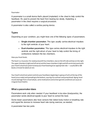
- 1. Pacemaker A pacemaker is a small device that's placed (implanted) in the chest to help control the heartbeat. It's used to prevent the heart from beating too slowly. Implanting a pacemaker in the chest requires a surgical procedure. A pacemaker is also called a cardiac pacing device. Types Depending on your condition, you might have one of the following types of pacemakers. Single chamber pacemaker. This type usually carries electrical impulses to the right ventricle of your heart. Dual chamber pacemaker. This type carries electrical impulses to the right ventricle and the right atrium of your heart to help control the timing of contractions between the two chambers. How yourheart beats The heart isa muscular,fist-sizedpumpwithfourchambers,twoon the leftside andtwoon the right. The upperchambers(rightand leftatria) andthe lowerchambers(rightandleftventricles) workwith your heart'selectrical systemtokeepyourheartbeatingatan appropriate rate — usually60 to 100 beatsa minute foradultsat rest. Your heart'selectrical systemcontrolsyourheartbeat,beginninginagroup of cellsat the top of the heart(sinusnode) andspreadingtothe bottom, causingitto contract and pumpblood.Aging,heart muscle damage froma heartattack, some medicationsandcertaingeneticconditionscancause an irregularheartrhythm. What a pacemaker does Pacemakers work only when needed. If your heartbeat is too slow (bradycardia), the pacemaker sends electrical signals to your heart to correct the beat. Some newer pacemakers also have sensors that detect body motion or breathing rate and signal the devices to increase heart rate during exercise, as needed. A pacemaker has two parts:
- 2. Pulse generator. This small metal container houses a battery and the electrical circuitry that controls the rate of electrical pulses sent to the heart. Leads (electrodes). One to three flexible, insulated wires are each placed in one or more chambers of the heart and deliver the electrical pulses to adjust the heart rate. However, some newer pacemakers don't require leads. These devices, called leadless pacemakers, are implanted directly into the heart muscle. Special precautions It's unlikelythatyourpacemakerwouldstopworkingproperlybecause of electrical interference.Still, you'll needtotake a fewprecautions: Cellphones.It'ssafe totalkon a cellphone,butkeepyourcellphone atleast6inches(15 centimeters) away fromyourpacemaker.Don'tkeepyourphone ina shirt pocket.Whentalkingonyourphone,hold it to the ear opposite the side where yourpacemakerwasimplanted. Securitysystems.Passingthroughanairportmetal detectorwon'tinterfere withyourpacemaker, althoughthe metal inthe pacemakercouldsoundthe alarm.But avoidlingeringnearor leaningagainst a metal-detectionsystem. To avoidpotential problems,carryanID card statingthatyou have a pacemaker. Medical equipment.Make sure all yourdoctorsand dentistsknow youhave a pacemaker.Certain medical procedures,suchasmagneticresonance imaging,CTscans,cancer radiationtreatment, electrocauterytocontrol bleedingduringsurgery,andshockwave lithotripsytobreakuplarge kidney stonesor gallstonescouldinterfere withyourpacemaker. Power-generatingequipment.Standatleast2 feet(61 centimeters) fromweldingequipment,high- voltage transformersormotor-generatorsystems.If youworkaroundsuch equipment,askyourdoctor aboutarranging a testin yourworkplace todetermine whetherthe equipmentaffectsyourpacemaker. Devicesthatare unlikelytointerfere withyourpacemakerincludemicrowaveovens,televisionsand remote controls,radios,toasters,electricblankets,electricshavers,andelectricdrills.