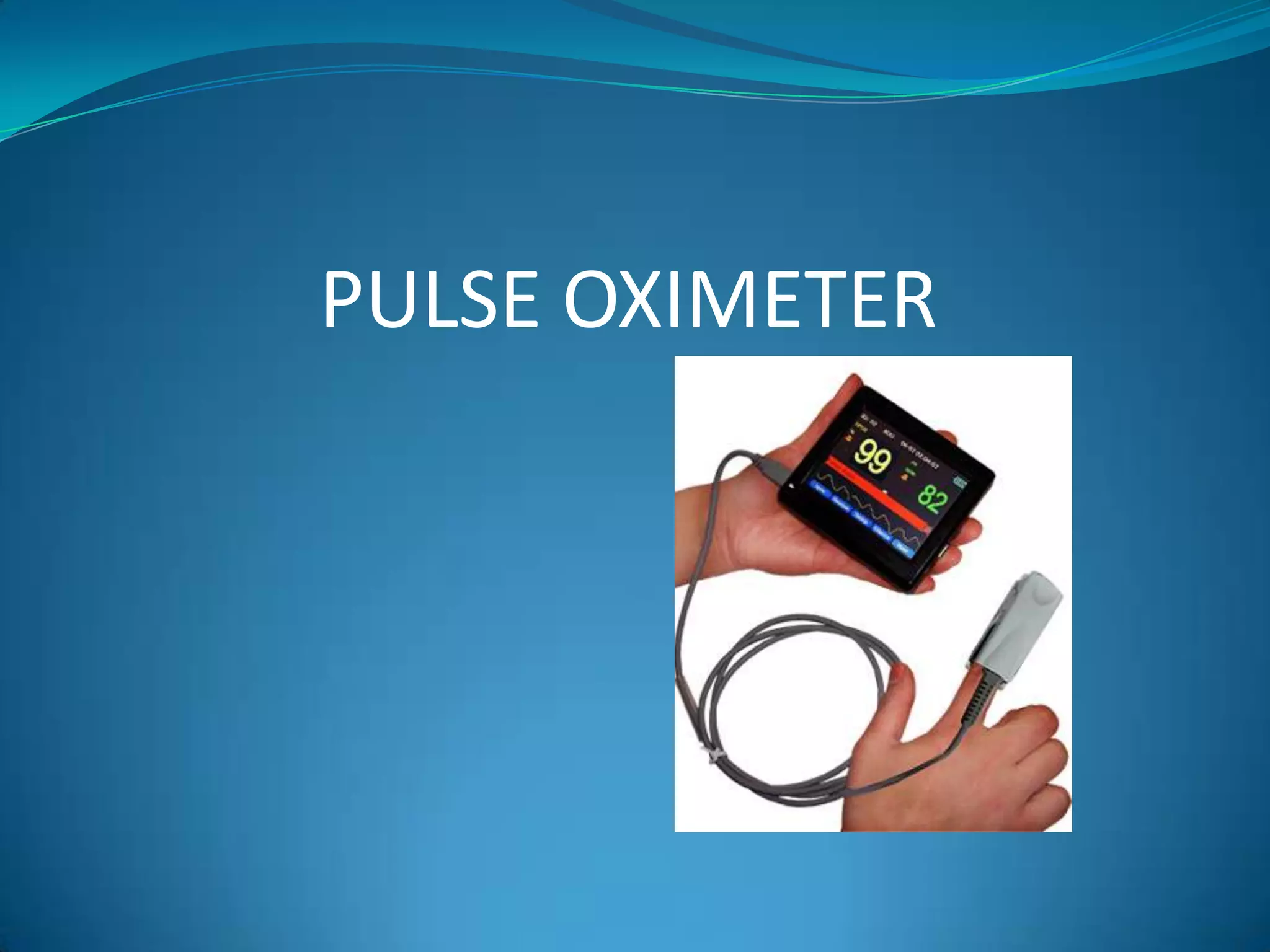
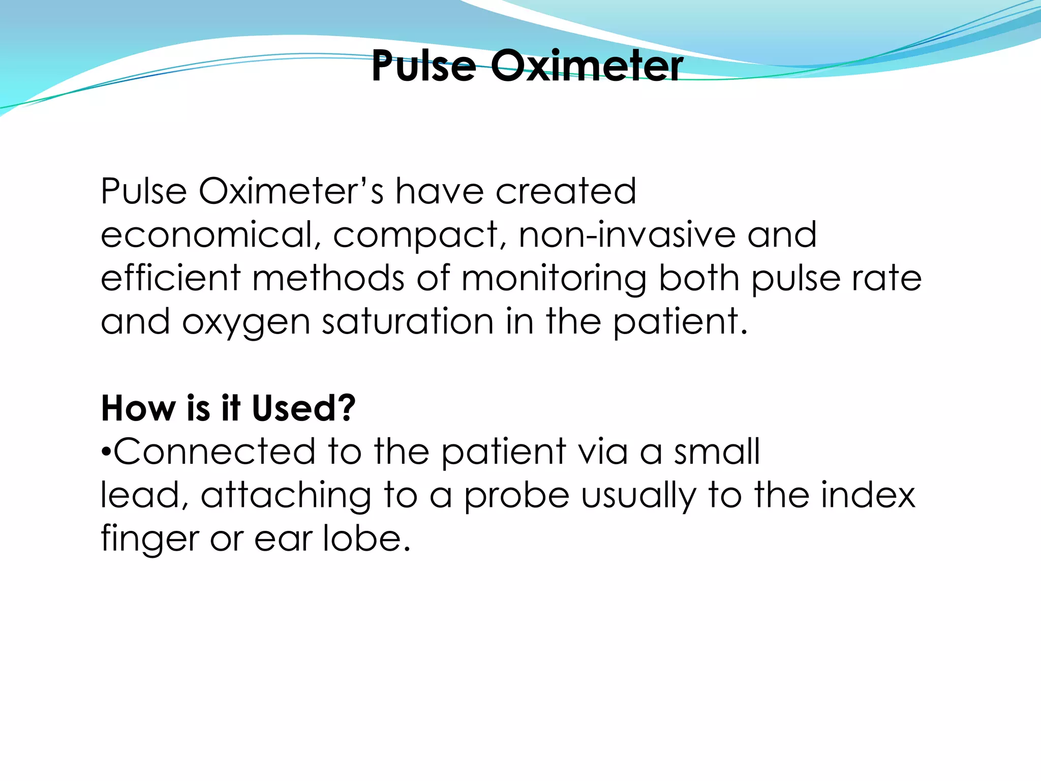
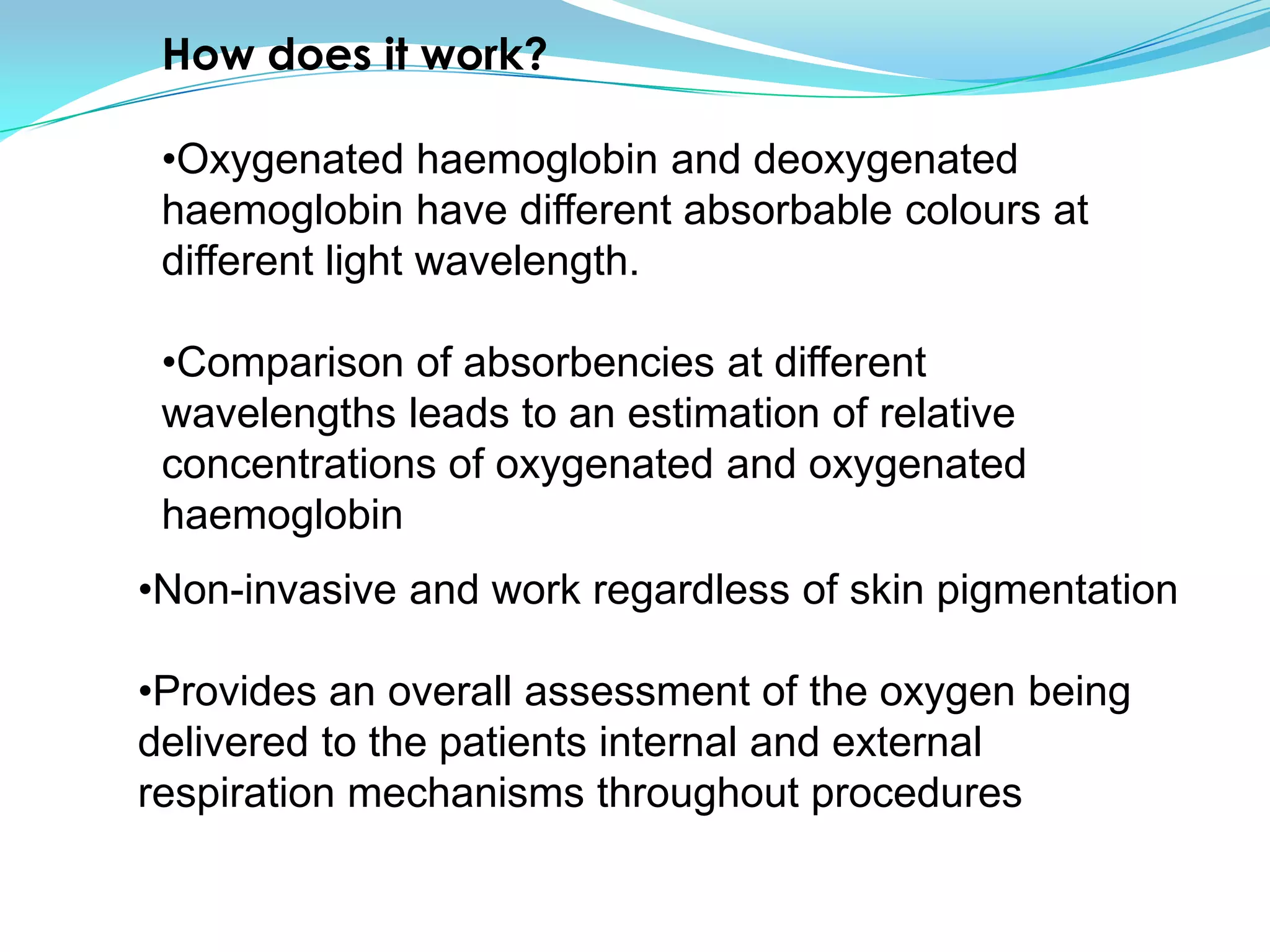
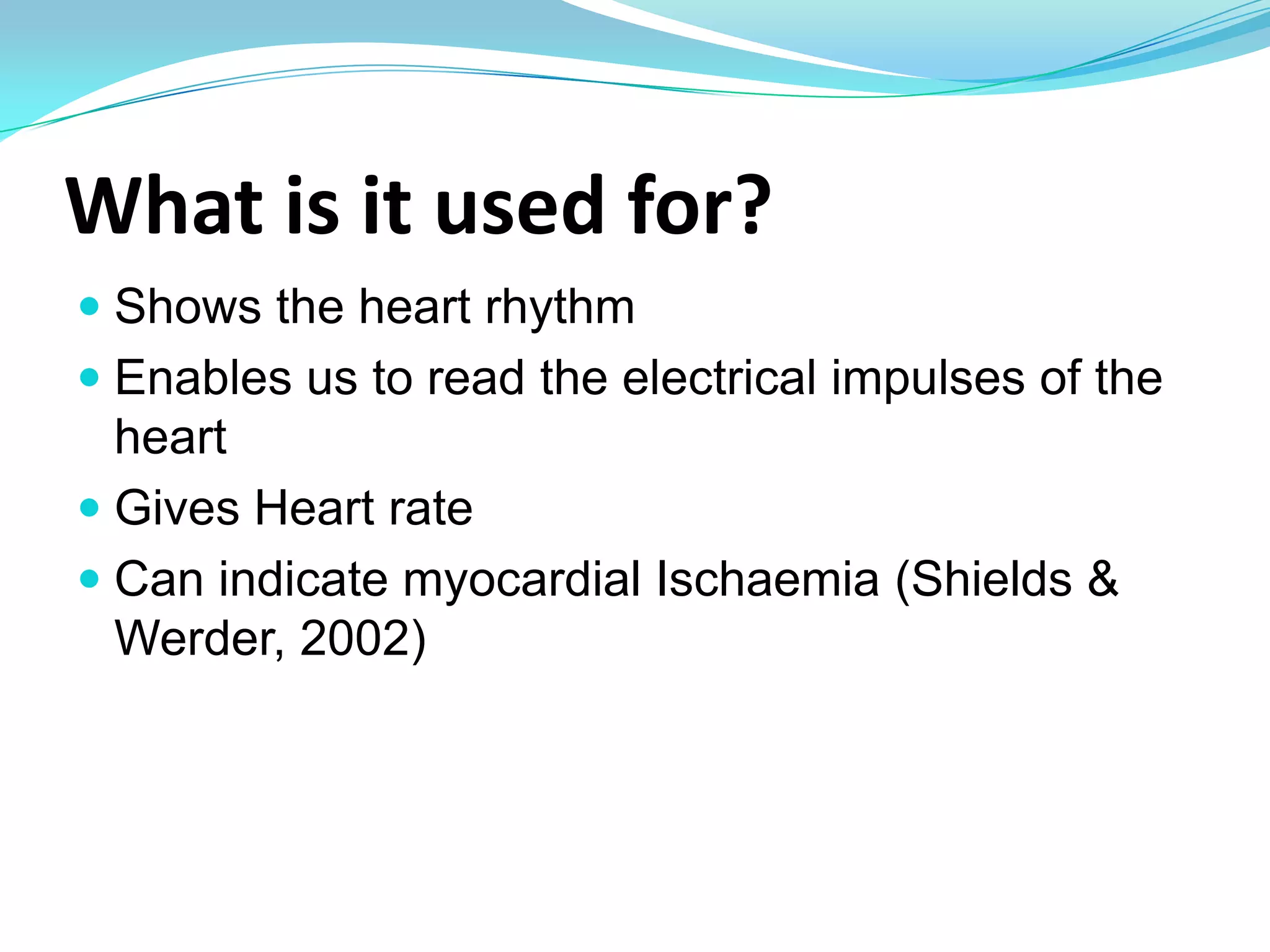
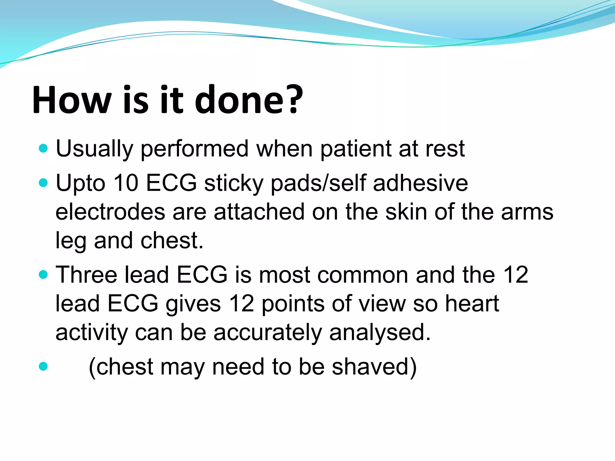
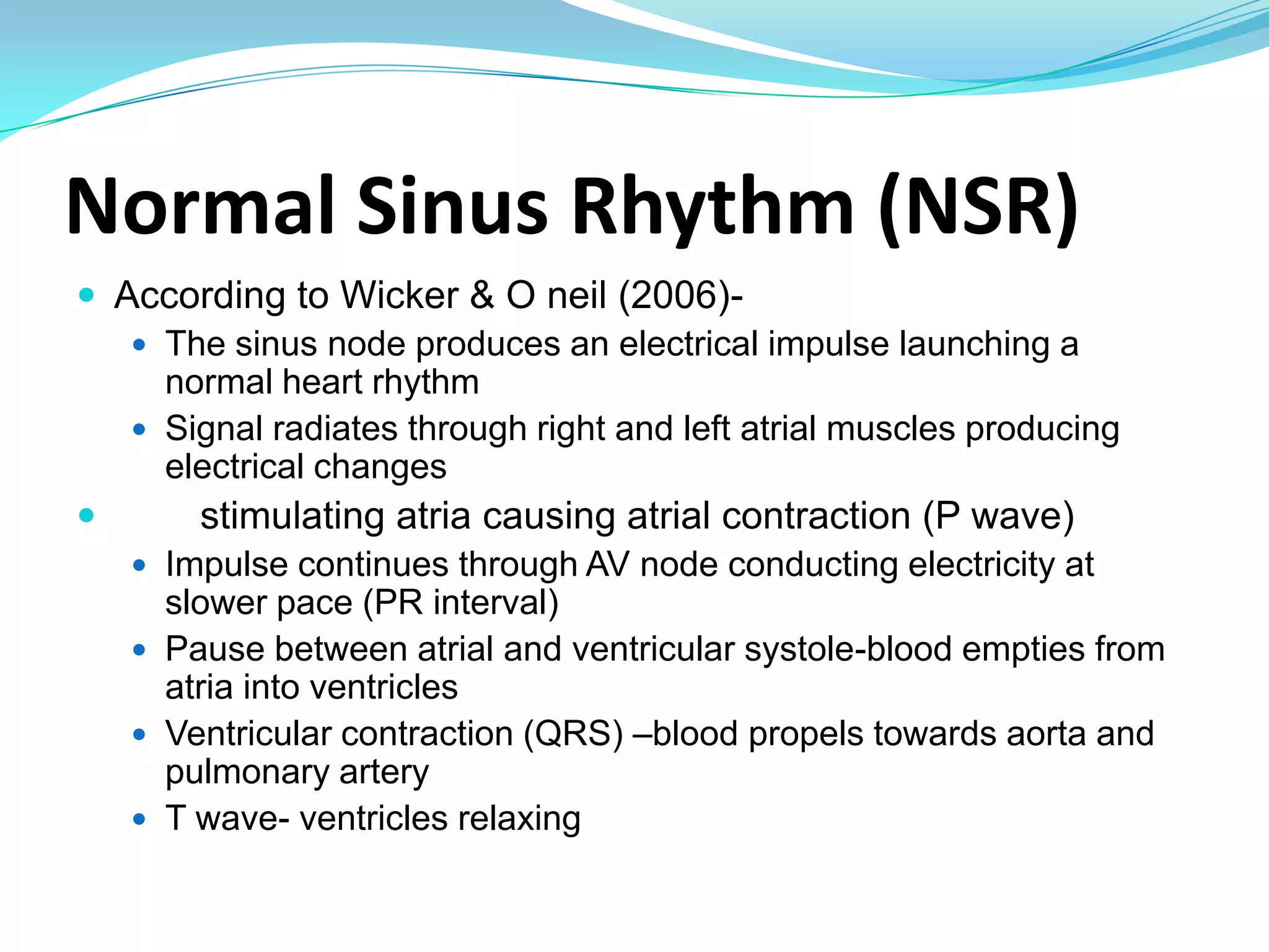
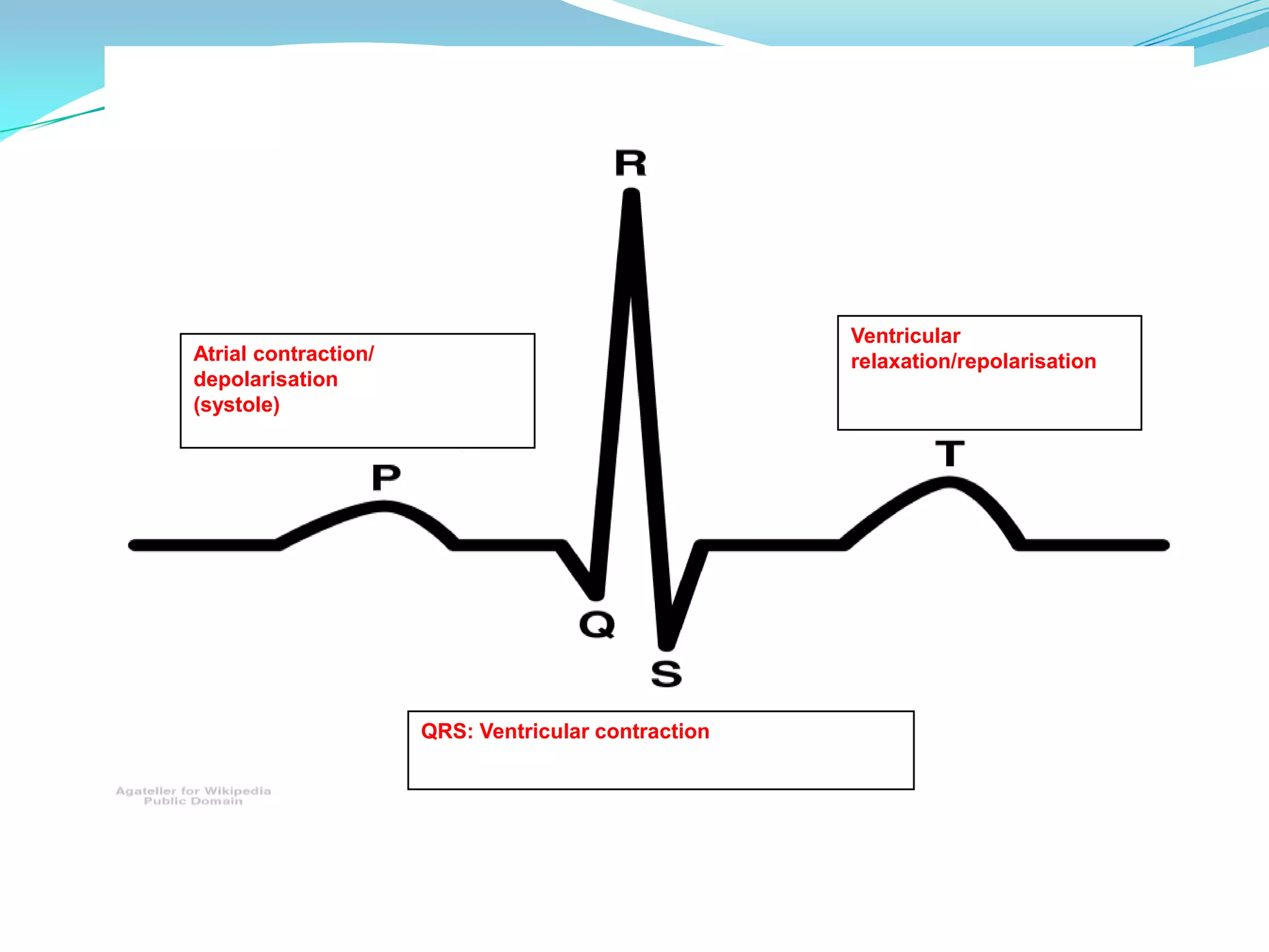
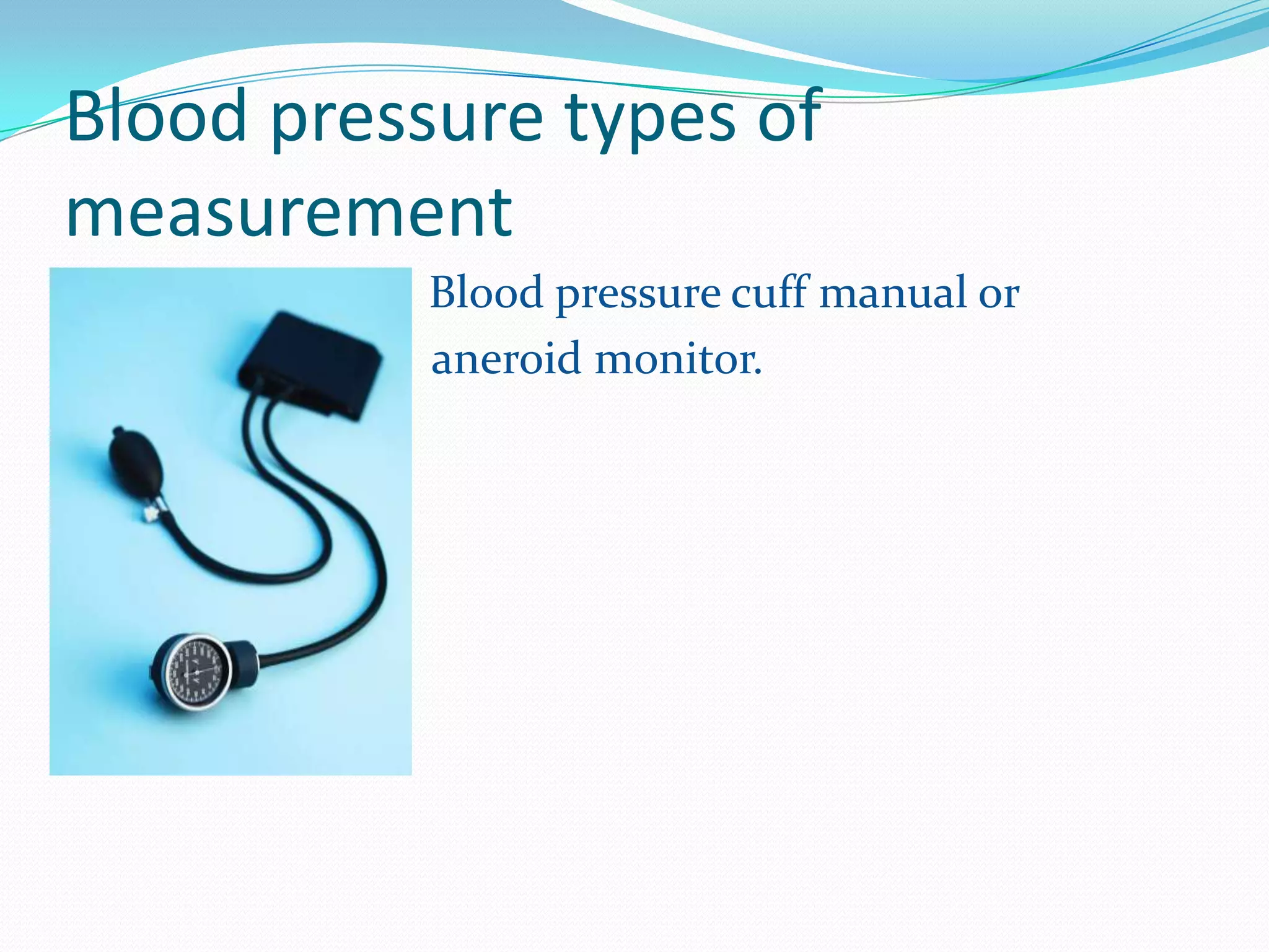
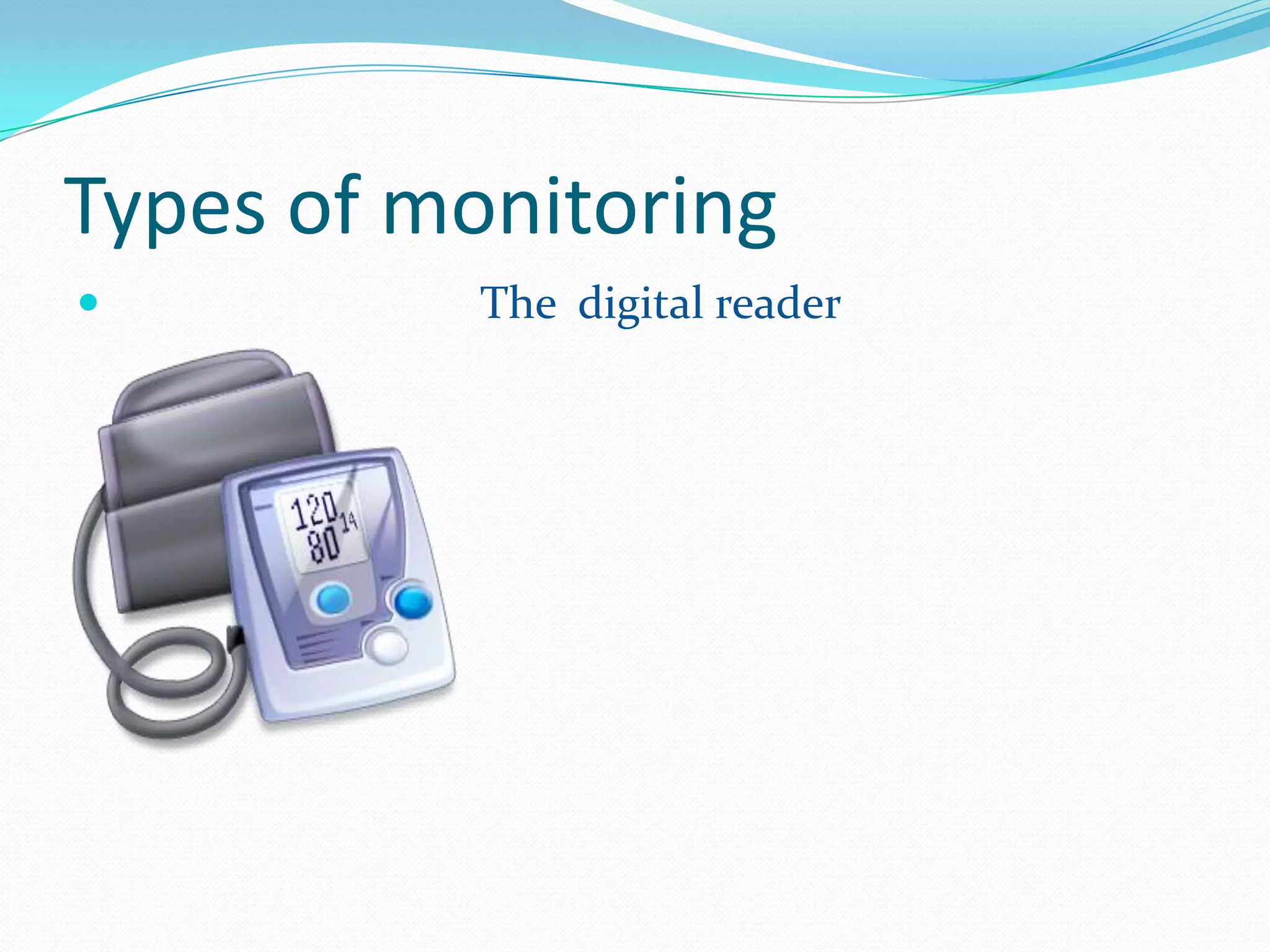
Pulse oximetry, ECG, and blood pressure monitoring are common ways to assess patients. A pulse oximeter uses light absorption to estimate oxygenated and deoxygenated hemoglobin levels non-invasively. An ECG attaches electrodes to record the heart's electrical activity and indicates rate, rhythm, and signs of ischemia. Blood pressure results from cardiac contraction and vascular resistance, and can be measured manually or via an arterial line that displays beat-to-beat variations to assess contractility, tone, and response to interventions.
8. What is it used for? Shows the heart rhythm Enables us to read the electrical impulses of the heart Gives Heart rate Can indicate myocardial Ischaemia (Shields & Werder, 2002)
9. How is it done? Usually performed when patient at rest Upto 10 ECG sticky pads/self adhesive electrodes are attached on the skin of the arms leg and chest. Three lead ECG is most common and the 12 lead ECG gives 12 points of view so heart activity can be accurately analysed. (chest may need to be shaved)
10. Normal Sinus Rhythm (NSR) According to Wicker & O neil (2006)- The sinus node produces an electrical impulse launching a normal heart rhythm Signal radiates through right and left atrial muscles producing electrical changes stimulating atria causing atrial contraction (P wave) Impulse continues through AV node conducting electricity at slower pace (PR interval) Pause between atrial and ventricular systole-blood empties from atria into ventricles Ventricular contraction (QRS) –blood propels towards aorta and pulmonary artery T wave- ventricles relaxing
12. This cardiac cycle repeats to create a rhythm Normal Heart rates: ADULT 60-80 BPM CHILD 100BPM INFANT 150BPM *Depends on health, mental state and BMI
13. Sinus Bradycardia Slow heart rate with normal sinus rhythm Can be benign but can be caused by beta blockers, hypothermia, stimulation of the vagus nerve, hypothyroidism etc Sinus Tachycardia Fast heart rate at normal rhythm Caused by shock, drug actions (e.g. Atropine), anxiety, hypovolaemia etc
14. Other Abnormal heart rhythms: Supraventricular tachycardia (SVT) impulse stimulating heart not coming from Sinus node, instead comes from tissue around AV node. HR up to 280 BPM Ventricular tachycardia tisues in ventricles casuing rapid irregular Heart rhythm Atrial Flutter Rapid heart rhythm. Abnormal. Impulse bypasses AV node
15. Other Abnormal heart rhythms: Atrioventricular block (AVB) Block in conduction between Sinus node and AV node. 2nd degree heart block- some signals from atria don’t reach ventricles= dropped beats 3rd degree= AV block means no impulses passing through AV node so ventricles create own rhythm Premature atrial contraction (PAC) Sinoatrial node fires early= early atrial contraction Premature ventricular contraction (PVC) Av node fires early- ventricles contract early
16. Other Abnormal heart rhythms: Atrial Fibrillation Electrical impulses fired irregularly from many sites in atria Asystole End of heartbeats, lack of electrical activity but not straight line so still some continuing residual activity
22. Why is it needed? To show beat to beat variation in the arterial trace. Other parameters that can be measured. Myocardial contractility. Vascular tone.
23. When would it be needed? Constantly changing clinical parameters in sick patients. Blood sampling- helpful multiple samples are needed. Acute interventions-major surgery.
24. Where is it used? Arm- radial, brachial and axillary arteries. Leg- dorsalis pedis, posterior tibialis and femoral. Transducer should be positioned level with heart to give accurate reading.
25. How is it measured? Transducer energy one form to another. Pressure change causes movement within transducer. This creates electrical current to be displayed on oscilloscope.
26. Placing the line Decide the anatomical site Palpate the artery. Clean area thoroughly-local anaesthetic? Seldinger technique- guidewires can be used to assist in insertion of the cannula up the lumen of the artery. Remove the needle, thread a cannula over the wire and up the artery before removing the wire.
27. Mark the arterial line to indicate it presence So as not to be mistaken for a venous line Ensure there is no air within the line Use heparinised saline to flush
It is essential to maintain the blood supply and the function of vital organsHigh blood pressure (hypertension) may indicate coronary heart disease, heart failure, stroke, kidney failure and many other health problemsLow bl pre hypotension my indicate heart problems endocrine or neurological disorders. Or may be an indication of dehydration It can vary with age typically the new born will have a reading of 80/50 while a constant rise will see a young adult have a blood pressure of 120/80, blood pressure as a rule will rise with ageForces. 1 is created by the heart as it pumps blood into the arteries 2 is the force of the arteries as they resist the blood flow
Arm is slightly flexed and the cuff placed around the bicep area, most cuffs have arrows pointing showing which where to place the cuff. Place a stethoscope just under the crease of the elbow. Ensuring the screw cap is tight you can inflate the cuff.Inflate the cuff until the radial pulse disappears Slowly unscrew the cap and let the air seep out slowly Listen for when blood passing through the arteris can be heard again and make a note of the number the pressure gauge is at this is the systolic or top number.When the blood can not be heard any more through the stethoscope this is the diastolic reading/ the bottom number
The cuff of this meter is placed in the same place as the others, but it digitally inflate and take the appropriate measurements it’s self