
Recommended
More Related Content
Similar to Bio-Medical Therapeutic of in Pacemaker& Respiratory
Similar to Bio-Medical Therapeutic of in Pacemaker& Respiratory (20)
Recently uploaded
Recently uploaded (20)
Bio-Medical Therapeutic of in Pacemaker& Respiratory
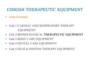
- 1. CBM368 THERAPEUTIC EQUIPMENT • Units Covered : • Unit 1 CARDIAC AND RESPIRATORY THERAPY EQUIPMENT • Unit 2 BIOMECHANICAL THERAPEUTIC EQUIPMENT • Unit 3 BODY CARE EQUIPMENT • Unit 4 DENTAL CARE EQUIPMENT • Unit 5 HEAT & PHOTON THERAPY EQUIPMENT
- 2. Unit 1 CARDIAC AND RESPIRATORY THERAPY EQUIPMENT • Cardiac Pacemaker: Internal and External Pacemaker- Programmable pacemakers. Cardiac Defibrillators: AC and DC Defibrillator- Internal and External Defibrillators – Protection Circuit, Defibrillator analyzers. Cardiac ablation catheter. Types of Ventilators – Pressure, Volume, and Time controlled. Basic principles of electromechanical, pneumatic and electronic ventilators, Patient Cycle Ventilators, Ventilator testing. Humidifiers, Nebulizers, Inhalators.
- 4. Cardiac and Respiratory System
- 6. Heart Disease • Coronary heart disease: Damage to the heart’s major blood arteries • Hypertension (high blood pressure) • Hypotension: Low blood pressure. • Cardiac arrest: A condition in which the heart stops beating, respiration stops, and the person loses consciousness. • Heart failure: A condition in which the heart fails to pump blood as efficiently as it should. It occurs when the heart does not pump the blood effectively • Arrhythmia: Irregular beating of the heart. • Bradycardia: Decreased heartbeat rate. • Tachycardia: Increased heartbeat rate • Ischemia: Restricted or reduced blood supply to heart muscles. • Angina pectoris: Acute chest pain caused due to insufficient blood supply to the heart muscle. • Coronary Thrombosis: It is a form of thrombosis (blood clots) in the right coronary arteries of the heart. • Stroke: The result of a disruption in the brain’s blood supply. • Congenital heart disease: A heart defect that appears before birth. • Blue baby: A condition in which a newborn baby’s skin turns blue. It is primarily caused by deficiencies in the heart, lungs, or blood
- 7. Symptoms of Heart Disease • heart problem symptoms listed below 1. Consistent chest pain 2. Shortness of breath 3. Irregular heartbeats 4. Dizziness or Light headedness 5. Feeling exhausted too often 6. Excessive sweating 7. Discomfort around the shoulders, the back, and the arm. 8. Swelling 9. Blue-tinged skin
- 21. A pacemaker is a device used to control an irregular heart rhythm. A pacemaker has flexible wires called leads. The wires are placed in one or more chambers of the heart. They deliver electrical signals to fix the heart rate. Some newer pacemakers don't need wires. Internal Pacemaker
- 27. Differences
- 29. Cardiac Defibrillators • Defibrillators are devices that apply an electric charge or current to the heart to restore a normal heartbeat. If the heart rhythm stops due to cardiac arrest, also known as sudden cardiac arrest (SCA), a defibrillator may help it start beating again. A sudden cardiac arrest is fatal unless treated right away with CPR (cardiopulmonary resuscitation) and a defibrillator.
- 30. Types of defibrillators • 1. Automated external defibrillators (AEDs) • 2. Implanted cardioverter defibrillators (ICDs) • 3. Wearable cardioverter defibrillators (WCDs) • Working Principle of defibrillators: • Defibrillators can detect sudden, dangerous heart rhythms or a cardiac arrest. If a defibrillator detects a cardiac arrest or a dangerous arrhythmia, it can send an electric charge to the heart to try to restore a normal heartbeat or rhythm.
- 31. Continue… Two types of defibrillators are showing below. 1. AC defibrillators 2. DC defibrillators
- 32. Diagram and Principle • AC Defibrillators An AC defibrillator is the oldest and simplest type. The construction of AC defibrillator is such that appropriate values are available for internal and external defibrillation. In AC defibrillation, a shock of 50 Hz a.c frequency is applied to the chest for a time of 0.25 to 1 second through electrodes. • Defibrillation continues until patient responds to the treatment. An AC defibrillator consists of a step-up transformer with primary and secondary winding, and two switches. A.C supply is given through switches and fuse to primary winding of the transformer. The timing circuit is connected with switch, which is used to preset the time for the defibrillator to deliver shock to the patient. • For safety reasons, secondary coil should be isolated from earth to avoid shock. To produce uniform and simultaneous contraction of heart muscles large currents are used for external defibrillation.
- 33. AC Defibrillators They are connected to the electrodes that delivers electric shock to the heart of the patient. Voltage value ranging between 250 V to 750 V is applied for AC external defibrillation. For internal fibrillation voltage values between 60 V to 250 V is applied.
- 34. DC Defibrillators • DC defibrillator does not produce side effects and produces normal heartbeat. Ventricular fibrillation is avoided when high-energy shock is passed through discharging capacitor that is exposed to heart or chest of the patient. DC defibrillator consists of auto transformer T1 that acts as primary of the high voltage transformer T2. • A diode rectifier rectifies the output voltage from T2. It is connected to vacuum type- high voltage over switch. At position A, switch is connected to one end of the capacitor. When connected in this position capacitor charges to a voltage. A foot switch present on the handle of the electrode is used to deliver shock to the patient. • Now the high voltage switch changes it position to B that makes the capacitor to discharge to the heart through electrodes. To slow down the discharge from the capacitor an inductor L is placed in one of the electrode lead. This L induces a counter voltage that reduces the capacitor discharge value.
- 35. Diagram of DC Defibrillators
- 36. Internal & External defibrillator • An implantable cardioverter-defibrillator (ICD) is a small battery-powered device placed in the chest. It detects and stops irregular heartbeats, also called arrhythmias. An ICD continuously checks the heartbeat. It delivers electric shocks, when needed, to restore a regular heart rhythm. • An AED, or automated external defibrillator, is used to help those experiencing sudden cardiac arrest. It's a sophisticated, yet easy-to-use, medical device that can analyze the heart's rhythm and, if necessary, deliver an electrical shock, or defibrillation, to help the heart re-establish an effective rhythm. • Internal Defibrillator External Defibrillator
- 39. Defibrillator analyzers • Defibrillator analyzers automate the inspection and preventive maintenance (IPM) testing of defibrillators. They need to be able to test at least four basic defibrillator performance characteristics: discharge energy, synchronized-mode operation, automated external defibrillation, and ECG monitoring.
- 40. Cardiac Ablation Catheter • A catheter ablation involves passing thin, flexible tubes, called catheters, through the blood vessels to the heart. The catheters record the heart's electrical activity and can pinpoint where the arrhythmia is coming from.
- 41. Continue… • Cardiac ablation is most often done using thin, flexible tubes called catheters that are inserted through a blood vessel. Less commonly, ablation is done during heart surgery. • Cardiac ablation is a treatment to stop or prevent irregular heartbeats, called arrhythmias. Depending on the type of irregular heartbeat, cardiac ablation may be one of the first treatments. • Risks •Bleeding or infection where the catheter was placed. •Blood vessel damage. •Heart valve damage. •A new irregular heartbeat or one that gets worse. •Slow heart rate, which may need a pacemaker to fix. •Blood clots in the legs or lungs. •Stroke or heart attack.
- 42. Continue… One of the following ablation methods is used to create small scars in the heart. The scars block the irregular heart rhythms: • Heat, called radiofrequency energy. • Extreme cold, called cryoablation. Catheter Insertion Process: 1.Inserts a small tube (called a sheath) through the skin and into a vein to create an opening. Usually, providers use a vein or an artery in your groin. Sometimes they go through veins in your arm or neck. 2.They insert the electrode catheters (thin tubes with wires) into the sheath. They thread the catheters through your vein or artery and up to your heart (using X-rays to guide them). 3.Uses the catheter to deliver hot or cold energy to the areas of your heart causing irregular rhythms. The catheter destroys the abnormal heart tissue to block those irregular rhythms. 4.Removes the catheter and sheath from your vein or artery.
- 43. Ventilators • A ventilator is a device that supports or recreates the process of breathing by pumping air into the lungs. Sometimes, people refer to it as a vent or breathing machine. • Ventilators play an important role in saving lives, in both hospitals and ambulances. People who require long-term ventilation can also use them at home. • Who needs a ventilator? People require ventilation if they are experiencing respiratory failure. There are many injuries and conditions that can cause respiratory failure, including •stroke •lung disease •spinal cord injury •polio •sudden cardiac arrest •head injury
- 44. Types of ventilator There are several ways a person can receive ventilator support. These include: Face mask ventilators- A face mask ventilator is a noninvasive method of supporting a person’s breathing and oxygen levels. To use one, a person wears a mask that fits over the nose and mouth while air blows into their airways and lungs. Mechanical ventilators - Mechanical ventilators are machines that take over the breathing process entirely. Doctors use these when a person cannot breathe on their own. • Mechanical ventilators work via a tube in a person’s throat, pumping air into the lungs and transporting carbon dioxide away. Manual resuscitator bags- Manual resuscitator bags are pieces of equipment that allow people to control the airflow of their ventilator with their hands. These devices consist of an empty bag, or “bladder,” that a person squeezes to pump air into the lungs. Tracheostomy ventilators - A tracheostomy is a procedure where a doctor creates an opening in the windpipe and inserts a tube, which allows air to flow in and out. This enables a person to breathe without using their nose or mouth. What are the 2 types of ventilators? Invasive ventilation with a tube inserted into the patient's airway, performed in the intensive care unit in the hospital. Noninvasive ventilation that can be used at home by people with respiratory difficulties.
- 45. Continue…
- 46. Continue…
- 48. Principle of Pneumatic System
- 49. Electronic Ventilators • The ventilator pushes a mixture of air and oxygen into the patient's lungs to get oxygen into the body. The ventilator can also hold a constant amount of low pressure, called positive end-expiratory pressure (PEEP). • A ventilator, sometimes called a mechanical ventilator, is a machine that helps you breathe when you're sick, injured, or sedated for an operation.
- 51. Humidifiers • A machine that puts moisture in the air. • Humidifiers are devices that add moisture to the air to prevent dryness that can cause irritation in many parts of the body.
- 52. Nebulizers • A nebulizer changes medication from a liquid to a mist so you can inhale it into your lungs. Nebulizers come in home (tabletop) and portable .
- 53. Inhalators • A device providing a mixture of oxygen and carbon dioxide for breathing that is used especially in conjunction with artificial respiration.