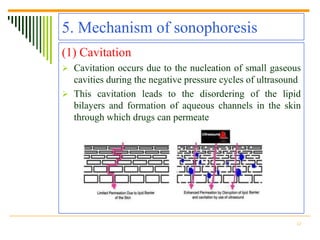
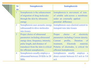
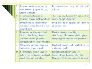
A
        Seminar on
  SONOPHORETIC
  DRUG DELIVERY
        Prepared By:
     Akshay.G.Patel
      M.PHARM (SEM-3)
         ROLL NO-5

DEPARTMENT OF PHARMCEUTICS
 NOOTAN PHARMACY COLLEGE
Contents

1. Introduction
2. Sonophoresis: a historical perspective
3. Generation of ultrasound
4. Biological effect of ultrasound
5. Mechanism of sonophoresis
6. Synergetic effect with other enhancers
7. Safety
8. Future trends
9. Applications of sonophoresis
10. Advantages
11. Limitations
12. Drugs used by sonophoretic drug delivery
13. Conclusion
14. References
                                               2
1. Introduction
 Definition: sonophoresis is the enhancement of
  migration of drug molecules through the skin by
  ultrasonic energy
 Sonophoresis occurs because ultrasound waves
  stimulate micro-vibrations within the skin epidermis
  and increase the overall kinetic energy of molecules
 When sound is emitted at a particular frequency, the
  sound waves disrupt the lipid bilayers
 The higher the frequency, the more dispersed the
  transmission


                                                     3
The skin
 Protective layer with large no. of dead cells, hence
  acts as barrier to penetration.
 Penetration varies with humidity, pigmentation, age,
  chemical status
 Of all layers Stratum Corneum (SC) offers
  maximum resistance. SC consists of keratinocytes
  and lipid bilayer
 Permeability can be increased by Chemicals,
  Electrical Fields or Ultrasound which disrupt lipid
  bilayer of SC and increase permeability.


                                                     4
2. Sonophoresis: a historical
perspective
 Sonophoresis was shown to enhance transdermal
  drug transport about 40 years ago by Fellinger and
  Schmidt who showed that application of ultrasound
  increases transport of hydrocortisone across the skin.
 ultrasound was also reported for local anesthetics
 Hofman and Moll who studied the percutaneous
  absorption of benzyl nicotinate.
 Bommannan et al. hypothesized that since the
  absorption coefficient of the skin varies directly with
  the ultrasound frequency

                                                        5
3. Generation of ultrasound
 Ultrasound is a sound wave possessing frequencies
  above 20 kHz .
 These waves are characterized by two main
  parameters, frequency and amplitude
 The waves used for sonophoresis which reduce the
  resistance offered by SC lie in the frequency range
  of 20 KHz to 20 MHz
 Ultrasound is generated with the help of a device
  called sonicator which is a AC electric signal
  generator. It produces a AC electric signal which is
  applied across a piezoelectric crystal i.e. transducer.

                                                        6
Figure 1: Ultrasonic generation system
  Pulse generator      Amplifier


        Gate           H. F. Generator
                       (20 KHz – 20MHz)


                                    Transducer
 Ultrasound gel
 + Drug                            Skin + Transducer interface


                                           Stratum cornium

                                           Skin


                                                                 7
Conti…
 Ultrasound is applied by bringing the transducer in
  contact with the skin.
 For sonophoretic delivery, the desired drug is
  dissolved in a solvent and applied to the skin.
 The coupling medium can be the same as the
  solvent used to dissolve the drug or it can be a
  commercial ultrasound coupling e.g. gel.
 It Helps to match impedence of tissue with the
  impedence of the transducer, so that the Ultrasound
  gets properly into the tissue.


                                                    8
Selection of ultrasound parameters
  (1) Ultrasound frequency
  a)     Therapeutic Frequency Ultrasound (1-3 MHz)
  b)     Low Frequency Ultrasound (Below 1MHz)
  c)     High Frequency Ultrasound (Above 3MHz)
  (2) Ultrasound intensity
       Various ultrasound intensities in the range of 0.1 to 2 W/cm2
  (3) Pulse length
      Ultrasound can be applied in a continuous or pulse mode.
       The pulse mode is frequently used because it reduces
       severity of side effects such as thermal effects.
      It was also found that urea permeability of cuprophane
       membrane increased from 6 to 56% as pulse length increased
       from 100 to 400 ms.
                                                                   9
4. Biological effect of ultrasound
 Significant   attention has thus been given to
   investigating the effects of ultrasound on biological
   tissues.
 Ultrasound affects biological tissues via three main
   effects
(1) Thermal effect may important when,
 The tissue has a high protein content
 A high intensity of continuous wave ultrasound is used
 Bone is included in the heated volume
 Vascularization is poor


                                                           10
Conti…
(2) Cavitation effect may important when,
 Low-frequency ultrasound is used
 Grassy fluids are exposed
 Small gas filled space are exposed
 The tissue temperature is higher than normal


(3) Acoustic streaming may important when,
 The medium has an acoustic impedance different from its
  surrounding
 The fluids in the biological medium is free to more
 Continuous wave application is used


                                                        11
5. Mechanism of sonophoresis
(1) Cavitation
 Cavitation occurs due to the nucleation of small gaseous
  cavities during the negative pressure cycles of ultrasound
 This cavitation leads to the disordering of the lipid
  bilayers and formation of aqueous channels in the skin
  through which drugs can permeate


                                                          12
Conti…
 The minimum ultrasound intensity required for the onset
  of cavitation, referred to as cavitation threshold
a) Cavitation inside skin
 cavitation bubbles near the keratinocytes–lipid bilayers
  interfaces may, in turn cause oscillations in the lipid
  bilayers, thereby causing structural disorder of the SC
  lipids
b) Cavitation out side skin
 cavitation bubbles can potentially play a role in the
  observed transdermal transport enhancement. these
  bubbles cause skin erosion, due to generation of shock
  waves, thereby enhancing transdermal transport


                                                            13
Conti…
(2) Convective transport
 Fluid velocities generated in this way may affect
  transdermal transport by inducing convective
  transport of the permeant across the skin.
 especially through hair follicles and sweat ducts


                                                  14
Conti…
(3) Mechanical stress
 Ultrasound is a longitudinal pressure wave inducing
  pressure variations in the skin, which, in turn, induce
  density variation
 Due to density variations, such as generation of
  cyclic stresses because of density changes that
  ultimately lead to fatigue of the medium
 Lipid bilayers, being self-assembled structures, can
  easily be disordered by these stresses, which result
  in an increase in the bilayers permeability


                                                       15
6. Synergistic effect with other enhancer
(1) Chemical enhancer
 Enhanced Partitioning
 Lipid Bilayer Disordering
 Keratin Denaturation
e.g. Application of SLS alone for 90 min induced about 3-
   fold increase in mannitol permeability, while application
   of ultrasound alone for 90 min induced about 8-fold
   enhancement. However, when combined, application of
   ultrasound from 1% SLS solution induced about 200-fold
   increase in skin permeability to mannitol


                                                          16
Conti…
(2) Iontophoresis
 Electrophoresis
 Lipid Bilayer Disordering
 Electroosmosis
e.g. The enhancement of heparin flux due to ultrasound +
  iontophoresis treatment was about 56-fold. This
  enhancement was higher than the sum of those obtained
  during ultrasound alone (3-fold) and iontophoresis alone
  (15-fold).
  Thus, the effect of ultrasound and iontophoresis on
  transdermal heparin transport was truly synergistic.


                                                        17
7.Safety
 The World Federation for Ultrasound in Medicine
  and Biology (WFUMB) has issued several
  publications related to safety of ultrasound
  bioeffects, addressing specifically thermal bioeffects
  and non-thermal bioeffects
 ultrasound affecting the structure of the skin: is it a
  reversible or irreversible change?
 What is the role of the free radicals that are
  generated during the cavitation process within the
  skin?


                                                       18
8. Future trends
(1) Vaccination
 In recent years, the potential for exploiting the skin for
  purposes of vaccination has received a great deal of
  attention
 One common strategy is to use an adjuvant, which is a
  compound used to enhance the immune response to
  vaccine compounds
 Ultrasound can be used to enhance skin permeability to
  both the adjuvant and the vaccine, and hence to facilitate
  their delivery to the target cells.


                                                          19
Conti…
(2) Gene therapy
 Gene therapy is a technique for correcting defective
  genes that are responsible for disease development, most
  commonly by replacing an „abnormal‟ disease-causing
  gene with the „normal‟ gene
 The most obvious candidate diseases for cutaneous gene
  therapy are the severe forms of particular genodermatoses
  (monogenic skin disorders), such as epidermolysis
  bullosa
 Other applications might be healing of cutaneous wounds
  such as severe burns and skin wounds of diabetic origin


                                                         20
9. Applications
1) Sonophoresis is used in the treatment of damaged skin.
2) Hormone Delivery.
3) In surgery it helps in dissection, connection, built-up and
    treatment of biological tissue.
4) Low-Frequency Ultrasonic Gene Delivery.
5) Sonophoresis is also very useful in drug enhancement in
    granulomas and tumors. Most cancer therapy drugs act
    intracellularly.
6) Ultrasound is used for Calcific Tendinitis of the Shoulder.
7) The dolphin therapy and sonophoretic model.
8) Ultrasound Helps in Treating Tennis Elbow and Tendon
    Problem.

                                                            21
10. Advantages
1) Avoids vagaries associated with gastrointestinal absorption due
   to pH, enzymatic activity, drug-food interactions etc.
2) Substitute oral administration when the route is unsuitable as in
   case of vomiting, diarrhea.
3) Avoids hepatic “first pass” effect.
4) Avoids the risks and inconveniences of parenteral therapy.
5) Reduces daily dosing, thus, improving patient compliance.
6) Extends the activity of drugs having short plasma half-life
   through the reservoir of drug present in the therapeutic delivery
   system and its controlled release characteristics.
7) Rapid termination of drug effect by removal of drug application
   from the surface of the skin.
8) Rapid identification of the medication in emergencies. (e.g..
   Non-responsive, unconscious, or comatose patient.)
                                                                  22
Conti…
9) Elimination of the hazards and difficulties of I.V. infusions or
   I.M. injections.
10) Enhance therapeutic efficacy, reduced side effects due to
   optimization of the blood concentration-time profile and
   elimination of pulse entry of drugs into the systemic
   circulation.
11) Provide predictable activity over extended duration of time and
   ability to approximate zero-order kinetics.
12) Improved control of the concentrations of drug with small
   therapeutic indices.
13) Minimize inter and intrapatient variation.
14) Suitability for self-administration.


                                                                 23
11. Limitations
1) Only limited number of potent drugs can be absorbed in
   therapeutic dose.
2) Many systemically effective therapeutic drugs produce skin
   irritation.
3) The drug must have some desirable physicochemical properties
   for penetration through stratum corneum.
4) If the drug dosage required for therapeutic value is more than
   10mg/day, the transdermal delivery will be very difficult.
5) The barrier function of the skin changes from one site to another
   on the same person, from person to person and with age


                                                                  24
12. Drug used by sonophoresis
(1) Sonophoresis with Corticosteroid
 Majority of studies on sonophoresis, ultrasound was used
   to enhanced the delivery of steroidal anti-inflammatory
   drugs (e.g. hydrocortisone).
(a) Fellinger & Schmid (1954) showed that ultrasound could
   carry hydrocortisone across a vascular membrane for the
   effective treatment of polyarthritis
(b) Newman et al. (1992) suggested that hydrocortisone
   sonophoresis is useful in the treatment of numerous
   musculo-skeletal injuries.


                                                        25
Conti…
(2) Sonophoresis with Salicylates
 In combination with ultrasound it is thought that
   Salicylate could be moved into deeper, subdermal tissues
   to help to reduce pain.
(a) Ciccone et al. (1991) reported on a study to evaluate
   ultrasound as an enhancer of topically applied Salicylates


                                                           26
Conti…
(3) Sonophoresis with Anesthetics
 The effectiveness of sonophoresis has been explored
   extensively for delivery of local anesthetics.
(a) McElnay (1985) and associates described a sonophoresis
   study using lignocain
(b) Moll (1979) studied the enhanced effects of topically
   applied Lidocaine (140.7 mg) and Decadron (3.75 mg).
   She obtained that 88% of the patients receiving
   sonophoresis with Decadron and Lidocaine obtained
   relief from their trigger point pain.


                                                        27
Conti...
(4) Sonophoresis with other Drugs
(a) Benson and colleagues (1987, 1989) studied ultrasound
   as an enhancer of benzydamine hydrochloride (3%) a
   nonsteroidal anti-inflammatory drug.
(b) Levy et al. (1989) studied the sonophoresis of D-
   mannitol, a diuretic.
(c) Romanenko (1992) used ultrasound with topically
   applied Amphotericin B.


                                                       28
Sonophoresis vs. iontophoresis
 Both   techniques    deliver   chemical   to
 biological tissues.


                                             29
Sr            Sonophoresis                            Iontophoresis
no
1  Sonophoresis is the enhancement        Iontophoresis is movement of ions
   of migration of drug molecules         of soluble salts across a membrane
   through the skin by ultrasonic         under an externally applied
   energy                                 potential difference
2  Sonophoresis uses acoustic energy      Iontophoresis uses electiral current to
   (ultrasound) to drive molecules        transport ions into tissues
   into tissues
3  Proper choice of ultrasound            Proper      choice     of   electricity
   parameters including ultrasound        parameters including Current density,
   energy dose, frequency, intensity,     Current      profile,   Duration     of
   pulse length, and distance of          treatment,      Electrode     material,
   transducer from the skin is critical   Polarity of electrodes, is critical for
   for efficient sonophoresis.            efficient Iontophoresis
4 Sonophoresis usually employs a          Iontophoresis usually employs a
   ultrasound between 20 KHz to 20        direct current between 0.5 mA to 5.0
   MHz                                    mA
                                                                            30
5   In sonophoresis drugs mixing        In Iontophoresis drug is mix with
    with a coupling agent like gel,     solvent
    cream, ointment
6   The main mechanism for              The main mechanism for transport of
    transport of drug is “Cavitation”   drug is “Electroporation”
7   Drug should be in aqueous or        Drug must be in aqueous and must be
    non aqueous and ionized or non      in ionized form
    ionized form
8   Enhanced partitioning, Lipid        Electrophoresis, Lipid bilayer
    bilayer disordering, Keratin        disordering, Electroosmosis etc. gives
    denaturation etc. gives the         the synergetic effect of Iontophoresis
    synergetic effect of sonophoresis
9   Ultrasound can be applied in a      Electrical current can be applied only in
    continuous or pulse mode            continuous mode
10 Sonophoresis mostly used for       Iontophoresis mostly used for
   delivery of corticosteroids, local hyperhydrosis diagnosis of cystic
   anesthetics and salicylates        fibrosis, metallic and non-metallic ions

                                                                            31
13. Conclusion
 Proper choice of ultrasound parameters including
  ultrasound energy dose, frequency, intensity, pulse
  length, and distance of transducer from the skin is
  critical for efficient sonophoresis.
 Various studies have indicated that application of
  ultrasound under conditions used for sonophoresis
  does not cause any permanent damage to the skin
 Ultrasound also works synergistically with several
  other     enhancers      including   chemicals  and
  iontophoresis.


                                                   32
14. References
 N.K.Jain, Sonophoresis: Biophysical of Transdermal Drug
  Delivery, Controlled and Novel Drug Delivery, 1st edition,
  1997, page. 208-235
 James Swarbrick, Transdermal Delivery: Sonophoresis,
  Encyclopedia of pharmaceutical technology, 3rd edition,
  Volume-6, 2007, page no. 3828-3842
 Mr. Ashish Pahade, Dr. Mrs. V.M.Jadhav, Dr. Mr. V.J.Kadam,
  Sonophoresis: an overview, International Journal of
  Pharmaceutical Science, 2010, Volume 3, Issue 2, page. 24-32
 http://www.dolphintherapy.ru/en/sonoforez.shtml


                                                            33
Thank you…


             34

Sonophorosis by akshay patel

  • 1.
    A Seminar on SONOPHORETIC DRUG DELIVERY Prepared By: Akshay.G.Patel M.PHARM (SEM-3) ROLL NO-5 DEPARTMENT OF PHARMCEUTICS NOOTAN PHARMACY COLLEGE
  • 2.
    Contents 1. Introduction 2. Sonophoresis:a historical perspective 3. Generation of ultrasound 4. Biological effect of ultrasound 5. Mechanism of sonophoresis 6. Synergetic effect with other enhancers 7. Safety 8. Future trends 9. Applications of sonophoresis 10. Advantages 11. Limitations 12. Drugs used by sonophoretic drug delivery 13. Conclusion 14. References 2
  • 3.
    1. Introduction  Definition:sonophoresis is the enhancement of migration of drug molecules through the skin by ultrasonic energy  Sonophoresis occurs because ultrasound waves stimulate micro-vibrations within the skin epidermis and increase the overall kinetic energy of molecules  When sound is emitted at a particular frequency, the sound waves disrupt the lipid bilayers  The higher the frequency, the more dispersed the transmission 3
  • 4.
    The skin  Protectivelayer with large no. of dead cells, hence acts as barrier to penetration.  Penetration varies with humidity, pigmentation, age, chemical status  Of all layers Stratum Corneum (SC) offers maximum resistance. SC consists of keratinocytes and lipid bilayer  Permeability can be increased by Chemicals, Electrical Fields or Ultrasound which disrupt lipid bilayer of SC and increase permeability. 4
  • 5.
    2. Sonophoresis: ahistorical perspective  Sonophoresis was shown to enhance transdermal drug transport about 40 years ago by Fellinger and Schmidt who showed that application of ultrasound increases transport of hydrocortisone across the skin.  ultrasound was also reported for local anesthetics  Hofman and Moll who studied the percutaneous absorption of benzyl nicotinate.  Bommannan et al. hypothesized that since the absorption coefficient of the skin varies directly with the ultrasound frequency 5
  • 6.
    3. Generation ofultrasound  Ultrasound is a sound wave possessing frequencies above 20 kHz .  These waves are characterized by two main parameters, frequency and amplitude  The waves used for sonophoresis which reduce the resistance offered by SC lie in the frequency range of 20 KHz to 20 MHz  Ultrasound is generated with the help of a device called sonicator which is a AC electric signal generator. It produces a AC electric signal which is applied across a piezoelectric crystal i.e. transducer. 6
  • 7.
    Figure 1: Ultrasonicgeneration system Pulse generator Amplifier Gate H. F. Generator (20 KHz – 20MHz) Transducer Ultrasound gel + Drug Skin + Transducer interface Stratum cornium Skin 7
  • 8.
    Conti…  Ultrasound isapplied by bringing the transducer in contact with the skin.  For sonophoretic delivery, the desired drug is dissolved in a solvent and applied to the skin.  The coupling medium can be the same as the solvent used to dissolve the drug or it can be a commercial ultrasound coupling e.g. gel.  It Helps to match impedence of tissue with the impedence of the transducer, so that the Ultrasound gets properly into the tissue. 8
  • 9.
    Selection of ultrasoundparameters (1) Ultrasound frequency a) Therapeutic Frequency Ultrasound (1-3 MHz) b) Low Frequency Ultrasound (Below 1MHz) c) High Frequency Ultrasound (Above 3MHz) (2) Ultrasound intensity Various ultrasound intensities in the range of 0.1 to 2 W/cm2 (3) Pulse length  Ultrasound can be applied in a continuous or pulse mode. The pulse mode is frequently used because it reduces severity of side effects such as thermal effects.  It was also found that urea permeability of cuprophane membrane increased from 6 to 56% as pulse length increased from 100 to 400 ms. 9
  • 10.
    4. Biological effectof ultrasound  Significant attention has thus been given to investigating the effects of ultrasound on biological tissues.  Ultrasound affects biological tissues via three main effects (1) Thermal effect may important when,  The tissue has a high protein content  A high intensity of continuous wave ultrasound is used  Bone is included in the heated volume  Vascularization is poor 10
  • 11.
    Conti… (2) Cavitation effectmay important when,  Low-frequency ultrasound is used  Grassy fluids are exposed  Small gas filled space are exposed  The tissue temperature is higher than normal (3) Acoustic streaming may important when,  The medium has an acoustic impedance different from its surrounding  The fluids in the biological medium is free to more  Continuous wave application is used 11
  • 12.
    5. Mechanism ofsonophoresis (1) Cavitation  Cavitation occurs due to the nucleation of small gaseous cavities during the negative pressure cycles of ultrasound  This cavitation leads to the disordering of the lipid bilayers and formation of aqueous channels in the skin through which drugs can permeate 12
  • 13.
    Conti…  The minimumultrasound intensity required for the onset of cavitation, referred to as cavitation threshold a) Cavitation inside skin  cavitation bubbles near the keratinocytes–lipid bilayers interfaces may, in turn cause oscillations in the lipid bilayers, thereby causing structural disorder of the SC lipids b) Cavitation out side skin  cavitation bubbles can potentially play a role in the observed transdermal transport enhancement. these bubbles cause skin erosion, due to generation of shock waves, thereby enhancing transdermal transport 13
  • 14.
    Conti… (2) Convective transport Fluid velocities generated in this way may affect transdermal transport by inducing convective transport of the permeant across the skin.  especially through hair follicles and sweat ducts 14
  • 15.
    Conti… (3) Mechanical stress Ultrasound is a longitudinal pressure wave inducing pressure variations in the skin, which, in turn, induce density variation  Due to density variations, such as generation of cyclic stresses because of density changes that ultimately lead to fatigue of the medium  Lipid bilayers, being self-assembled structures, can easily be disordered by these stresses, which result in an increase in the bilayers permeability 15
  • 16.
    6. Synergistic effectwith other enhancer (1) Chemical enhancer  Enhanced Partitioning  Lipid Bilayer Disordering  Keratin Denaturation e.g. Application of SLS alone for 90 min induced about 3- fold increase in mannitol permeability, while application of ultrasound alone for 90 min induced about 8-fold enhancement. However, when combined, application of ultrasound from 1% SLS solution induced about 200-fold increase in skin permeability to mannitol 16
  • 17.
    Conti… (2) Iontophoresis  Electrophoresis Lipid Bilayer Disordering  Electroosmosis e.g. The enhancement of heparin flux due to ultrasound + iontophoresis treatment was about 56-fold. This enhancement was higher than the sum of those obtained during ultrasound alone (3-fold) and iontophoresis alone (15-fold). Thus, the effect of ultrasound and iontophoresis on transdermal heparin transport was truly synergistic. 17
  • 18.
    7.Safety  The WorldFederation for Ultrasound in Medicine and Biology (WFUMB) has issued several publications related to safety of ultrasound bioeffects, addressing specifically thermal bioeffects and non-thermal bioeffects  ultrasound affecting the structure of the skin: is it a reversible or irreversible change?  What is the role of the free radicals that are generated during the cavitation process within the skin? 18
  • 19.
    8. Future trends (1)Vaccination  In recent years, the potential for exploiting the skin for purposes of vaccination has received a great deal of attention  One common strategy is to use an adjuvant, which is a compound used to enhance the immune response to vaccine compounds  Ultrasound can be used to enhance skin permeability to both the adjuvant and the vaccine, and hence to facilitate their delivery to the target cells. 19
  • 20.
    Conti… (2) Gene therapy Gene therapy is a technique for correcting defective genes that are responsible for disease development, most commonly by replacing an „abnormal‟ disease-causing gene with the „normal‟ gene  The most obvious candidate diseases for cutaneous gene therapy are the severe forms of particular genodermatoses (monogenic skin disorders), such as epidermolysis bullosa  Other applications might be healing of cutaneous wounds such as severe burns and skin wounds of diabetic origin 20
  • 21.
    9. Applications 1) Sonophoresisis used in the treatment of damaged skin. 2) Hormone Delivery. 3) In surgery it helps in dissection, connection, built-up and treatment of biological tissue. 4) Low-Frequency Ultrasonic Gene Delivery. 5) Sonophoresis is also very useful in drug enhancement in granulomas and tumors. Most cancer therapy drugs act intracellularly. 6) Ultrasound is used for Calcific Tendinitis of the Shoulder. 7) The dolphin therapy and sonophoretic model. 8) Ultrasound Helps in Treating Tennis Elbow and Tendon Problem. 21
  • 22.
    10. Advantages 1) Avoidsvagaries associated with gastrointestinal absorption due to pH, enzymatic activity, drug-food interactions etc. 2) Substitute oral administration when the route is unsuitable as in case of vomiting, diarrhea. 3) Avoids hepatic “first pass” effect. 4) Avoids the risks and inconveniences of parenteral therapy. 5) Reduces daily dosing, thus, improving patient compliance. 6) Extends the activity of drugs having short plasma half-life through the reservoir of drug present in the therapeutic delivery system and its controlled release characteristics. 7) Rapid termination of drug effect by removal of drug application from the surface of the skin. 8) Rapid identification of the medication in emergencies. (e.g.. Non-responsive, unconscious, or comatose patient.) 22
  • 23.
    Conti… 9) Elimination ofthe hazards and difficulties of I.V. infusions or I.M. injections. 10) Enhance therapeutic efficacy, reduced side effects due to optimization of the blood concentration-time profile and elimination of pulse entry of drugs into the systemic circulation. 11) Provide predictable activity over extended duration of time and ability to approximate zero-order kinetics. 12) Improved control of the concentrations of drug with small therapeutic indices. 13) Minimize inter and intrapatient variation. 14) Suitability for self-administration. 23
  • 24.
    11. Limitations 1) Onlylimited number of potent drugs can be absorbed in therapeutic dose. 2) Many systemically effective therapeutic drugs produce skin irritation. 3) The drug must have some desirable physicochemical properties for penetration through stratum corneum. 4) If the drug dosage required for therapeutic value is more than 10mg/day, the transdermal delivery will be very difficult. 5) The barrier function of the skin changes from one site to another on the same person, from person to person and with age 24
  • 25.
    12. Drug usedby sonophoresis (1) Sonophoresis with Corticosteroid  Majority of studies on sonophoresis, ultrasound was used to enhanced the delivery of steroidal anti-inflammatory drugs (e.g. hydrocortisone). (a) Fellinger & Schmid (1954) showed that ultrasound could carry hydrocortisone across a vascular membrane for the effective treatment of polyarthritis (b) Newman et al. (1992) suggested that hydrocortisone sonophoresis is useful in the treatment of numerous musculo-skeletal injuries. 25
  • 26.
    Conti… (2) Sonophoresis withSalicylates  In combination with ultrasound it is thought that Salicylate could be moved into deeper, subdermal tissues to help to reduce pain. (a) Ciccone et al. (1991) reported on a study to evaluate ultrasound as an enhancer of topically applied Salicylates 26
  • 27.
    Conti… (3) Sonophoresis withAnesthetics  The effectiveness of sonophoresis has been explored extensively for delivery of local anesthetics. (a) McElnay (1985) and associates described a sonophoresis study using lignocain (b) Moll (1979) studied the enhanced effects of topically applied Lidocaine (140.7 mg) and Decadron (3.75 mg). She obtained that 88% of the patients receiving sonophoresis with Decadron and Lidocaine obtained relief from their trigger point pain. 27
  • 28.
    Conti... (4) Sonophoresis withother Drugs (a) Benson and colleagues (1987, 1989) studied ultrasound as an enhancer of benzydamine hydrochloride (3%) a nonsteroidal anti-inflammatory drug. (b) Levy et al. (1989) studied the sonophoresis of D- mannitol, a diuretic. (c) Romanenko (1992) used ultrasound with topically applied Amphotericin B. 28
  • 29.
    Sonophoresis vs. iontophoresis Both techniques deliver chemical to biological tissues. 29
  • 30.
    Sr Sonophoresis Iontophoresis no 1 Sonophoresis is the enhancement Iontophoresis is movement of ions of migration of drug molecules of soluble salts across a membrane through the skin by ultrasonic under an externally applied energy potential difference 2 Sonophoresis uses acoustic energy Iontophoresis uses electiral current to (ultrasound) to drive molecules transport ions into tissues into tissues 3 Proper choice of ultrasound Proper choice of electricity parameters including ultrasound parameters including Current density, energy dose, frequency, intensity, Current profile, Duration of pulse length, and distance of treatment, Electrode material, transducer from the skin is critical Polarity of electrodes, is critical for for efficient sonophoresis. efficient Iontophoresis 4 Sonophoresis usually employs a Iontophoresis usually employs a ultrasound between 20 KHz to 20 direct current between 0.5 mA to 5.0 MHz mA 30
  • 31.
    5 In sonophoresis drugs mixing In Iontophoresis drug is mix with with a coupling agent like gel, solvent cream, ointment 6 The main mechanism for The main mechanism for transport of transport of drug is “Cavitation” drug is “Electroporation” 7 Drug should be in aqueous or Drug must be in aqueous and must be non aqueous and ionized or non in ionized form ionized form 8 Enhanced partitioning, Lipid Electrophoresis, Lipid bilayer bilayer disordering, Keratin disordering, Electroosmosis etc. gives denaturation etc. gives the the synergetic effect of Iontophoresis synergetic effect of sonophoresis 9 Ultrasound can be applied in a Electrical current can be applied only in continuous or pulse mode continuous mode 10 Sonophoresis mostly used for Iontophoresis mostly used for delivery of corticosteroids, local hyperhydrosis diagnosis of cystic anesthetics and salicylates fibrosis, metallic and non-metallic ions 31
  • 32.
    13. Conclusion  Properchoice of ultrasound parameters including ultrasound energy dose, frequency, intensity, pulse length, and distance of transducer from the skin is critical for efficient sonophoresis.  Various studies have indicated that application of ultrasound under conditions used for sonophoresis does not cause any permanent damage to the skin  Ultrasound also works synergistically with several other enhancers including chemicals and iontophoresis. 32
  • 33.
    14. References  N.K.Jain,Sonophoresis: Biophysical of Transdermal Drug Delivery, Controlled and Novel Drug Delivery, 1st edition, 1997, page. 208-235  James Swarbrick, Transdermal Delivery: Sonophoresis, Encyclopedia of pharmaceutical technology, 3rd edition, Volume-6, 2007, page no. 3828-3842  Mr. Ashish Pahade, Dr. Mrs. V.M.Jadhav, Dr. Mr. V.J.Kadam, Sonophoresis: an overview, International Journal of Pharmaceutical Science, 2010, Volume 3, Issue 2, page. 24-32  http://www.dolphintherapy.ru/en/sonoforez.shtml 33
  • 34.