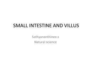
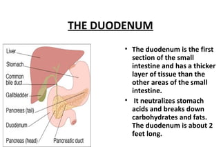
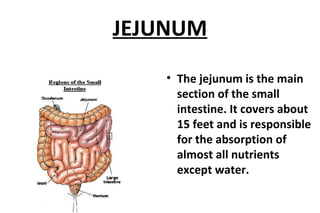
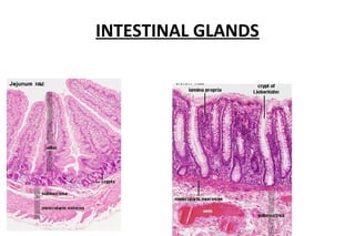
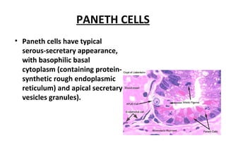
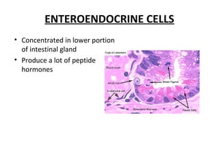
SMALL INTESTINE AND VILLUS 
Sathyananthinee.s 
Natural science
INTRODUCTION 
• small intestine is a tube of about 2.5 cm wide. 
• As it is too long, it lies coiled and folded in the 
abdominal cavity. 
• Food particles move only very slowly through this small 
intestine. 
• There are a number of blood vessels in the small 
intestine. 
• Within these infolding finger like projections called villi. 
• These villi enhance the absorptive capacity of the small 
intestine
SMALL INTESTINE 
• The small intestine is 
divided 
• duodenum 
• jejunum 
• ileum.
THE DUODENUM 
• The duodenum is the first 
section of the small 
intestine and has a thicker 
layer of tissue than the 
other areas of the small 
intestine. 
• It neutralizes stomach 
acids and breaks down 
carbohydrates and fats. 
The duodenum is about 2 
feet long.
JEJUNUM 
• The jejunum is the main 
section of the small 
intestine. It covers about 
15 feet and is responsible 
for the absorption of 
almost all nutrients 
except water.
ILEUM 
• The ileum is the last 
section of the small 
intestine and spans 
about 6 feet. Its function 
is to absorb water and 
vitamins.
PLICAE CIRCULARES 
• (valves of Kerkering) are macroscopically 
visible, crescent-shaped folds of the 
mucosa and sub mucosa. 
• permanent structures, i.e. their 
presence does not depend on the 
state of distension of the small 
intestine. 
• are absent from the first few 
centimeters of the duodenum and the 
distal part of the ileum. 
• well developed in the jejunum. 
• increase the surface area of the 
mucosa
MUCOSA
INTESTINAL VILLI 
• The entire intestinal mucosa 
forms intestinal villi (about one 
mm long), which increase the 
surface area by a factor of ten. 
• The surface of the villi is 
formed by a simple columnar 
epithelium.
ABSORPTION OF NUTRIENTS 
• Within each Digested fats are transported through small 
vessels called lacteals. 
• The digested fats are transported into the lymphatic 
system, and from there into the bloodstream. 
• villus is a network of tiny blood vessels called capillaries. 
• All nutrients, except digested fats, enter the bloodstream 
through the capillaries.
Villi and Microvilli
INTESTINAL GLANDS
INTESTINAL CELLS
GOBLET CELLS 
• The apical end of each 
goblet cell is occupied by 
a large mass of mucus, 
which compresses 
adjacent cells. 
• The nucleus toward the 
basal end of the cell. 
• Attached by junctional 
complexes (evidenced in 
light microscopy as the 
"terminal bar") to 
adjacent absorptive 
cells .
PANETH CELLS 
• Paneth cells are secretary 
epithelial cells located at 
the ends of intestinal 
crypts. 
• The function for these 
cells is secretion of anti-bacterial 
proteins into the 
crypt lumen, thereby 
providing protection for 
the stem cells which line 
the crypt walls.
PANETH CELLS 
• Paneth cells have typical 
serous-secretary appearance, 
with basophilic basal 
cytoplasm (containing protein-synthetic 
rough endoplasmic 
reticulum) and apical secretary 
vesicles granules).
ENTEROENDOCRINE CELLS 
• Concentrated in lower portion 
of intestinal gland 
• Produce a lot of peptide 
hormones
CONCLUSION 
• The digestion of food is completed in the small intestine. 
• The absorption of food particles take place mainly in the 
small intestine. 
• The structure of the small intestine is suitable for the 
absorption of food particles. 
• Finger like project into the lumen of small intestine are 
called villi.
REFERENCE 
• Dr. B.B. Arora and A.K Sabharwal(2010): A text book of 
biology std .XII, Modern publication 
• Dr . E. Valsala Kumar (2004): A text book of botany , 
Trivandrum publication 
• Kumar Pushkar and Dr A.P.Singh (2011): A text book of CSIR-UGC 
Life science, Upkar publication
Small intestine

Small intestine

  • 1.
    SMALL INTESTINE ANDVILLUS Sathyananthinee.s Natural science
  • 2.
    INTRODUCTION • smallintestine is a tube of about 2.5 cm wide. • As it is too long, it lies coiled and folded in the abdominal cavity. • Food particles move only very slowly through this small intestine. • There are a number of blood vessels in the small intestine. • Within these infolding finger like projections called villi. • These villi enhance the absorptive capacity of the small intestine
  • 3.
    SMALL INTESTINE •The small intestine is divided • duodenum • jejunum • ileum.
  • 4.
    THE DUODENUM •The duodenum is the first section of the small intestine and has a thicker layer of tissue than the other areas of the small intestine. • It neutralizes stomach acids and breaks down carbohydrates and fats. The duodenum is about 2 feet long.
  • 5.
    JEJUNUM • Thejejunum is the main section of the small intestine. It covers about 15 feet and is responsible for the absorption of almost all nutrients except water.
  • 6.
    ILEUM • Theileum is the last section of the small intestine and spans about 6 feet. Its function is to absorb water and vitamins.
  • 7.
    PLICAE CIRCULARES •(valves of Kerkering) are macroscopically visible, crescent-shaped folds of the mucosa and sub mucosa. • permanent structures, i.e. their presence does not depend on the state of distension of the small intestine. • are absent from the first few centimeters of the duodenum and the distal part of the ileum. • well developed in the jejunum. • increase the surface area of the mucosa
  • 8.
  • 9.
    INTESTINAL VILLI •The entire intestinal mucosa forms intestinal villi (about one mm long), which increase the surface area by a factor of ten. • The surface of the villi is formed by a simple columnar epithelium.
  • 10.
    ABSORPTION OF NUTRIENTS • Within each Digested fats are transported through small vessels called lacteals. • The digested fats are transported into the lymphatic system, and from there into the bloodstream. • villus is a network of tiny blood vessels called capillaries. • All nutrients, except digested fats, enter the bloodstream through the capillaries.
  • 11.
  • 12.
  • 13.
  • 14.
    GOBLET CELLS •The apical end of each goblet cell is occupied by a large mass of mucus, which compresses adjacent cells. • The nucleus toward the basal end of the cell. • Attached by junctional complexes (evidenced in light microscopy as the "terminal bar") to adjacent absorptive cells .
  • 15.
    PANETH CELLS •Paneth cells are secretary epithelial cells located at the ends of intestinal crypts. • The function for these cells is secretion of anti-bacterial proteins into the crypt lumen, thereby providing protection for the stem cells which line the crypt walls.
  • 16.
    PANETH CELLS •Paneth cells have typical serous-secretary appearance, with basophilic basal cytoplasm (containing protein-synthetic rough endoplasmic reticulum) and apical secretary vesicles granules).
  • 17.
    ENTEROENDOCRINE CELLS •Concentrated in lower portion of intestinal gland • Produce a lot of peptide hormones
  • 18.
    CONCLUSION • Thedigestion of food is completed in the small intestine. • The absorption of food particles take place mainly in the small intestine. • The structure of the small intestine is suitable for the absorption of food particles. • Finger like project into the lumen of small intestine are called villi.
  • 19.
    REFERENCE • Dr.B.B. Arora and A.K Sabharwal(2010): A text book of biology std .XII, Modern publication • Dr . E. Valsala Kumar (2004): A text book of botany , Trivandrum publication • Kumar Pushkar and Dr A.P.Singh (2011): A text book of CSIR-UGC Life science, Upkar publication