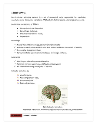
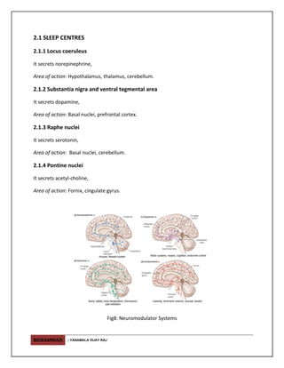
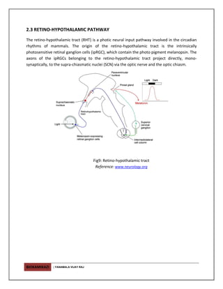
BIOKAMIKAZI | YANAMALA VIJAY RAJ
1 SLEEP WAVES
RAS (reticular activating system) is a set of connected nuclei responsible for regulating
wakefulness and sleep wake transitions. RAS has both cholinergic and adrenergic components.
Anatomical components of RAS are
• Mid-brain reticular formation,
• Dorsal hypo-thalamus,
• Thalamic intra laminar nuclei,
• Tegmentum.
Cholinergic
 Neuro-transmitters having quaternary ammonium salts,
 Present in acetylcholine and functions with inositol and basic constituent of lecithin,
 Prevents fat deposition in liver,
 Parasympathetic system communicates via cholinergic pathway.
Adrenergic
 Working on adrenaline or nor-adrenaline,
 Adrenalic nervous system as part of autonomous system,
 No role in modulating activity of RAS neurons.
Reticular formation by
Visual impulse,
Ascending sensory tract,
Auditory impulse ,
Descending motor.
Fig7: Reticular formation
Reference: http://www.daviddarling.info/encyclopedia/R/reticular_formation.html
BIOKAMIKAZI | YANAMALA VIJAY RAJ
2.1 SLEEP CENTRES
2.1.1 Locus coeruleus
It secrets norepinephrine,
Area of action: Hypothalamus, thalamus, cerebellum.
2.1.2 Substantia nigra and ventral tegmental area
It secrets dopamine,
Area of action: Basal nuclei, prefrontal cortex.
2.1.3 Raphe nuclei
It secrets serotonin,
Area of action: Basal nuclei, cerebellum.
2.1.4 Pontine nuclei
It secrets acetyl-choline,
Area of action: Fornix, cingulate gyrus.
Fig8: Neuromodulator Systems
BIOKAMIKAZI | YANAMALA VIJAY RAJ
2.1.5 Medullary synchronization area
May stimulate the Raphe nuclei,
Area of action: nuc. tractus solitarius level.
2.1.6 Encephalic sleep areas
Area of action:
I. Rostral of hypothalamus, especially the supra-chiasmatic area,
II. Intra-laminar and anterior thalamic nuclei.
2.1.7 Basal forebrain sleep area
Area of action: Pre-optic area and Broca’s diagonal band.
2.2 Major centers for sleep and factors interfering sleep
Location Function
Thalamus Cortical activation
Sleep spindle
EEG synchronization
Hypo-thalamus Sleep/watch switch
SCN Circadian clock
Brain-stem Ascending cortical activation
REM/ SWS switch
Factor Function
adenosine Inhibits cholinergic neurons of RAS which
stimulate cortex
PgD2 (Prosta-glandin D2) Increase sleep tendency
Hypothalamus secrets it
PgE2 (Prosta-glandin E2) Secreted by hypothalamus
IL-1 Immunological role
Sleep inducing factor
Muramil peptide Cytokine activation
BIOKAMIKAZI | YANAMALA VIJAY RAJ
2.3 RETINO-HYPOTHALAMIC PATHWAY
The retino-hypothalamic tract (RHT) is a photic neural input pathway involved in the circadian
rhythms of mammals. The origin of the retino-hypothalamic tract is the intrinsically
photosensitive retinal ganglion cells (ipRGC), which contain the photo pigment melanopsin. The
axons of the ipRGCs belonging to the retino-hypothalamic tract project directly, mono-
synaptically, to the supra-chiasmatic nuclei (SCN) via the optic nerve and the optic chiasm.
Fig9: Retino-hypothalamic tract
Reference: www.neurology.org

Sleep action centres

  • 1.
    BIOKAMIKAZI | YANAMALAVIJAY RAJ 1 SLEEP WAVES RAS (reticular activating system) is a set of connected nuclei responsible for regulating wakefulness and sleep wake transitions. RAS has both cholinergic and adrenergic components. Anatomical components of RAS are • Mid-brain reticular formation, • Dorsal hypo-thalamus, • Thalamic intra laminar nuclei, • Tegmentum. Cholinergic  Neuro-transmitters having quaternary ammonium salts,  Present in acetylcholine and functions with inositol and basic constituent of lecithin,  Prevents fat deposition in liver,  Parasympathetic system communicates via cholinergic pathway. Adrenergic  Working on adrenaline or nor-adrenaline,  Adrenalic nervous system as part of autonomous system,  No role in modulating activity of RAS neurons. Reticular formation by Visual impulse, Ascending sensory tract, Auditory impulse , Descending motor. Fig7: Reticular formation Reference: http://www.daviddarling.info/encyclopedia/R/reticular_formation.html
  • 2.
    BIOKAMIKAZI | YANAMALAVIJAY RAJ 2.1 SLEEP CENTRES 2.1.1 Locus coeruleus It secrets norepinephrine, Area of action: Hypothalamus, thalamus, cerebellum. 2.1.2 Substantia nigra and ventral tegmental area It secrets dopamine, Area of action: Basal nuclei, prefrontal cortex. 2.1.3 Raphe nuclei It secrets serotonin, Area of action: Basal nuclei, cerebellum. 2.1.4 Pontine nuclei It secrets acetyl-choline, Area of action: Fornix, cingulate gyrus. Fig8: Neuromodulator Systems
  • 3.
    BIOKAMIKAZI | YANAMALAVIJAY RAJ 2.1.5 Medullary synchronization area May stimulate the Raphe nuclei, Area of action: nuc. tractus solitarius level. 2.1.6 Encephalic sleep areas Area of action: I. Rostral of hypothalamus, especially the supra-chiasmatic area, II. Intra-laminar and anterior thalamic nuclei. 2.1.7 Basal forebrain sleep area Area of action: Pre-optic area and Broca’s diagonal band. 2.2 Major centers for sleep and factors interfering sleep Location Function Thalamus Cortical activation Sleep spindle EEG synchronization Hypo-thalamus Sleep/watch switch SCN Circadian clock Brain-stem Ascending cortical activation REM/ SWS switch Factor Function adenosine Inhibits cholinergic neurons of RAS which stimulate cortex PgD2 (Prosta-glandin D2) Increase sleep tendency Hypothalamus secrets it PgE2 (Prosta-glandin E2) Secreted by hypothalamus IL-1 Immunological role Sleep inducing factor Muramil peptide Cytokine activation
  • 4.
    BIOKAMIKAZI | YANAMALAVIJAY RAJ 2.3 RETINO-HYPOTHALAMIC PATHWAY The retino-hypothalamic tract (RHT) is a photic neural input pathway involved in the circadian rhythms of mammals. The origin of the retino-hypothalamic tract is the intrinsically photosensitive retinal ganglion cells (ipRGC), which contain the photo pigment melanopsin. The axons of the ipRGCs belonging to the retino-hypothalamic tract project directly, mono- synaptically, to the supra-chiasmatic nuclei (SCN) via the optic nerve and the optic chiasm. Fig9: Retino-hypothalamic tract Reference: www.neurology.org