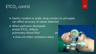
EMS Equipment Review
MARCH 2015 CE
CONDELL MEDICAL CENTER EMS SYSTEM
CE
IDPH SITE CODE #107200E-1215
PREPARED BY: SHARON HOPKINS, RN, BSN, EMT-P
1
Objectives
Upon successful completion of this module, the EMS
provider will be able to:
 List indications for use of a variety of EMS equipment used
in the field.
 Manage a group of peers in setting up and applying a
variety of equipment used in the field.
 Evaluate the effectiveness of application of a variety of EMS
equipment in a practical setting.
2
Objectives cont’d
 Actively participate in review of selected Region X
SOP’s as related to the topics presented.
 Actively participate in review of the process of
transmission of 12 lead EKG’s using department
specific equipment.
 Actively participate in reviewing the operation of your
department monitor/defibrillator, pacing capacity,
synchronized cardioversion
and defibrillation at the paramedic level.
3
Objectives cont’d
 Actively participate in HARE/Saeger traction
application.
 Successfully complete the post quiz with a score of
80% or better.
4
Equipment and Patient Interventions
 There comes responsibility when using equipment in the
delivery of patient care. You need to:
 recognize what the problem is to know what to do
 be able to distinguish what the appropriate intervention(s) is/are
 understand how to properly apply and use the equipment
chosen
 recognize when the intervention is working as well as not
accomplishing the goal
 know what documentation must be done with each piece of
equipment used in patient care
 be knowledgeable regarding the cleaning and returning to
service for each piece of equipment
5
Capnography Background
 A continuous, non-invasive monitoring tool
 Measures level of CO2 at end of exhalation
 Quantitative results provides a number
 Assesses respiratory status thru-out respiratory cycle
 Provides current, at the moment, breath-to-breath
information on patient status
 Results measured as mmHg of CO2
 Normal 35 – 45 mmHg
6
Capnography Information
 Numeric value provides end tidal (end of breath) CO2
level
 Waveform is a picture representation of the CO2
value exhaled with each breath
 Airway status reflected in:
 ETCO2 value (mmHg)
 Waveform picture
 Respiratory rate
7
Definitions
 Ventilation
 Process of breathing; eliminating CO2 from body
 Respiration
 Exchange of gasses at alveoli level
 Oxygenation
 Getting O2 to tissues; measured by pulse oximetry
 Diffusion
 Process by which gas moves between alveoli and pulmonary
capillaries (gases move from area of high concentration to areas of
low concentrations)
8
Capnography Usefulness
 Provides information on how effectively the body is:
 Producing CO2 (metabolism)
 Transporting CO2 (perfusion)
 Exhaling CO2 (ventilations)
 Goal – attain/maintain CO2 levels 35 – 45 mmHg
9
Capnography Usefulness cont’d
 Confirms and monitors advanced airway placement
 Indicates effectiveness of chest compressions
 Blood must circulate through lungs to off-load CO2 for it to be
exhaled
 Levels expected to minimally be >10mmHg during CPR
 Indicates return of spontaneous circulation (ROSC)
 Sudden, sustained rise in levels toward 35-45 mmHg
 Allows early interventions to be started
10
Capnography Usefulness cont’d
 Monitor asthma & COPD conditions and response to bronchodilator
therapy
 Detect increased respiratory depression and hypoventilation
 Tiring accessory muscles
 Neuromuscular disease effect on respiratory center
 Change in level of consciousness – alcohol/drug overdose, head
trauma, sedation/analgesia
 Seizure activity &/or post ictal period
11
Capnography Waveform
 A-B – respiratory baseline
 B-C expiratory upslope
 C-D expiratory plateau
 D – end of exhalation
point of measurement
 D-E – inspiratory downslope
12
Capnography Waveforms
 Hypoventilation
 CO2 retained so values

 Hyperventilation
 CO2 eliminated so
values 
13
Capnography Waveforms
 Asthma attack or COPD
 Difficulty exhaling evidenced by slow, gradual upslope
14
Capnography Waveforms
 Apnea or loss of
advanced airway
- flat line
15
ETCO2 Detector
 End tidal (end of breath) CO2 detector
 Qualitative device
 Indicates presence/absence of detectable CO2 exhaled via pH
sensitive paper
 Does not provide specific measurement of numeric value
Color scale estimates CO2 level
 Able to change as detected levels change
 May take up to 6 breaths to wash enough CO2 out for proper
measurement
16
ETCO2 cont’d
 Gastric content or acidic drug contact on pH paper
can affect accuracy of values detected
 When perfusion decreased (shock,
arrest) ETCO2 reflects change in
pulmonary blood flow and CO2 level
 Does not reflect ventilation status
17
Altered CO2 Levels
  CO2 level
 Shock, cardiac arrest, pulmonary embolism,
bronchospasm, complete airway obstruction
  CO2 level
 Hypoventilation, respiratory depression, hyperthermia
18
CO2 Influence on Circulation
  CO2 in blood (hypoventilation)
Cerebral vasodilation  increase in intracranial
pressure (ICP) due to increased blood flow to the
brain
  CO2 in blood (hyperventilation)
Cerebral vasoconstriction  decrease in fresh blood
flow to brain; decrease in levels of adequate oxygen
and glucose negatively affect function of brain
19
ETCO2 Result Interpretation
 Yellow – yes, CO2 is being detected in exhaled breath
 Tan – poor perfusion or ventilation status
 First evaluate placement of airway device
 Continue to trouble shoot
 Blue or purple – no CO2 being detected
 First evaluate placement of airway device
 Continue to trouble shoot
20
Trouble Shooting Advanced Airway
Placement – “DOPE”
 D – displacement of tube (i.e.: into esophagus)
 Chest rise and fall?
 Gastric sounds?
 Bilateral breath sounds?
 O – obstruction
 P – pneumothorax
 E – equipment failure
 Faulty cuff
21
Esophageal Detector Device - EDD
 A modified bulb syringe
 Simple means of evaluating for missed endotracheal intubation
 Squeeze bulb, attach to end of endotracheal tube
 Bulb re-expands = tube in trachea
 Bulb does not re-expand or does so slowly – collapsing sides of
esophagus onto tube preventing air from filling EDD – consider
esophageal placement
22
EDD cont’d
 Need to interrupt ventilations to use device
 Evaluate results of technique used with results of all other steps
of confirmation – could be extenuating reason why you get false
negatives
23
Defibrillators
 Electrical capacitor that stores energy
 Biphasic defibrillators provide waveforms that use
less DC energy than monophasic machines
 Energy flows in one direction and then reverses
 Therefore, possible decrease in tissue damage
 Survival rates increase if early CPR provided with
prompt defibrillation attempt as soon as possible
after collapse
24
Defibrillation
 Early defibrillation critical to survival from sudden
cardiac arrest
 Most frequent initial rhythm in arrest is VF
 Treatment for VF is defib (defibrillation)
 Probability of successful defibrillation diminishes over
time
 VF deteriorates to asystole over time
 Check with your vendor to know your biphasic
device’s recommended energy settings
25
Ventricular Fibrillation as Presenting
Rhythm
 Best chance of survival in public
 Early activation of EMS
 CPR initiated very soon after collapse
 Early application of AED or other defibrillation attempt
 Current passes though fibrillating heart to depolarize
heart cells to allow them to uniformly repolarize
 Allows dominant pacemaker (SA node) to take over
electrical control
 Goal – resume organized electrical activity
26
Influences on Success of Defibrillation
 Time from onset of VF – shorter time  survival
 Condition of myocardium
 Less success in presence of hypoxia, acidosis,
hypothermia, electrolyte imbalance, drug toxicity
 Pad size
 Larger pads felt to be more effective and cause less
myocardial damage; should not overlap
Ideal size for adults10-13 cm (4 -5 inches)
Ideal size for peds 4.5 cm (roughly 3 inches)
27
Influences cont’d
 Pad / skin interface
 Need to  the resistance
Greater the resistance the less energy delivered to the
heart and the greater the heat production at the skin
surface
 Pad contact
 Max contact with skin; no air bubbles breaking contact;
no pads touching or overlapping
 Avoiding placement of pads over bone
 Bone is poor conductor of electricity
28
Pad Placement
Operator Choice
 Anterior /posterior
 1 pad over apex of heart, under left
breast
 1 pad under left scapula in line with
anterior pad
 Anterior/anterior (apex)
 Anterior pad on right upper sternum just below
clavicle
 Apex pad below left nipple in anterior axillary line over apex
of heart
29
Pad Placement cont’d
 DO NOT place pads
 Over sternum – bone poor conductor of electricity
 Over pacemaker or AICD – deflects energy; could
damage the implanted device
Place at least one inch away from device
 Over topical medication patches – deflects energy
30
Defibrillation
 Indications
 VF, pulseless VT
 Contraindications
 Failure to demonstrate one of the above rhythms
 Asystole – defibrillation places a patient into asystole
for the dominant pacemaker to take over
 PEA – electrical activity not a problem; needs
mechanical response fixed
31
Defibrillation
 Equipment
 Monitor/defibrillator
 Defibrillating pads
Example: PadPro
Defibrillation/pacing/cardioversion/monitoring electrodes
Most come with conductive gel already applied in center
of pad
32
Defibrillation Safety
 CPR is performed just until the defibrillator is ready
 Confirm O2 not blowing across patient’s chest wall – hold
away from the patient when not using the BVM
 Physically look all around (“nose to toes”)
 Clearly yell out “all clear”
 Deliver energy
 Immediately resume CPR
33
Return of Spontaneous Circulation
ROSC
 After 2 minutes of resumed CPR, evaluate the rhythm
 If an organized rhythm is viewed on the monitor,
THEN check for a pulse
 If no pulse, rhythm is PEA
 Resume CPR
Adult 1 and 2 man CPR 30:2
Infant and child 1 man CPR 30:2
Infant and child 2 man CPR 15:2
34
Indications to Activate Cooling
Protocol Post ROSC
 Presumed cardiac arrest
 NOT indicated for respiratory or traumatic arrest
 Remains unconscious and unresponsive
 ROSC present at least 5 minutes
 Systolic B/P >90 with or without pressor agent use
(i.e.: Dopamine)
 Airway has been secured
35
ROSC Contraindications
 Major head trauma or traumatic arrest
 Recent major surgery within past 14 days
 Systemic infection (i.e.: septic shock)
 Coma from other causes
 Active bleeding
 Isolated respiratory arrest
 Hypothermia (34o C/93.2o F) already present
36
Induction of ROSC
 Place ice paks in the axilla, neck and groin
 Areas where blood vessels tend to be superficial
 Place ice pak over IV site
 If patient begins to shiver, contact Medical Control
 Anticipate order for Valium to stop the shivering
 Shivering will generate heat and therefore increase
body temperature
37
Vasopressor - Dopamine
 Stimulates alpha, beta, and dopaminergic receptors
based on dose provided
 Starting dose 5mcg/kg/min IVPB up to 20
mcg/kg/min
 Take patient’s weight and drop last number
 Minus 2 from number left
 Left with rate to run IVPB in drops per minute
 Ex: 150 pounds; drop “0”
15 – 2 = 13 drops per minute
38
Dopamine cont’d
 Dopaminergic effects at 2 mcg/kg/min
 Renal vasodilation to improve blood flow to kidneys
 Keep kidneys working, the body keeps working
 Beta effects 5 – 10 mcg/kg/min
 Increases strength of myocardial contraction – squeeze
more blood out of ventricles
 Alpha effects at >20 mcg/kg/min
 Severe vasoconstriction that diminishes blood flow to all
tissues
39
AED (Automated External Defibrillator)
Function
 AED’s will
 Analyze rhythms
 Deliver a shock if indicated
Ventricular fibrillation (VF)
Monomorphic and polymorphic VT if rate and R wave
morphology exceed preset values
 Will not deliver a synchronized shock
 Can indicate loose electrodes / poor electrode
contact
40
AED Use in Pediatrics
 Pediatric attenuator used to deliver lower energy doses to
children (built into cables with peds pads)
 1-8 year old
 Use pediatric pads if available
 No attenuator (peds pads)available, use standard AED pads
 < 1 year old
 Manual defibrillator preferred
 If no manual defibrillator, use peds pads with attenuator
 No peds pads, use AED pads available
41
AED Use With CPR
 Do NOT interrupt CPR to apply pads
 Apply pads while CPR in progress
 Do not touch patient during analysis phase
 Can provide compressions during charging phase
 No O2 flow across patient body during defibrillation
attempt
 Call and look “ALL CLEAR” prior to each defibrillation
attempt
 Immediately resume CPR
42
Transition From AED To Defibrillator
 Upon arrival at scene, if AED ready to discharge, utilize
AED
Do not interrupt operation of device
 During 2 minutes of CPR, can switch from AED use to
monitor/defibrillator
 Immediately resume CPR after delivery of each
defibrillation attempt regardless of equipment used
43
Synchronized Cardioversion
 A controlled form of defibrillation using a lower energy level
that interrupts underlying reentrant pathway
 Used with organized rhythms and in presence of a pulse
 Monitor interprets QRS cycle and energy delivered during R
wave
 Less vulnerable area of QRS
 Downslope of T wave is relative refractory area
Minimal stimulant could generate rhythm into VF
44
Indications Synchronized Cardioversion
 Unstable tachyarrhythmias
 SVT
 Rapid atrial fibrillation or flutter
Hazard of breaking loose a blood clot in the atria and
resulting in a stroke
 Ventricular tachycardia
Note: polymorphic VT NOT likely to respond to
synchronized cardioversion – no defined R wave
45
Synchronized Cardioversion Procedure
 Apply pads
 Anterior/anterior or anterior/posterior position
 Sedate if possible
 This is a painful procedure!
 Versed 2 mg IVP/IO; repeated every 2 minutes; max 10
mg (desired effect – sedation!)
 Consider pain management
 Fentanyl 1 mcg/kg IVP/IN/IO; may repeat in 5 minutes
to max of 200 mcg total dose
46
Sync Procedure cont’d
 Activate “sync” button
 Verify R wave is being flagged/identified
 Choose energy setting starting at the lowest watt setting
 100j, 200j, 300j, 360j
 Verify O2 not blowing across chest wall
 Look (nose to toes) and call “ALL CLEAR”
 Press and hold sync buttons until energy discharged
 Momentary delay waiting to identify the R wave
47
Sync Procedure cont’d
 If synchronized cardioversion needs to be repeated,
need to reset the “sync” button
 Safety that machine will default to defibrillation mode
after every discharge of energy
 If VF occurs, verify sync mode is off and defibrillate
patient without delay
48
Transcutaneous Pacemaker - TCP
 Electrical cardiac pacing across the skin
 TCP is a painful non-invasive procedure so sedation will
most likely be necessary
 Indications
 Symptomatic bradycardia
Hypotensive
Hypoperfusing
 Evaluate level of consciousness and B/P for most reliable
indicators of patient condition/stability
49
TCP Procedure
 Apply pads
 (-) over apex of heart, anterior chest wall
 (+) mid upper back below left scapula
 Set desired heart rate (80)
 Confirm sensitivity at auto/demand
 Begin mA current at 0
 Turn pacer on
50
TCP Procedure
cont’d
 Slowly increase output until ventricular capture
 Spike followed by widened QRS
 Reassess vital signs and pain level
 Document settings – mA and rate
 Reassess need for sedation and analgesia
 Valium 2 mg IVP/IO over 2 minutes; repeat every 2
minutes until max of 10 mg total dose
 Fentanyl 1 mcg/kg IVP/IO/IN ; can repeat dose in 5
minutes with max total of 200 mcg
51
Critical Thinking Skill and TCP
 In setting of acute MI, consider contacting Medical
Control
 May want to decrease heart rate of TCP just enough to
maintain perfusion
 Want to avoid increasing the work load on the heart
by automatically selecting 80 as the heart rate
 Increasing work load on heart may increase the size of
the infarction
52
What would you do…
 You applied the TCP for a symptomatic bradycardia
 You had a paced rhythm
 You notice the following rhythm strip change – what is the
rhythm and what would you do?
 Reassess patient; increase mA; consider need for CPR
Failure to capture
53
Defibrillation During Pacing Mode
 Check your device for specifics
 When in the pacing mode and the need to
defibrillate occurs, for some models, you may have to
turn off the pacing mode
 If pacing must be resumed, reset all levels
54
12 Lead EKG’s
 A graphic recording of electrical activity in the heart
 Must evaluate the pulse to determine mechanical response
 Single lead (i.e.: lead II) evaluates cardiac rhythms
 12 lead views can diagnose an acute MI
 Early interpretation of 12 lead EKG  early diagnosing 
early reperfusion & restoring blood flow to ischemic tissues
55
Acute MI
 Death of portion of heart muscle from prolonged
deprivation of oxygenated blood
 Heart’s demand exceeds supply of oxygen over
extended period of time
 Often associated with atherosclerosis process
 Location and size of infarct depends on vessel
involved and site of obstruction
 Left ventricle most common site
56
Evolution of Acute MI
 Ischemia – initial lack of oxygen
 ST depression can be reversible
 Injury to myocardial tissue
 ST elevation can be reversible
 Death/infarction
 Necrotic tissue can lead to scar formation
 Irreversible process
 Can leave a positive Q wave marker in leads affected
57
58
AMI Process
 Ring of ischemic tissue surrounds infarcted
myocardium
 Collateral circulation may develop
 Ischemic area often site of arrhythmia development
59
Complications of AMI
 Arrhythmia most common
 VF most lethal
 Most common cause of sudden death within one hour of onset
of signs and symptoms
 Destruction of myocardial muscle mass can lead to CHF due
to impairment of pumping capability
 Cardiogenic shock may develop if heart function is inefficient
and inadequate
 Ventricular aneurysm can develop due to damaged wall of
heart – can rupture causing instant death
60
Patient Assessment
 Pain most common chief complaint
 Lasts more than 30 minutes
 Not relieved by rest or NTG
 Tired and weak most often complaint in elderly, long
standing diabetic and women
 Determine responses to OPQRST assessment
 Activity at onset, provocation/palliation
(worsens/improves), quality in their words, radiation,
severity on 0 -10 scale, time of onset
61
EMS Action
 Apply monitor
 Examine underlying rhythm – document rhythm
 Obtain 12 lead EKG
 Evaluate for ST segment elevation
If elevation, in what group of leads?
If depressed, look for reciprocal elevation
 Watch for development of arrhythmias
62
Proper Placement EKG Chest Leads 63
Groups of Acute MI by Leads 64
Identifying Groups of ST Elevation 65
Why Aspirin???
 Inhibits platelets from aggregating/collecting at site of plaque
rupture inside vessel wall
 Decreases morbidity and mortality rate
 Chewed to increase breakdown and absorption time of
medication
 Patients on daily aspirin already have elevated and acceptable
blood levels of aspirin – don’t have to supplement a dose if
absolutely sure they took one today
 Always better to give full dose than to risk skipping any dose
(just in case of skipped dose)
66
12 Lead EKG Procedure
 Obtain rhythm strip
 Interpret, report and document rhythm
 Obtain 12 lead EKG
 Identified with patient age, sex, department name in
preparation for transmission
 Review for ST elevation pattern
 Report to Medical Control what you see, then read
word for word interpretation on 12 lead EKG printout
67
12 Lead EKG Documentation
 Interpret the rhythm strip and document on patient
care run report
 Document presence or absence of ST elevation
 If elevation, report and document in which leads
 Provide copy of rhythm strip and 12 lead EKG to ED
secretary for placement on patient’s medical record
68
CPAP
 Continuous positive airway pressure
 Effective therapy for acute CHF – pulmonary
edema
 Can avert the need for intubation and mechanical
ventilation if applied early enough
 Maintains constant pressure within the airway and
through-out the respiratory cycle
 Keeps alveoli open and expanded
 Increases surface space for diffusion of gases
69
CPAP cont’d
 Buys time for other therapies (i.e.:
medications) to work
 Precaution
Too much pressure can inhibit
ventricular filling decreasing
cardiac output
B/P can drop
70
CPAP Indications
 Stable pulmonary edema
 Alert; systolic B/P >90mmHg
 COPD with wheezing
 First contact Medical Control for orders
 For unstable pulmonary edema (altered mental
status, systolic B/P <90 mmHg), contact Medical
Control to discuss use of CPAP
 Reminder: all therapies used in pulmonary edema
have potential to drop the B/P
71
CPAP Contraindications
 Respiratory arrest or apnea
 Pneumothorax or trauma to chest wall
 Tracheostomy present
 Can’t get tight fit over trach stoma
 Actively vomiting
72
CPAP Procedure
 Sit patient upright
 Assess and obtain baseline vital signs
 Begin O2 via non-rebreather mask while setting up
equipment
 Administer first dose NTG
 Used as venodilator to decrease blood return to heart
(decreases pre-load)
73
CPAP Flow Safe II Procedure
 Assemble CPAP Flow Safe II
 Attach proximal end of O2 tubing with manometer to
port in mask
 Attach distal end of tubing to O2 source
 Secure face mask snugly to patient’s face using head
harness
 Adjust O2 flow – 13-14 lpm for 10 cm H2O
 Continue administration of medications
74
CPAP Procedure cont’d
 Lasix 40 mg IVP (80mg if on med at home) as a diuretic
 If systolic B/P remains >90 mmHg
 Morphine 2 mg IVP slowly over 2 minutes
 May repeat 2 mg every 2 minutes as needed to max of 10 mg
 Used to decrease anxiety and for benefit of vasodilation
 If patient shows deterioration during CPAP treatment,
remove CPAP, consider intubation, inform Medical Control
75
CPAP Tidbits
 Be prepared to coach patient through first few minutes
of CPAP use until positive effects begin
 Patient is already frightened
 Patient may feel suffocated with the mask on
 Exhaling against the resistance is tough at first
 

76
HARE and Saeger Traction
 Indicated for isolated mid-femur fractures
 Reduces muscle spasm and therefore pain level
 Reduces risk of bones overriding
 Contraindications
 Open fracture
Do not want to draw contamination into the wound
 Hip, knee, or pelvic fractures
Increased risk of nervous or vascular complications
77
Preparing for Traction Application
 Assess motor/sensory/circulation before and after
splinting
 Can you move this/can you feel that?
 Mark pulses once found – easier to find the site on
reassessment
 Compare to uninjured side
 Apply manual traction until mechanical traction in
place
78
HARE Application
 Measure and adjust splint
 Support distal end of splint on backboard
 Apply distal ankle hitch while maintaining manual
traction
 Position traction under injured extremity
 Secure proximal end to groin area
 Apply hook to ankle hitch
 Replace manual traction with mechanical traction
79
HARE Traction
 Adjust straps avoiding over the knee and over the injured site
80
Saeger Traction Application
 Support leg and maintain gentle traction
 Use uninjured leg to measure and adjust splint length
 Place splint inside injure leg; padded bar snug against pelvis
in groin (watch pressure areas!!!)
 Attach strap to thigh
 Attach padded hitch to foot and ankle
 Extend splint until correct tension obtained
 Apply elastic straps to secure leg to splint
81
Saeger Splint
 Do not place straps over fracture site
 Release manual
traction
 Reassess distal pulse, motor,
and sensory
82
Pain Control With Use of Traction
 Fentanyl 1 mcg/kg IVP/IN/IO
 May repeat same dose in 5 minutes
 Max total dose of 200 mcg
 As a CNS depressant, watch the respiratory status
 If respiratory depression occurs, begin to support
ventilations via BVM
 1 Breath every 5 – 6 seconds
 Document 10 -12 breaths per minute assisted
 Narcan 2 mg IVP/IN/IO can be used to reverse respiratory
depression due to opioid use
83
Cleaning of Traction Splints
 Rinse off gross contaminant
 Wet down all surfaces with Cavicide wipes
 Let device air dry
 Confirm all straps are accounted for and repackage
device in preparation for next patient
84
Combat Application Tourniquet - CAT
 Indications
 Uncontrollable hemorrhage when usual
means have failed
 Contraindications
 Non-compressable site
 Equipment
 Tourniquet with attached rod
85
CAT - Procedure
 Apply tourniquet proximal to bleeding site as distal
as possible; preferably over bare skin
 Pull band very tight and securely fasten band back on
itself
 Twist rod until bright red bleeding has stopped
 Or until distal pulses are eliminated
 Place rod inside clip; locking into place
 Secure straps over clip holding rod
86
CAT – Potential Problems
 Inability to control bleeding
 Continue with direct pressure
 Prepare to apply a second CAT
 Apply QuikClot dressing if available
Must be applied directly over wound site for
impregnated material to be effective
87
CAT – Documentation Pearls
 Reason CAT was applied
 Time and site of CAT application
 Results post intervention
 Consideration of administration of pain medication
 Fentanyl 1 mcg/kg IVP/IN/IO
May repeat in 5 minutes, same dose
Max 200 mcg total dosing
88
Midazolam - Versed
 Potent, rapid onset, short acting benzodiazepine
 Onset 3-5 minutes
 Duration 20-30 minutes
 Used as sedative and hypnotic
 Has amnesic properties and reduces anxiety
 Amnesia of recent past (antegrade) useful to inhibit
unpleasant reminders of procedures
 Low toxicity and high rate of effectiveness
89
Indications for Versed Per Region X SOP’s
 Sedation prior to synchronized cardioversion
 Useful to maintain sedation post drug assisted
intubation procedure
 Suppresses seizure activity
 IN route allows safer delivery method
 Decreases severe anxiety and apprehension
90
Precautions With Versed
 Crosses placental barrier – could cause respiratory
depression in newly born infant
 Elderly more sensitive to effects; metabolize med
more slowly
 Toxicity increases when mixed with CNS depressants
(alcohol, opioids like Fentanyl, tricyclic
antidepressants)
 Toxicity may be higher in patients with COPD
91
Side Effects of Versed
 Respiratory depression
 Drowsiness
 Hypotension
 When administering, have a BVM readily available
 Be prepared to assist respirations
 1 breath every 5 – 6 seconds
 Document 10 – 12 breaths per minute assisted
92
Fentanyl
 Synthetic opiate analgesic for pain control
 Shorter acting than morphine
 Onset immediate when administered IVP
 Peak effect 3 5 minutes
 Lasts 30 – 60 minutes
 Does not affect blood pressure like Morphine does
93
Dosing For Fentanyl per Region X SOP’s
 Adult
 1 mcg/kg IN/IVP/IO
 May repeat same dose in 5 minutes
 Max total dose 200 mcg
 Pediatrics
 0.5 mcg/kg IVP/IN/IO
 May repeat same dose in 5 minutes
 Max total dose 200 mcg
94
Precautions With Fentanyl
 Crosses the placental barrier – could cause respiratory
depression in newly born infant
 Monitor respiratory rate, SpO2 levels, and level of
consciousness
 Have BVM available to counteract potential respiratory
depression
 1 breath every 5 – 6 seconds
 Document 10 – 12 respirations per minute
95
Cleaning of Equipment – After
Every Patient Use
 In general, each piece of equipment in contact with a patient MUST
be cleaned between each patient use
 Gross contaminant must be removed
 Surfaces need to remain wet and allowed to air dry
 All cables need to be wiped down (i.e.: EKG, B/P, pulse ox)
 Cables drag across contaminated surfaces A LOT!!!
 B/P cuffs need to be wiped down
 Pulse ox sensors need to be cleaned following manufacturer
recommendations
96
Department Review of Equipment
 Review set up of capnography monitoring
 Review operation of monitor/defibrillator for
defibrillation, synchronized cardioversion, and TCP
 Review procedures for transmission of 12 lead EKG to
receiving hospital
 In teams, apply the HARE or Saeger traction device to
a peer
97
Bibliography
 Bledsoe, B., Porter, R., Cherry, R. Paramedic Care Principles & Practices,
4th edition. Brady. 2013.
 Campbell, J., International Trauma Life Support for Emergency Care
Providers. 7th Edition. Pearson. 2012.
 McDonald, J. ALS Skills Review. AAOS. Jones and Bartlett. 2009.
 Mistovich, J., Karren, K. Prehospital Emergency Care 9th Edition. Brady.
2010.
 Pediatric Education for Prehospital Professionals 3rd Edition. American
Academy of Pediatrics. 2014.
 Region X SOP’s; IDPH Approved January 6, 2012.
 www.MARescue.com
98

march2015-ppt.ppt

  • 1.
    EMS Equipment Review MARCH2015 CE CONDELL MEDICAL CENTER EMS SYSTEM CE IDPH SITE CODE #107200E-1215 PREPARED BY: SHARON HOPKINS, RN, BSN, EMT-P 1
  • 2.
    Objectives Upon successful completionof this module, the EMS provider will be able to:  List indications for use of a variety of EMS equipment used in the field.  Manage a group of peers in setting up and applying a variety of equipment used in the field.  Evaluate the effectiveness of application of a variety of EMS equipment in a practical setting. 2
  • 3.
    Objectives cont’d  Activelyparticipate in review of selected Region X SOP’s as related to the topics presented.  Actively participate in review of the process of transmission of 12 lead EKG’s using department specific equipment.  Actively participate in reviewing the operation of your department monitor/defibrillator, pacing capacity, synchronized cardioversion and defibrillation at the paramedic level. 3
  • 4.
    Objectives cont’d  Activelyparticipate in HARE/Saeger traction application.  Successfully complete the post quiz with a score of 80% or better. 4
  • 5.
    Equipment and PatientInterventions  There comes responsibility when using equipment in the delivery of patient care. You need to:  recognize what the problem is to know what to do  be able to distinguish what the appropriate intervention(s) is/are  understand how to properly apply and use the equipment chosen  recognize when the intervention is working as well as not accomplishing the goal  know what documentation must be done with each piece of equipment used in patient care  be knowledgeable regarding the cleaning and returning to service for each piece of equipment 5
  • 6.
    Capnography Background  Acontinuous, non-invasive monitoring tool  Measures level of CO2 at end of exhalation  Quantitative results provides a number  Assesses respiratory status thru-out respiratory cycle  Provides current, at the moment, breath-to-breath information on patient status  Results measured as mmHg of CO2  Normal 35 – 45 mmHg 6
  • 7.
    Capnography Information  Numericvalue provides end tidal (end of breath) CO2 level  Waveform is a picture representation of the CO2 value exhaled with each breath  Airway status reflected in:  ETCO2 value (mmHg)  Waveform picture  Respiratory rate 7
  • 8.
    Definitions  Ventilation  Processof breathing; eliminating CO2 from body  Respiration  Exchange of gasses at alveoli level  Oxygenation  Getting O2 to tissues; measured by pulse oximetry  Diffusion  Process by which gas moves between alveoli and pulmonary capillaries (gases move from area of high concentration to areas of low concentrations) 8
  • 9.
    Capnography Usefulness  Providesinformation on how effectively the body is:  Producing CO2 (metabolism)  Transporting CO2 (perfusion)  Exhaling CO2 (ventilations)  Goal – attain/maintain CO2 levels 35 – 45 mmHg 9
  • 10.
    Capnography Usefulness cont’d Confirms and monitors advanced airway placement  Indicates effectiveness of chest compressions  Blood must circulate through lungs to off-load CO2 for it to be exhaled  Levels expected to minimally be >10mmHg during CPR  Indicates return of spontaneous circulation (ROSC)  Sudden, sustained rise in levels toward 35-45 mmHg  Allows early interventions to be started 10
  • 11.
    Capnography Usefulness cont’d Monitor asthma & COPD conditions and response to bronchodilator therapy  Detect increased respiratory depression and hypoventilation  Tiring accessory muscles  Neuromuscular disease effect on respiratory center  Change in level of consciousness – alcohol/drug overdose, head trauma, sedation/analgesia  Seizure activity &/or post ictal period 11
  • 12.
    Capnography Waveform  A-B– respiratory baseline  B-C expiratory upslope  C-D expiratory plateau  D – end of exhalation point of measurement  D-E – inspiratory downslope 12
  • 13.
    Capnography Waveforms  Hypoventilation CO2 retained so values   Hyperventilation  CO2 eliminated so values  13
  • 14.
    Capnography Waveforms  Asthmaattack or COPD  Difficulty exhaling evidenced by slow, gradual upslope 14
  • 15.
    Capnography Waveforms  Apneaor loss of advanced airway - flat line 15
  • 16.
    ETCO2 Detector  Endtidal (end of breath) CO2 detector  Qualitative device  Indicates presence/absence of detectable CO2 exhaled via pH sensitive paper  Does not provide specific measurement of numeric value Color scale estimates CO2 level  Able to change as detected levels change  May take up to 6 breaths to wash enough CO2 out for proper measurement 16
  • 17.
    ETCO2 cont’d  Gastriccontent or acidic drug contact on pH paper can affect accuracy of values detected  When perfusion decreased (shock, arrest) ETCO2 reflects change in pulmonary blood flow and CO2 level  Does not reflect ventilation status 17
  • 18.
    Altered CO2 Levels  CO2 level  Shock, cardiac arrest, pulmonary embolism, bronchospasm, complete airway obstruction   CO2 level  Hypoventilation, respiratory depression, hyperthermia 18
  • 19.
    CO2 Influence onCirculation   CO2 in blood (hypoventilation) Cerebral vasodilation  increase in intracranial pressure (ICP) due to increased blood flow to the brain   CO2 in blood (hyperventilation) Cerebral vasoconstriction  decrease in fresh blood flow to brain; decrease in levels of adequate oxygen and glucose negatively affect function of brain 19
  • 20.
    ETCO2 Result Interpretation Yellow – yes, CO2 is being detected in exhaled breath  Tan – poor perfusion or ventilation status  First evaluate placement of airway device  Continue to trouble shoot  Blue or purple – no CO2 being detected  First evaluate placement of airway device  Continue to trouble shoot 20
  • 21.
    Trouble Shooting AdvancedAirway Placement – “DOPE”  D – displacement of tube (i.e.: into esophagus)  Chest rise and fall?  Gastric sounds?  Bilateral breath sounds?  O – obstruction  P – pneumothorax  E – equipment failure  Faulty cuff 21
  • 22.
    Esophageal Detector Device- EDD  A modified bulb syringe  Simple means of evaluating for missed endotracheal intubation  Squeeze bulb, attach to end of endotracheal tube  Bulb re-expands = tube in trachea  Bulb does not re-expand or does so slowly – collapsing sides of esophagus onto tube preventing air from filling EDD – consider esophageal placement 22
  • 23.
    EDD cont’d  Needto interrupt ventilations to use device  Evaluate results of technique used with results of all other steps of confirmation – could be extenuating reason why you get false negatives 23
  • 24.
    Defibrillators  Electrical capacitorthat stores energy  Biphasic defibrillators provide waveforms that use less DC energy than monophasic machines  Energy flows in one direction and then reverses  Therefore, possible decrease in tissue damage  Survival rates increase if early CPR provided with prompt defibrillation attempt as soon as possible after collapse 24
  • 25.
    Defibrillation  Early defibrillationcritical to survival from sudden cardiac arrest  Most frequent initial rhythm in arrest is VF  Treatment for VF is defib (defibrillation)  Probability of successful defibrillation diminishes over time  VF deteriorates to asystole over time  Check with your vendor to know your biphasic device’s recommended energy settings 25
  • 26.
    Ventricular Fibrillation asPresenting Rhythm  Best chance of survival in public  Early activation of EMS  CPR initiated very soon after collapse  Early application of AED or other defibrillation attempt  Current passes though fibrillating heart to depolarize heart cells to allow them to uniformly repolarize  Allows dominant pacemaker (SA node) to take over electrical control  Goal – resume organized electrical activity 26
  • 27.
    Influences on Successof Defibrillation  Time from onset of VF – shorter time  survival  Condition of myocardium  Less success in presence of hypoxia, acidosis, hypothermia, electrolyte imbalance, drug toxicity  Pad size  Larger pads felt to be more effective and cause less myocardial damage; should not overlap Ideal size for adults10-13 cm (4 -5 inches) Ideal size for peds 4.5 cm (roughly 3 inches) 27
  • 28.
    Influences cont’d  Pad/ skin interface  Need to  the resistance Greater the resistance the less energy delivered to the heart and the greater the heat production at the skin surface  Pad contact  Max contact with skin; no air bubbles breaking contact; no pads touching or overlapping  Avoiding placement of pads over bone  Bone is poor conductor of electricity 28
  • 29.
    Pad Placement Operator Choice Anterior /posterior  1 pad over apex of heart, under left breast  1 pad under left scapula in line with anterior pad  Anterior/anterior (apex)  Anterior pad on right upper sternum just below clavicle  Apex pad below left nipple in anterior axillary line over apex of heart 29
  • 30.
    Pad Placement cont’d DO NOT place pads  Over sternum – bone poor conductor of electricity  Over pacemaker or AICD – deflects energy; could damage the implanted device Place at least one inch away from device  Over topical medication patches – deflects energy 30
  • 31.
    Defibrillation  Indications  VF,pulseless VT  Contraindications  Failure to demonstrate one of the above rhythms  Asystole – defibrillation places a patient into asystole for the dominant pacemaker to take over  PEA – electrical activity not a problem; needs mechanical response fixed 31
  • 32.
    Defibrillation  Equipment  Monitor/defibrillator Defibrillating pads Example: PadPro Defibrillation/pacing/cardioversion/monitoring electrodes Most come with conductive gel already applied in center of pad 32
  • 33.
    Defibrillation Safety  CPRis performed just until the defibrillator is ready  Confirm O2 not blowing across patient’s chest wall – hold away from the patient when not using the BVM  Physically look all around (“nose to toes”)  Clearly yell out “all clear”  Deliver energy  Immediately resume CPR 33
  • 34.
    Return of SpontaneousCirculation ROSC  After 2 minutes of resumed CPR, evaluate the rhythm  If an organized rhythm is viewed on the monitor, THEN check for a pulse  If no pulse, rhythm is PEA  Resume CPR Adult 1 and 2 man CPR 30:2 Infant and child 1 man CPR 30:2 Infant and child 2 man CPR 15:2 34
  • 35.
    Indications to ActivateCooling Protocol Post ROSC  Presumed cardiac arrest  NOT indicated for respiratory or traumatic arrest  Remains unconscious and unresponsive  ROSC present at least 5 minutes  Systolic B/P >90 with or without pressor agent use (i.e.: Dopamine)  Airway has been secured 35
  • 36.
    ROSC Contraindications  Majorhead trauma or traumatic arrest  Recent major surgery within past 14 days  Systemic infection (i.e.: septic shock)  Coma from other causes  Active bleeding  Isolated respiratory arrest  Hypothermia (34o C/93.2o F) already present 36
  • 37.
    Induction of ROSC Place ice paks in the axilla, neck and groin  Areas where blood vessels tend to be superficial  Place ice pak over IV site  If patient begins to shiver, contact Medical Control  Anticipate order for Valium to stop the shivering  Shivering will generate heat and therefore increase body temperature 37
  • 38.
    Vasopressor - Dopamine Stimulates alpha, beta, and dopaminergic receptors based on dose provided  Starting dose 5mcg/kg/min IVPB up to 20 mcg/kg/min  Take patient’s weight and drop last number  Minus 2 from number left  Left with rate to run IVPB in drops per minute  Ex: 150 pounds; drop “0” 15 – 2 = 13 drops per minute 38
  • 39.
    Dopamine cont’d  Dopaminergiceffects at 2 mcg/kg/min  Renal vasodilation to improve blood flow to kidneys  Keep kidneys working, the body keeps working  Beta effects 5 – 10 mcg/kg/min  Increases strength of myocardial contraction – squeeze more blood out of ventricles  Alpha effects at >20 mcg/kg/min  Severe vasoconstriction that diminishes blood flow to all tissues 39
  • 40.
    AED (Automated ExternalDefibrillator) Function  AED’s will  Analyze rhythms  Deliver a shock if indicated Ventricular fibrillation (VF) Monomorphic and polymorphic VT if rate and R wave morphology exceed preset values  Will not deliver a synchronized shock  Can indicate loose electrodes / poor electrode contact 40
  • 41.
    AED Use inPediatrics  Pediatric attenuator used to deliver lower energy doses to children (built into cables with peds pads)  1-8 year old  Use pediatric pads if available  No attenuator (peds pads)available, use standard AED pads  < 1 year old  Manual defibrillator preferred  If no manual defibrillator, use peds pads with attenuator  No peds pads, use AED pads available 41
  • 42.
    AED Use WithCPR  Do NOT interrupt CPR to apply pads  Apply pads while CPR in progress  Do not touch patient during analysis phase  Can provide compressions during charging phase  No O2 flow across patient body during defibrillation attempt  Call and look “ALL CLEAR” prior to each defibrillation attempt  Immediately resume CPR 42
  • 43.
    Transition From AEDTo Defibrillator  Upon arrival at scene, if AED ready to discharge, utilize AED Do not interrupt operation of device  During 2 minutes of CPR, can switch from AED use to monitor/defibrillator  Immediately resume CPR after delivery of each defibrillation attempt regardless of equipment used 43
  • 44.
    Synchronized Cardioversion  Acontrolled form of defibrillation using a lower energy level that interrupts underlying reentrant pathway  Used with organized rhythms and in presence of a pulse  Monitor interprets QRS cycle and energy delivered during R wave  Less vulnerable area of QRS  Downslope of T wave is relative refractory area Minimal stimulant could generate rhythm into VF 44
  • 45.
    Indications Synchronized Cardioversion Unstable tachyarrhythmias  SVT  Rapid atrial fibrillation or flutter Hazard of breaking loose a blood clot in the atria and resulting in a stroke  Ventricular tachycardia Note: polymorphic VT NOT likely to respond to synchronized cardioversion – no defined R wave 45
  • 46.
    Synchronized Cardioversion Procedure Apply pads  Anterior/anterior or anterior/posterior position  Sedate if possible  This is a painful procedure!  Versed 2 mg IVP/IO; repeated every 2 minutes; max 10 mg (desired effect – sedation!)  Consider pain management  Fentanyl 1 mcg/kg IVP/IN/IO; may repeat in 5 minutes to max of 200 mcg total dose 46
  • 47.
    Sync Procedure cont’d Activate “sync” button  Verify R wave is being flagged/identified  Choose energy setting starting at the lowest watt setting  100j, 200j, 300j, 360j  Verify O2 not blowing across chest wall  Look (nose to toes) and call “ALL CLEAR”  Press and hold sync buttons until energy discharged  Momentary delay waiting to identify the R wave 47
  • 48.
    Sync Procedure cont’d If synchronized cardioversion needs to be repeated, need to reset the “sync” button  Safety that machine will default to defibrillation mode after every discharge of energy  If VF occurs, verify sync mode is off and defibrillate patient without delay 48
  • 49.
    Transcutaneous Pacemaker -TCP  Electrical cardiac pacing across the skin  TCP is a painful non-invasive procedure so sedation will most likely be necessary  Indications  Symptomatic bradycardia Hypotensive Hypoperfusing  Evaluate level of consciousness and B/P for most reliable indicators of patient condition/stability 49
  • 50.
    TCP Procedure  Applypads  (-) over apex of heart, anterior chest wall  (+) mid upper back below left scapula  Set desired heart rate (80)  Confirm sensitivity at auto/demand  Begin mA current at 0  Turn pacer on 50
  • 51.
    TCP Procedure cont’d  Slowlyincrease output until ventricular capture  Spike followed by widened QRS  Reassess vital signs and pain level  Document settings – mA and rate  Reassess need for sedation and analgesia  Valium 2 mg IVP/IO over 2 minutes; repeat every 2 minutes until max of 10 mg total dose  Fentanyl 1 mcg/kg IVP/IO/IN ; can repeat dose in 5 minutes with max total of 200 mcg 51
  • 52.
    Critical Thinking Skilland TCP  In setting of acute MI, consider contacting Medical Control  May want to decrease heart rate of TCP just enough to maintain perfusion  Want to avoid increasing the work load on the heart by automatically selecting 80 as the heart rate  Increasing work load on heart may increase the size of the infarction 52
  • 53.
    What would youdo…  You applied the TCP for a symptomatic bradycardia  You had a paced rhythm  You notice the following rhythm strip change – what is the rhythm and what would you do?  Reassess patient; increase mA; consider need for CPR Failure to capture 53
  • 54.
    Defibrillation During PacingMode  Check your device for specifics  When in the pacing mode and the need to defibrillate occurs, for some models, you may have to turn off the pacing mode  If pacing must be resumed, reset all levels 54
  • 55.
    12 Lead EKG’s A graphic recording of electrical activity in the heart  Must evaluate the pulse to determine mechanical response  Single lead (i.e.: lead II) evaluates cardiac rhythms  12 lead views can diagnose an acute MI  Early interpretation of 12 lead EKG  early diagnosing  early reperfusion & restoring blood flow to ischemic tissues 55
  • 56.
    Acute MI  Deathof portion of heart muscle from prolonged deprivation of oxygenated blood  Heart’s demand exceeds supply of oxygen over extended period of time  Often associated with atherosclerosis process  Location and size of infarct depends on vessel involved and site of obstruction  Left ventricle most common site 56
  • 57.
    Evolution of AcuteMI  Ischemia – initial lack of oxygen  ST depression can be reversible  Injury to myocardial tissue  ST elevation can be reversible  Death/infarction  Necrotic tissue can lead to scar formation  Irreversible process  Can leave a positive Q wave marker in leads affected 57
  • 58.
  • 59.
    AMI Process  Ringof ischemic tissue surrounds infarcted myocardium  Collateral circulation may develop  Ischemic area often site of arrhythmia development 59
  • 60.
    Complications of AMI Arrhythmia most common  VF most lethal  Most common cause of sudden death within one hour of onset of signs and symptoms  Destruction of myocardial muscle mass can lead to CHF due to impairment of pumping capability  Cardiogenic shock may develop if heart function is inefficient and inadequate  Ventricular aneurysm can develop due to damaged wall of heart – can rupture causing instant death 60
  • 61.
    Patient Assessment  Painmost common chief complaint  Lasts more than 30 minutes  Not relieved by rest or NTG  Tired and weak most often complaint in elderly, long standing diabetic and women  Determine responses to OPQRST assessment  Activity at onset, provocation/palliation (worsens/improves), quality in their words, radiation, severity on 0 -10 scale, time of onset 61
  • 62.
    EMS Action  Applymonitor  Examine underlying rhythm – document rhythm  Obtain 12 lead EKG  Evaluate for ST segment elevation If elevation, in what group of leads? If depressed, look for reciprocal elevation  Watch for development of arrhythmias 62
  • 63.
    Proper Placement EKGChest Leads 63
  • 64.
    Groups of AcuteMI by Leads 64
  • 65.
    Identifying Groups ofST Elevation 65
  • 66.
    Why Aspirin???  Inhibitsplatelets from aggregating/collecting at site of plaque rupture inside vessel wall  Decreases morbidity and mortality rate  Chewed to increase breakdown and absorption time of medication  Patients on daily aspirin already have elevated and acceptable blood levels of aspirin – don’t have to supplement a dose if absolutely sure they took one today  Always better to give full dose than to risk skipping any dose (just in case of skipped dose) 66
  • 67.
    12 Lead EKGProcedure  Obtain rhythm strip  Interpret, report and document rhythm  Obtain 12 lead EKG  Identified with patient age, sex, department name in preparation for transmission  Review for ST elevation pattern  Report to Medical Control what you see, then read word for word interpretation on 12 lead EKG printout 67
  • 68.
    12 Lead EKGDocumentation  Interpret the rhythm strip and document on patient care run report  Document presence or absence of ST elevation  If elevation, report and document in which leads  Provide copy of rhythm strip and 12 lead EKG to ED secretary for placement on patient’s medical record 68
  • 69.
    CPAP  Continuous positiveairway pressure  Effective therapy for acute CHF – pulmonary edema  Can avert the need for intubation and mechanical ventilation if applied early enough  Maintains constant pressure within the airway and through-out the respiratory cycle  Keeps alveoli open and expanded  Increases surface space for diffusion of gases 69
  • 70.
    CPAP cont’d  Buystime for other therapies (i.e.: medications) to work  Precaution Too much pressure can inhibit ventricular filling decreasing cardiac output B/P can drop 70
  • 71.
    CPAP Indications  Stablepulmonary edema  Alert; systolic B/P >90mmHg  COPD with wheezing  First contact Medical Control for orders  For unstable pulmonary edema (altered mental status, systolic B/P <90 mmHg), contact Medical Control to discuss use of CPAP  Reminder: all therapies used in pulmonary edema have potential to drop the B/P 71
  • 72.
    CPAP Contraindications  Respiratoryarrest or apnea  Pneumothorax or trauma to chest wall  Tracheostomy present  Can’t get tight fit over trach stoma  Actively vomiting 72
  • 73.
    CPAP Procedure  Sitpatient upright  Assess and obtain baseline vital signs  Begin O2 via non-rebreather mask while setting up equipment  Administer first dose NTG  Used as venodilator to decrease blood return to heart (decreases pre-load) 73
  • 74.
    CPAP Flow SafeII Procedure  Assemble CPAP Flow Safe II  Attach proximal end of O2 tubing with manometer to port in mask  Attach distal end of tubing to O2 source  Secure face mask snugly to patient’s face using head harness  Adjust O2 flow – 13-14 lpm for 10 cm H2O  Continue administration of medications 74
  • 75.
    CPAP Procedure cont’d Lasix 40 mg IVP (80mg if on med at home) as a diuretic  If systolic B/P remains >90 mmHg  Morphine 2 mg IVP slowly over 2 minutes  May repeat 2 mg every 2 minutes as needed to max of 10 mg  Used to decrease anxiety and for benefit of vasodilation  If patient shows deterioration during CPAP treatment, remove CPAP, consider intubation, inform Medical Control 75
  • 76.
    CPAP Tidbits  Beprepared to coach patient through first few minutes of CPAP use until positive effects begin  Patient is already frightened  Patient may feel suffocated with the mask on  Exhaling against the resistance is tough at first    76
  • 77.
    HARE and SaegerTraction  Indicated for isolated mid-femur fractures  Reduces muscle spasm and therefore pain level  Reduces risk of bones overriding  Contraindications  Open fracture Do not want to draw contamination into the wound  Hip, knee, or pelvic fractures Increased risk of nervous or vascular complications 77
  • 78.
    Preparing for TractionApplication  Assess motor/sensory/circulation before and after splinting  Can you move this/can you feel that?  Mark pulses once found – easier to find the site on reassessment  Compare to uninjured side  Apply manual traction until mechanical traction in place 78
  • 79.
    HARE Application  Measureand adjust splint  Support distal end of splint on backboard  Apply distal ankle hitch while maintaining manual traction  Position traction under injured extremity  Secure proximal end to groin area  Apply hook to ankle hitch  Replace manual traction with mechanical traction 79
  • 80.
    HARE Traction  Adjuststraps avoiding over the knee and over the injured site 80
  • 81.
    Saeger Traction Application Support leg and maintain gentle traction  Use uninjured leg to measure and adjust splint length  Place splint inside injure leg; padded bar snug against pelvis in groin (watch pressure areas!!!)  Attach strap to thigh  Attach padded hitch to foot and ankle  Extend splint until correct tension obtained  Apply elastic straps to secure leg to splint 81
  • 82.
    Saeger Splint  Donot place straps over fracture site  Release manual traction  Reassess distal pulse, motor, and sensory 82
  • 83.
    Pain Control WithUse of Traction  Fentanyl 1 mcg/kg IVP/IN/IO  May repeat same dose in 5 minutes  Max total dose of 200 mcg  As a CNS depressant, watch the respiratory status  If respiratory depression occurs, begin to support ventilations via BVM  1 Breath every 5 – 6 seconds  Document 10 -12 breaths per minute assisted  Narcan 2 mg IVP/IN/IO can be used to reverse respiratory depression due to opioid use 83
  • 84.
    Cleaning of TractionSplints  Rinse off gross contaminant  Wet down all surfaces with Cavicide wipes  Let device air dry  Confirm all straps are accounted for and repackage device in preparation for next patient 84
  • 85.
    Combat Application Tourniquet- CAT  Indications  Uncontrollable hemorrhage when usual means have failed  Contraindications  Non-compressable site  Equipment  Tourniquet with attached rod 85
  • 86.
    CAT - Procedure Apply tourniquet proximal to bleeding site as distal as possible; preferably over bare skin  Pull band very tight and securely fasten band back on itself  Twist rod until bright red bleeding has stopped  Or until distal pulses are eliminated  Place rod inside clip; locking into place  Secure straps over clip holding rod 86
  • 87.
    CAT – PotentialProblems  Inability to control bleeding  Continue with direct pressure  Prepare to apply a second CAT  Apply QuikClot dressing if available Must be applied directly over wound site for impregnated material to be effective 87
  • 88.
    CAT – DocumentationPearls  Reason CAT was applied  Time and site of CAT application  Results post intervention  Consideration of administration of pain medication  Fentanyl 1 mcg/kg IVP/IN/IO May repeat in 5 minutes, same dose Max 200 mcg total dosing 88
  • 89.
    Midazolam - Versed Potent, rapid onset, short acting benzodiazepine  Onset 3-5 minutes  Duration 20-30 minutes  Used as sedative and hypnotic  Has amnesic properties and reduces anxiety  Amnesia of recent past (antegrade) useful to inhibit unpleasant reminders of procedures  Low toxicity and high rate of effectiveness 89
  • 90.
    Indications for VersedPer Region X SOP’s  Sedation prior to synchronized cardioversion  Useful to maintain sedation post drug assisted intubation procedure  Suppresses seizure activity  IN route allows safer delivery method  Decreases severe anxiety and apprehension 90
  • 91.
    Precautions With Versed Crosses placental barrier – could cause respiratory depression in newly born infant  Elderly more sensitive to effects; metabolize med more slowly  Toxicity increases when mixed with CNS depressants (alcohol, opioids like Fentanyl, tricyclic antidepressants)  Toxicity may be higher in patients with COPD 91
  • 92.
    Side Effects ofVersed  Respiratory depression  Drowsiness  Hypotension  When administering, have a BVM readily available  Be prepared to assist respirations  1 breath every 5 – 6 seconds  Document 10 – 12 breaths per minute assisted 92
  • 93.
    Fentanyl  Synthetic opiateanalgesic for pain control  Shorter acting than morphine  Onset immediate when administered IVP  Peak effect 3 5 minutes  Lasts 30 – 60 minutes  Does not affect blood pressure like Morphine does 93
  • 94.
    Dosing For Fentanylper Region X SOP’s  Adult  1 mcg/kg IN/IVP/IO  May repeat same dose in 5 minutes  Max total dose 200 mcg  Pediatrics  0.5 mcg/kg IVP/IN/IO  May repeat same dose in 5 minutes  Max total dose 200 mcg 94
  • 95.
    Precautions With Fentanyl Crosses the placental barrier – could cause respiratory depression in newly born infant  Monitor respiratory rate, SpO2 levels, and level of consciousness  Have BVM available to counteract potential respiratory depression  1 breath every 5 – 6 seconds  Document 10 – 12 respirations per minute 95
  • 96.
    Cleaning of Equipment– After Every Patient Use  In general, each piece of equipment in contact with a patient MUST be cleaned between each patient use  Gross contaminant must be removed  Surfaces need to remain wet and allowed to air dry  All cables need to be wiped down (i.e.: EKG, B/P, pulse ox)  Cables drag across contaminated surfaces A LOT!!!  B/P cuffs need to be wiped down  Pulse ox sensors need to be cleaned following manufacturer recommendations 96
  • 97.
    Department Review ofEquipment  Review set up of capnography monitoring  Review operation of monitor/defibrillator for defibrillation, synchronized cardioversion, and TCP  Review procedures for transmission of 12 lead EKG to receiving hospital  In teams, apply the HARE or Saeger traction device to a peer 97
  • 98.
    Bibliography  Bledsoe, B.,Porter, R., Cherry, R. Paramedic Care Principles & Practices, 4th edition. Brady. 2013.  Campbell, J., International Trauma Life Support for Emergency Care Providers. 7th Edition. Pearson. 2012.  McDonald, J. ALS Skills Review. AAOS. Jones and Bartlett. 2009.  Mistovich, J., Karren, K. Prehospital Emergency Care 9th Edition. Brady. 2010.  Pediatric Education for Prehospital Professionals 3rd Edition. American Academy of Pediatrics. 2014.  Region X SOP’s; IDPH Approved January 6, 2012.  www.MARescue.com 98