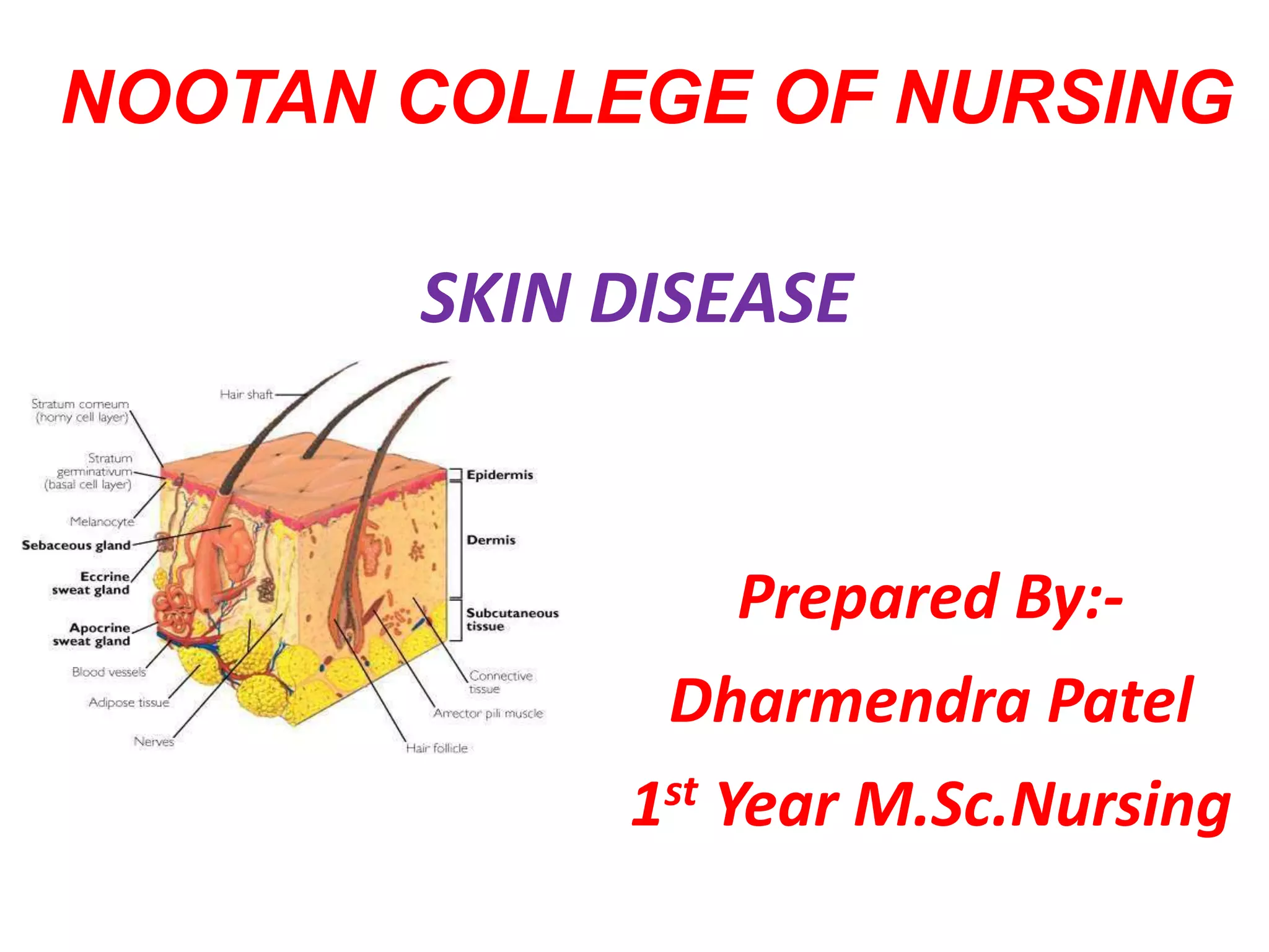
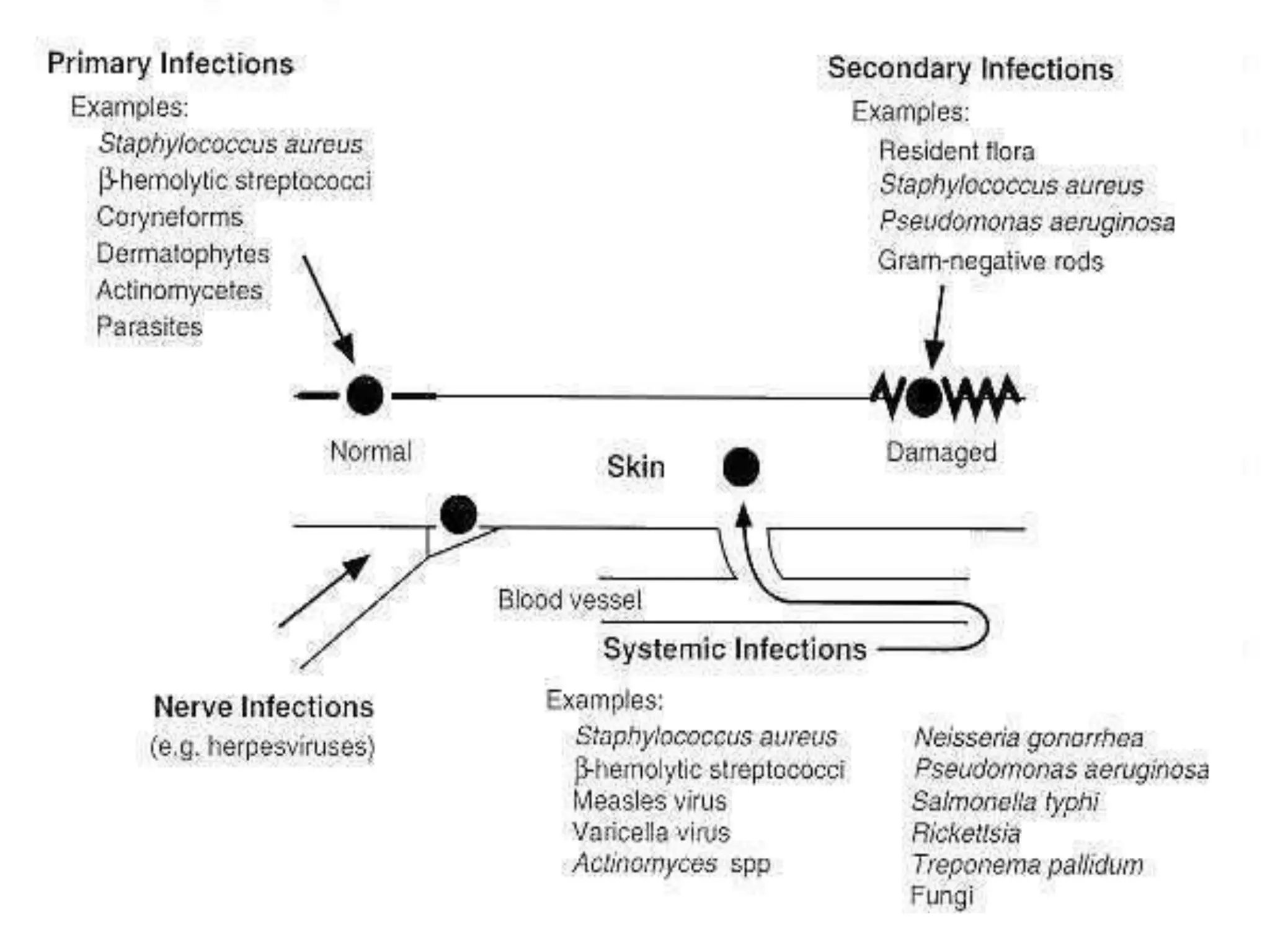
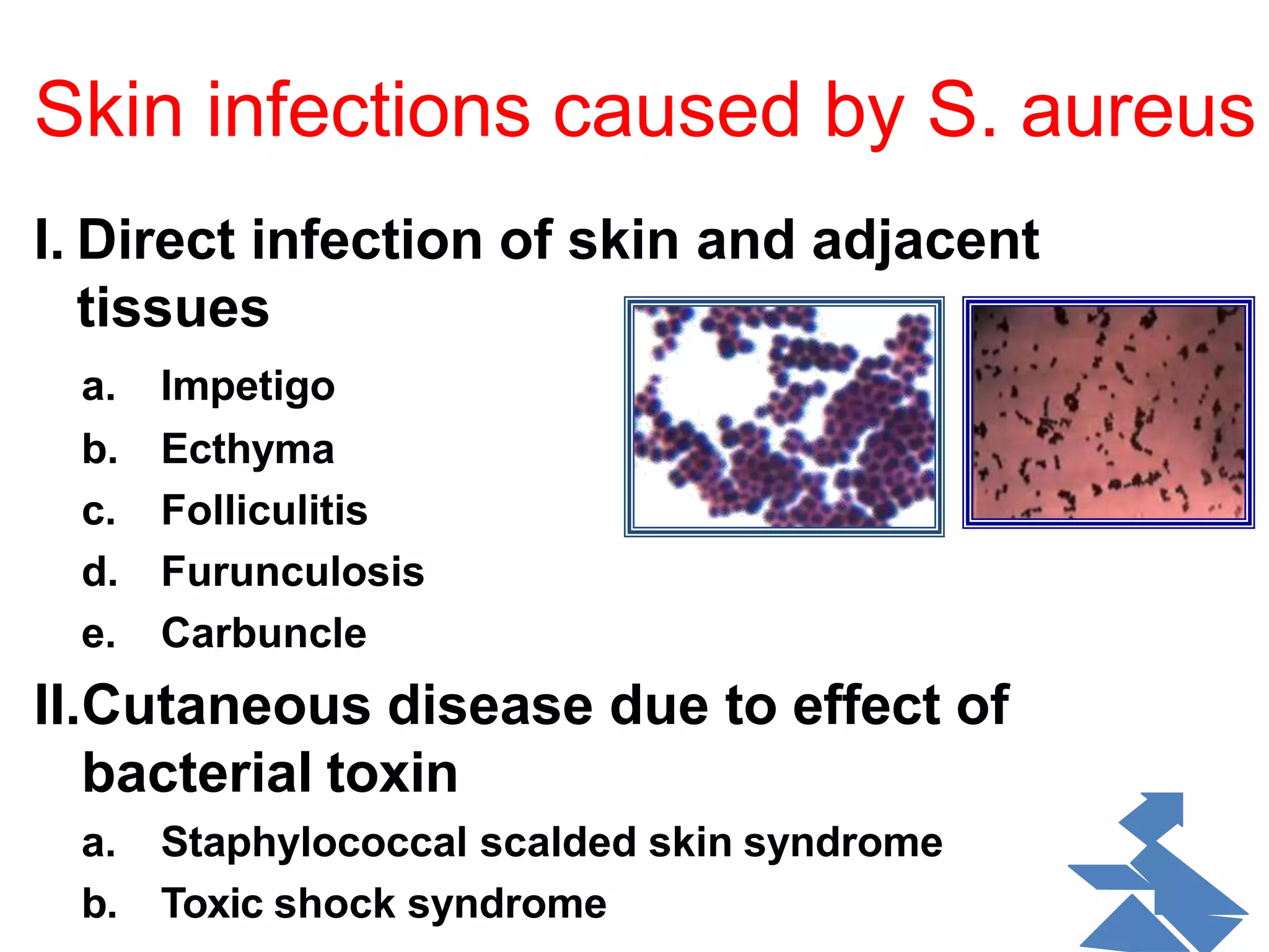
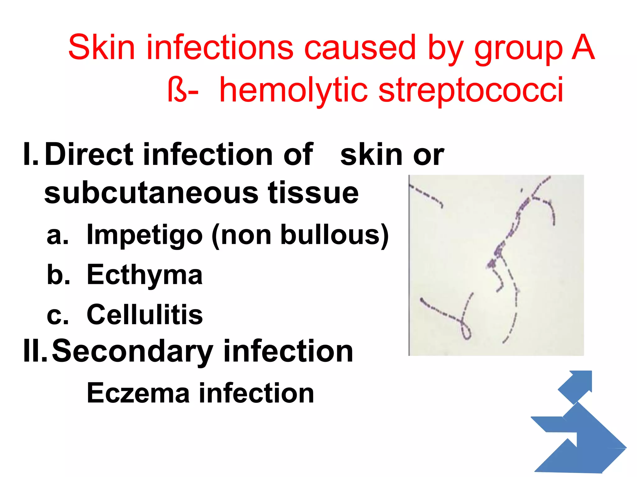
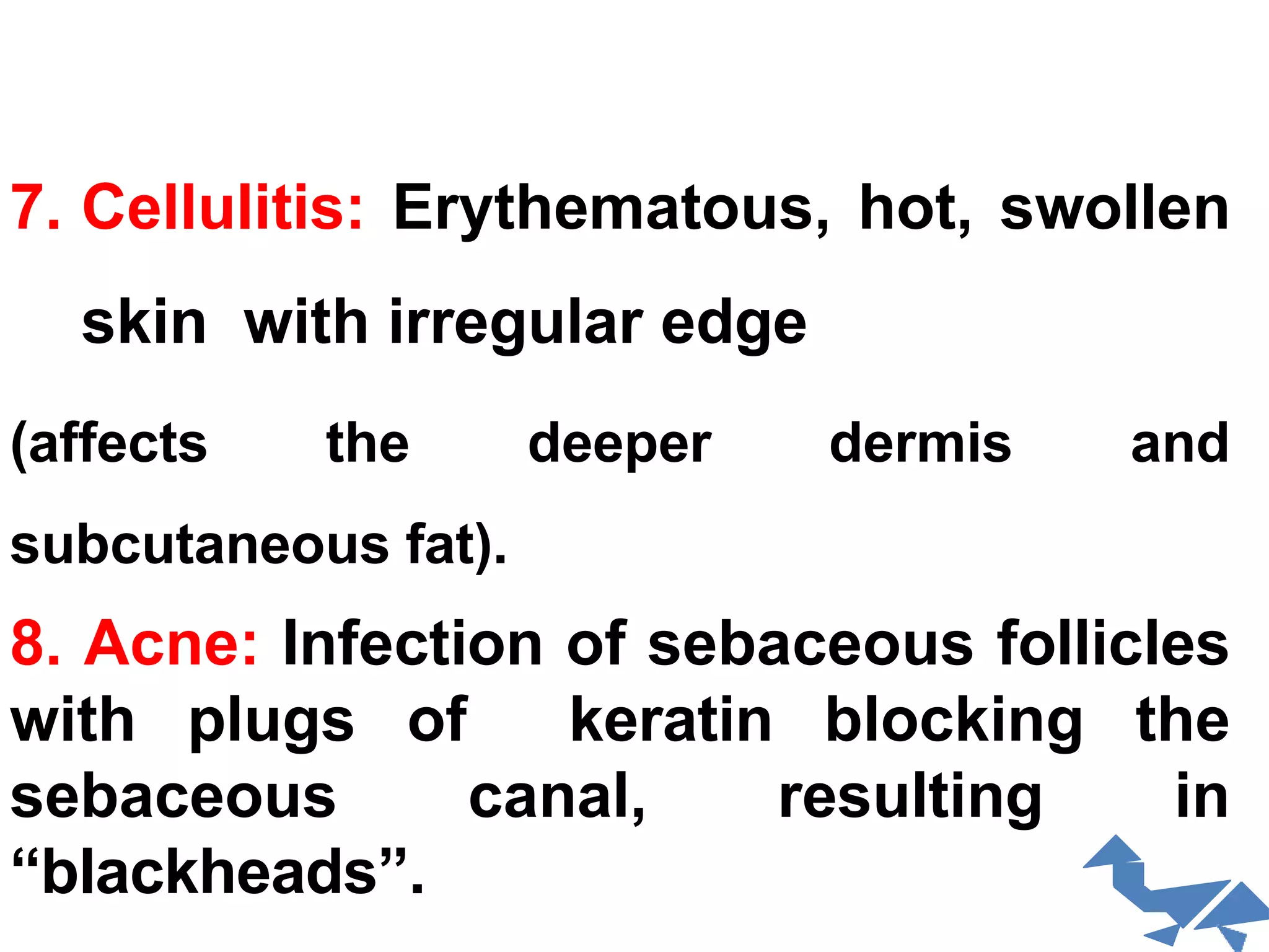
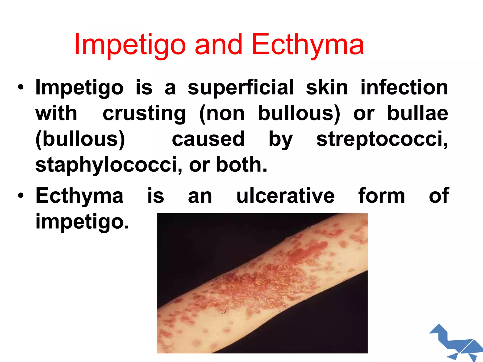
NOOTAN COLLEGE OF NURSING
Prepared By:-
Dharmendra Patel
1st Year M.Sc.Nursing
SKIN DISEASE
Introduction:-
Pathological changes may arise
in epidermis, dermis and subcutaneous tissue. The
pattern of changes may allow a diagnosis to be made
or it may be non-specific. The appearance of many
skin diseases vary at different stages of their
development and may be altered by attempted
treatment and secondary changes such as
scratching or infection.
Route of infection
Primary Infections
• caused by a single pathogen, usually affect normal
skin.
• Impetigo, folliculitis, and boils are common types.
• The most common primary skin pathogens are
S aureus β-hemolytic streptococci, and
coryneform bacteria.
• Organisms usually enter through a break in the
skin.
Secondary Infections
• Secondary infections occur in skin that is
already diseased.
• Because of the underlying disease, the clinical
picture and course of these infections vary.
Skin infections caused by S. aureus
I. Direct infection of skin and adjacent
tissues
a. Impetigo
b. Ecthyma
c. Folliculitis
d. Furunculosis
e. Carbuncle
II.Cutaneous disease due to effect of
bacterial toxin
a. Staphylococcal scalded skin syndrome
b. Toxic shock syndrome
Skin infections caused by group A
ß- hemolytic streptococci
I.Direct infection of skin or
subcutaneous tissue
a. Impetigo (non bullous)
b. Ecthyma
c. Cellulitis
II.Secondary infection
Eczema infection
Skin and Soft Tissue Infections
1.Impetigo: Initially a vesicular infection that
rapidly evolves into pustules that rupture, with
dried discharge forming honey-colored crust on
an erythematous base.
2.Ecthyma (Pustules): Begin as vesicles
that rupture, creating circular erythematous
lesions with adherent crusts.
3.Folliculitis: Inflammation at the opening of
the hair follicle that causes
erythematouspapules and pustules
surrounding individual hairs.
4. Furuncle: Deep-seated inflammatory
nodule with a pustular center that
develops around a hair follicle. (painful,
localized, abscess).
5.Carbuncle: Involvement of several
adjacent follicles, with pus discharging
from multiple follicular orifices.
6.Cutaneous abscesses:
Painful, fluctuant, red, tender swelling, on
which may rest a pustule.
7. Cellulitis: Erythematous, hot, swollen
skin with irregular edge
(affects the deeper dermis and
subcutaneous fat).
8. Acne: Infection of sebaceous follicles
with plugs of keratin blocking the
sebaceous canal, resulting in
“blackheads”.
Impetigo and Ecthyma
• Impetigo is a superficial skin infection
with crusting (non bullous) or bullae
(bullous) caused by streptococci,
staphylococci, or both.
• Ecthyma is an ulcerative form of
impetigo.
IMPETIGO (non-bullous)
IMPETIGO (bullous)
Non-bullous impetigo is a
superficial skin infection that
manifests as clusters of
vesicles or pustules that
rupture and develop a
honey-colored crust.
Bullous impetigo is a superficial
skin infection that manifests as
clusters of vesicles or pustules
that enlarge rapidly to form
bullae. The bullae burst and
expose larger bases, which
become covered with honey-
colored varnish or crust.
Impetigo(Bullous)Impetigo (Non-Bullous)
Ecthyma is a skin infection similar to
impetigo, but more deeply invasive. Usually
caused by a streptococcus infection,
ecthyma goes through the outer layer
(epidermis) to the deeper layer (dermis) of
skin, possibly causing scars.
Folliculitis
• Folliculitis is a bacterial infection of hair
follicles.
• It is usually caused by Staphylococcus aureus
but occasionally Pseudomonas aeruginosa
(hot-tub folliculitis)
• The bacteria is commonly found in
contaminated whirlpools, hot tubs or
physiotherapy pools
• Children tend to get hot tub folliculitis more
• Hot-tub folliculitis occurs because of
inadequate treatment of water with chlorine
or bromine.
FOLLICULITIS
Superficial erythematous
papules & crusts of hair
follicles in the beard area ,
aggravated by shaving.
Folliculitis manifests as superficial pustules or
inflammatory nodules surrounding hair follicles.
Furuncles and Carbuncles
• Furuncles are skin abscesses caused by
staphylococcal infection, which involve a
hair follicle and surrounding tissue.
• Carbuncles are clusters of furuncles
connected subcutaneously, causing
deeper suppuration and scarring. They
are smaller and more superficial than
subcutaneous abscesses
FURUNCLE
CARBUNCLE
Staph aureus - Large, inflammatory plaque studded
with multiple pustules, some have ruptured,
draining pus, on the nape of the neck. This very
painful area is surrounded by erythema and edema,
extends down to fascia, formed from a confluence
of many furuncles.
Carbuncles
Cellulitis
• Cellulitis is an acute bacterial infection of the
skin and subcutaneous tissue most often
caused by streptococci or staphylococci.
• Some people are at risk for infection by other
types of bacteria. They include people with
weak immune system, and those who handle
fish, meat, poultry, or soil without using
gloves.
• Cellulitis can occur anywhere on the body. In
adults, it often occurs on the legs, face, or
arms. In children, it is most common on the
face or around the anus. An infection on the
face could lead to a dangerous eye infection.
CELLULITIS
Portal of entry of infection is
seen on the lateral thigh with
necrosis of skin; infection has
extended mainly proximally
from this site.
Cutaneous Abscess
• A cutaneous abscess is a localized
collection of pus in the skin and may
occur on any skin surface.
Acne
• Most common skin disease in humans
• Propionibacterium acnes: Gram +ve rod ,causative
agent
• Pathogenesis: bacteria digest sebum , Attracts neutrophils
- Neutrophil digestive enzymes cause lesions, “pus
pockets”
• Obstruction of sebaceous follicles (oil glands)
• Open comedones or closed comedones
• Usually on the face, chest, back
• Risk factors:
– Stressful events (hormonal changes)
– Friction acne
– Oil based cosmetics
– NO correlation between chocolate, chips or
Acne
Acne Treatments
• Topical or oral antibiotics.
• Benzoyl peroxide dries plugged
follicles, kills microbes
• Tetracycline (antibiotic)
• Accutane – inhibits sebum formation
HIDRADENITIS SUPPURATIVA
Hidradenitis suppurativa is a chronic,
suppurative recurring inflammatory disease
of apocrine gland follicles.
Commoner in females specially after
puppetry.
Sites: axillae, around the nipples,
under the
breast, perineum, groin, buttocks, n eck and
scalp.
 lesions:
nodules, abcesses, scarring,sinus tract
formation.
HIDRADENITIS SUPPURATIVA
Laboratory Diagnosis of Bacterial
Infections of Skin
Specimen collection:
1. Skin biopsy
2. Skin swab
3. Pus swab
4. Nasal swab
Suspected organisms
• Impetigo: Group A Streptococcus,
Staphylococcus aureus
• Folliculitis:Staphylococcus aureus,
Pseudomonas aeruginosa
• Furuncles: Staphylococcus aureus
• Carbuncles: Staphylococcus aureus
• Cellulitis: Group A Streptococcus,
Staphylococcus aureus, Hemophilus
influenzae
1. Clinical specimen: Scrape the base of the skin
lesion with a swab.
•Gram stain: Gram (+) cocci in clusters (or in
chains)
2.Culture: Blood Agar (for both), Mannitol Salt Agar
(for staph)
• Identification: catalase , Coagulase , mannitol
fermentation
•On blood agar : both S. aureus& strept pyogens
produces beta hemolysis.
Catalasa testTube Coagulase test
3. Mannitol salts agar (MSA):high
salt(7.5) inhibits the grow of most
other organisms, S. aureus ferments
mannitol, the acid produced turns the
colonies yellow.
Principles of therapy of pyoderma
• Good personal hygiene
• Management of predisposing factors
• Aluminum chloride, a drying agent, inhibits
overgrowth of opportunistic bacteria in foot,
perineal, and axillary areas.
• Keratinolytic agents (e.g., topical salicylates)
remove hyperkeratotic lesions that harbor
pathogens, improving the exposure of the
infected skin surface to other topical
treatments
Systemic
• Treatment of disease like DM
• Nutritional deficiency
• Immunodeficiency
Principles of therapy of pyoderma
• Local therapy
– Cleaning with soap-water and weak
KMN04 solution
– Removal of crusts with KMN04 solution
– Application of antibacterial creams or
ointments
• Systemic therapy
– Antibiotics
Topical Treatment
• Topical antibiotics contain a combination of:
neomycin, bacitracin, and polymyxin.
• Some newer preparations contain:
mupirocin, gramicidin, or erythromycin, and
others combine these antibiotics with
steroids.
Tinea (ringworm) infections:-
Trichophyton, Microsporon and
Epidermophyton species are responsible for
this group of dermatophyte infections.
Trichophyton rubrum, T. mentagrophytes and
Epidermophyton floccosum are the most
common causes of dermatophyte infection in
humans. Microsporon canis caught from
dogs, cats or children causes tinea capitis in
children and, uncommonly, other types of
ringworm infection.
Clinical classification:
• Tinea corporis - ringworm infection of the body
• Tinea pedis - ringworm infection of the foot
• Tinea cruris - ringworm infection of the groin
• Tinea unguium - ringworm infection of the nails
• Tinea capitis - ringworm infection of the head,scalp,
eyebrows, eyelashes
• Tinea favosa - ringworm infection of the scalp
• Tinea manuum - ringworm infection of the hand
• Tinea barbae - ringworm infection of the beard’
Tinea corporis:
This is ringworm of the skin of the body
or limbs. Pruntic, round or annular, red,
scaling, well-marginated patches are typical.
It has to be distinguished from patches of
eczema or psoriasis by history and the
presence of mycelium in the scales. Any of
the species may cause this condition. When
an animal species is responsible (e.g. T.
verrucosum), the affected skin is very
inflamed and pustular and heals
spontaneously after a few weeks
Tinea pedis:
•Ringworm infection of the feet may be:
•Vesicular, with itchy vesicles occurring on
the sides of the feet on a background of
erythema;
•Plantar, in which the sole is red and
scaling; or
•Interdigital, in which the skin between the
fourth and fifth toes in particular is scaling
and macerated.
Tinea pedis is very common and
particularly so in young and middle-aged
men, who often contract it from communal
changing rooms. It tends to be itchy and
persistent. T. rubrum, in particular, but also
T. mentagrophytes and E. floccosum cause
the infection.
Needs To Be Used in dermatophyte.
•Griseofulvin (500 mg b.d.) is only active in
ringworm infections and has a low incidence of
serious side effects.
•Ketoconazole (200 mg daily) is active in both
yeast and dermatophyte infec- tions.
•Terbinafine (250 mg daily) is indicated for
dermatophyte infections only. Serious side
effects are uncommon.
• These agents are administered for 2–6 weeks
except for griseofulvin, which, when given for
tinea unguium of the toenails, may need to be
given for 6–12 months.
DERMATITIS:-
Definition:-
Dermatitis, also known as eczema, is a group of
diseases that results in inflammation of the
skin.These diseases are characterized
byitchiness, red skin, and a rash.
Type Definition
Frequency/a
ge group
Remarks
Atopic
dermatitis
extremely itchy disorder
Very
common,
mostly
occurs in
infants and
the very
young
Cause
unknown, but
appears to be
immunological
ly mediated
Seborrhoeic
dermatitis
common eczematous
disorder that
characteristically occurs in
hairy areas,
on the flexures and on the
central parts
of the trunk, and is now
believed to be
at least in part due to
overgrowth of the
Very
common in
all age
groups
Probably has a
microbial
cause, with
overgrowth of
normal skin
flora being
responsible
Discoid
eczema
Discoid eczema is a quite
common eczematous disorder
of unknown cause, distin-
guished by the appearance of
reddened, scaling, rounded
areas on the arms and legs.
Uncommon,
mainly in
middle-aged
individuals
Cause unknown
Lichen
simplex
chronicus
This is an intensely pruritic rash,
sharply localized to one or a few
sites, which is characterized by
thickening and exaggeration of
the skin surface markings
Quite common,
mainly in young
and middle-aged
adults
Initial cause
appears to be a
localized itch
causing an ‘itch–
scratch cycle’
Venous
eczema
Venous eczema occurs on
the lower legs and is the
result of chronic venous
hypertension
Common in the
age group that
has
gravitational
syndrome
Multiple causes,
a common
variety is
allergic contact
dermatitis to
medicaments
used
Allergic
contact
dermatitis
Allergic contact dermatitis
is an eczematous rash that
develops after contact with
an agent to which delayed
(cellular) hypersensitivity
has developed.
Common in all
adult age
groups except
the very old
Delayed
hypersensitivity
response to a
specific agent
Primary
irritant
contact
dermatitis
Primary irritant dermatitis
is an eczematous rash that
results from direct contact
with toxic ‘irritating’
materials.
Very common
in all adult
age groups
except the
very elderly
Both mechanical
and chemical
trauma
responsible
Signs and symptom:
•Range from skin rashes to bumpy rashes or
including blisters.
•Redness of the skin, swelling,itching and skin
lesions with sometimes oozing and scarring.
•The area of the skin on which the symptoms
appear tends to be different with every type of
dermatitis, whether on
the neck, wrist, forearm, thigh or ankle.
•Irritant contact dermatitis is usually
more painful than itchy.
•Atopic dermatitis vary from person to
person, the most common symptoms are
dry, itchy, red skin. Typical affected skin
areas include the folds of the arms, the back
of the knees, wrists, face and hands.
•Perioral dermatitis refers to a red bumpy
rash around the mouth.
•Seborrheic dermatitis, on the other hand,
tend to appear gradually, from dry or greasy
scaling of the scalp (dandruff) to scaling of
facial areas, sometimes with itching, but
without hair loss.
Diagnosis:-
•History and physical examination.
•Skin biopsy
•Patch tests are used in the diagnosis of
allergic contact dermatitis.
Management:-
Lifestyle:
•Products that contain dyes, perfumes, or
peanuts should not be use.
•Occlusive dressings at night may be useful.
•People can wear clothing designed to manage
the itching, scratching and peeling.
Moisturizers:
•Moisturizing agents (also known as emollients)
are recommended at least once or twice a day.
Oilier formulations appear to be better and
water-based formulations are not recommended.
Medications:-
Corticosteroids
•hydrocortisone
•clobetasol propionate
Immunosuppressants:
Light therapy:
• Light therapy using ultraviolet light has tentative support
but the quality of the evidence is not very good. A number
of different types of light may be used
including UVA and UVB; in some forms of treatment, light
sensitive chemicals such as psoralen are also used.
Overexposure to ultraviolet light carries its own risks,
particularly that of skin cancer.
Alternative medicine:
• Limited evidence suggests that acupuncture may reduce
itching in those affected by atopic dermatitis. There is
insufficient evidence to support the use of zinc, selenium,
vitamin D, vitamin E, pyridoxine (vitamin B6), sea buckthorn
oil, hempseed oil, sunflower oil, or fish oil as dietary
supplements.
PSORIASIS:-
Psoriasis is a long-lasting autoimmune
disease characterized by patches of
abnormal skin. These skin patches are
typically red, itchy, and scaly. Psoriasis
varies in severity from small, localized
patches to complete body coverage.
Causes:-
Genetics:
 Family history of the disease.
 Monozygotic twins show concordance for psoriasis.
Lifestyle:
 Chronic infections.
 Stress.
 Changes in season and climate.
 Skin dryness.
 Excessive alcohol consumption.
 Cigarette smoking.
 Obesity.
Microbes:
• Staphylococcus aureus, Malassezia, and Candida
albicans.
Medications:
• Drug-induced psoriasis may occur with
• Beta blockers.
• Lithium.
• Antimalarial medications.
• Non-steroidal anti-inflammatory drugs.
• Terbinafine.
• Calcium channel blockers.
Pathophysiology :-
Abnormally excessive and rapid growth of
the epidermal layer of the skin.
Abnormal production of skin cells (especially
during wound repair) of pathological events in
psoriasis.
It’s stem from the premature maturation
of keratinocytes
Induced by an inflammatory cascade in
the dermis involving dendritic
cells, macrophages, and T cells.
These secreted inflammatory signals are
believed to stimulate keratinocytes to proliferate.
Regulatory cytokine interleukin-10 process will
be occurs
Common Sites Affected By Psoriasis:-
Can affect any part of the body –
•typically scalp.
•Elbow.
•knees and
•sacrum.
Differential diagnosis and clinical clues to guide differentiation
Psoriasis presentation Differential diagnosis Clinical differentiation
Stable plaque  Eczema (discoid and chronic
lichenified).
 Hypertrophic lichen planus.
 Bowen's disease.
 Weeping in active discoid eczema;
lichenification in lichen simplex, scales in
lichen simplex are not as loosely attached
and silvery as in psoriasis. Violaceous
plaques mostly located on the shins,
scales are not as loosely attached and
silvery.
 Long standing solitary thin
 plaque without the characteristic silvery
scale in psoriasis.
Guttate  Pityriasis rosea.
 Secondary syphilis.
 Pityriasis lichenoides
chronicus (PLC).
 Oval papulosquamous plaques with long
axis of lesions aligned in a fir tree
distribution mostly on the trunk.
 Palm and sole involvement, mucosal
lesions, systemic symptom,
lymphadenopathy.
 More polymorphic in PLC, mica scale and
Oblaten sign
Unstable  Eczema
 Tinea incognito
 Among the unstable psoriatic lesions,
some may have otherwise typical
morphology of psoriasis; history of using
potent topical steroid or systemic steroid.
 Less plaque like and angry looking in
Erythrodermic  Other causes of
erythroderma such as
eczema/dermatitis,
cutaneous T cell
lymphoma,drug reaction,
pityriasis rubra pilaris
(PRP), pemphigus
foliaceus.
 History of psoriasis; nail
changes of psoriasis; history of
eczema or exposure to
allergens; painful keratoderma,
diffuse alopecia and leonine face
in T cell lymphoma; starts as
morbilliform rash in drug
reaction; island of sparing,
perifollicular erythema and
orange hue in PRP; impetigo-like
blisters and erosions followed
by collarettes of scale or scale-
like crusts in pemphigus
foliaceus
Pustular  Generalised: acute
generalised exanthematous
pustulosis.
 Subcorneal
pustularDermatosis.
 Localised palmoplantar
 pompholyx; tinea pedis.
 Sudden onset after taking drug;
no past history of skin diseases;
self -limited.
 Predilection for axillae and
around the groins.
 Vesicular lesions instead of
pustular lesions.
Scalp  Seborrhoeic dermatitis
 Tinea capitis
 Discernible plaque or patch in
psoriasis
 Bald patches in tinea with
broken hair stump
Inverse  Candidiasis
 Tinea
 Extramammary Paget's
disease
 Satellite lesions with scalloped
scale in candida intertrigo.
 Faintly discernible active
margin in tinea.
 More wet looking and with
erosion in extramammary
Paget's disease.
Nail  Onychomycosis  Chalky white subungual
hyperkeratosis or superficial or
proximal subungual whitish
discolouration; the oil drop
sign and thimble pitting in nail
psoriasis
Diagnostic test:-
• A diagnosis of psoriasis is usually based on the
appearance of the skin. Skin characteristics typical for
psoriasis are scaly, erythematous plaques, papules, or
patches of skin that may be painful and itch.
• The differential diagnosis of psoriasis includes
dermatological conditions similar in appearance such
as discoid eczema, seborrhoeic eczema, pityriasis
rosea (may be confused with guttate psoriasis), nail
fungus (may be confused with nail psoriasis).
• A skin biopsy
Skin from a biopsy will show clubbed epidermal
projections that interdigitate with dermis on microscopy.
Management:-
Topical agents
• Topical corticosteroid preparations are the most
effective agents when used continuously for 8 weeks.
• Vitamin D analogues such as paricalcitol were
found to be superior to placebo. Combination therapy
with vitamin D and a corticosteroid was superior to
either treatment alone and vitamin D was found to be
superior to coal tar for chronic plaque psoriasis.
• Moisturizers and emollients such as mineral
oil, petroleum jelly, calcipotriol, and decubal (an oil-in-
water emollient) were found to increase the clearance
of psoriatic plaques.
•The emollient salicylic acid is structurally similar
to para-aminobenzoic acid (PABA), commonly
found in sunscreen, and is known to interfere with
phototherapy in psoriasis. Coconut oil, when used
as an emollient in psoriasis, has been found to
decrease plaque clearance with phototherapy.
• Medicated creams and ointments applied
directly to psoriatic plaques can help reduce
inflammation, remove built-up scale, reduce skin
turnover, and clear affected skin of plaques
UV phototherapy:
Phototherapy in the form of sunlight has long been used
for psoriasis. UVB Wavelengths of 311–313 nanometers are
most effective, and special lamps have been developed for
this application. The exposure time should be controlled to
avoid over exposure and burning of the skin. The UVB lamps
should have a timer that will turn off the lamp when the time
ends. The amount of light used is determined by a person's
skin type. Increased rates of cancer from treatment appear to
be small. Narrow band UVB light (NBUVB) phototherapy has
been demonstrated to have similar efficacy to Psoralen and
ultraviolet A phototherapy(PUVA).
Systemic agents
•Methotrexate and ciclosporin are drugs that suppress
the immune system; retinoids are synthetic forms
of vitamin A. These agents are also regarded as first-
line treatments for psoriatic erythroderma.
•Oral corticosteroids should not be used, for they can
severely flare psoriasis upon their discontinuation.
Surgery:
• Limited evidence suggests removal of the
tonsils may benefit people with chronic plaque
psoriasis, guttate psoriasis, and palmoplantar
pustulosis
Diet:
Diet recommendations include consumption
of cold water fish (preferably wild fish, not
farmed) such as salmon, herring, and mackerel;
extra virgin olive oil; legumes; vegetables; fruits;
and whole grains; and avoid consumption of
alcohol, red meat, and dairy products. The
effect of consumption of caffeine (including
coffee, black tea, mate, and dark chocolate)
remains to be determined.
Nursing diagnosis:-
•Risk for infection related to loss of the
protective barrier of skin and mucus membrane
•Deficient fluid volume and electrolyte losses
related to loss of fluid from denuded skin.
•Acute pain of skin related to blistering and
erosions.
•Impaired skin integrity related to ruptured
blister area of the skin.
•Disturbed body image related to appearance of
the skin.
•Anxiety related to physical appearance of the
skin and prognosis.
Skin disease ppt for nursing student

Skin disease ppt for nursing student

  • 1.
    NOOTAN COLLEGE OFNURSING Prepared By:- Dharmendra Patel 1st Year M.Sc.Nursing SKIN DISEASE
  • 2.
    Introduction:- Pathological changes mayarise in epidermis, dermis and subcutaneous tissue. The pattern of changes may allow a diagnosis to be made or it may be non-specific. The appearance of many skin diseases vary at different stages of their development and may be altered by attempted treatment and secondary changes such as scratching or infection.
  • 3.
  • 4.
    Primary Infections • causedby a single pathogen, usually affect normal skin. • Impetigo, folliculitis, and boils are common types. • The most common primary skin pathogens are S aureus β-hemolytic streptococci, and coryneform bacteria. • Organisms usually enter through a break in the skin. Secondary Infections • Secondary infections occur in skin that is already diseased. • Because of the underlying disease, the clinical picture and course of these infections vary.
  • 6.
    Skin infections causedby S. aureus I. Direct infection of skin and adjacent tissues a. Impetigo b. Ecthyma c. Folliculitis d. Furunculosis e. Carbuncle II.Cutaneous disease due to effect of bacterial toxin a. Staphylococcal scalded skin syndrome b. Toxic shock syndrome
  • 7.
    Skin infections causedby group A ß- hemolytic streptococci I.Direct infection of skin or subcutaneous tissue a. Impetigo (non bullous) b. Ecthyma c. Cellulitis II.Secondary infection Eczema infection
  • 8.
    Skin and SoftTissue Infections 1.Impetigo: Initially a vesicular infection that rapidly evolves into pustules that rupture, with dried discharge forming honey-colored crust on an erythematous base. 2.Ecthyma (Pustules): Begin as vesicles that rupture, creating circular erythematous lesions with adherent crusts. 3.Folliculitis: Inflammation at the opening of the hair follicle that causes erythematouspapules and pustules surrounding individual hairs.
  • 9.
    4. Furuncle: Deep-seatedinflammatory nodule with a pustular center that develops around a hair follicle. (painful, localized, abscess). 5.Carbuncle: Involvement of several adjacent follicles, with pus discharging from multiple follicular orifices. 6.Cutaneous abscesses: Painful, fluctuant, red, tender swelling, on which may rest a pustule.
  • 10.
    7. Cellulitis: Erythematous,hot, swollen skin with irregular edge (affects the deeper dermis and subcutaneous fat). 8. Acne: Infection of sebaceous follicles with plugs of keratin blocking the sebaceous canal, resulting in “blackheads”.
  • 11.
    Impetigo and Ecthyma •Impetigo is a superficial skin infection with crusting (non bullous) or bullae (bullous) caused by streptococci, staphylococci, or both. • Ecthyma is an ulcerative form of impetigo.
  • 12.
  • 13.
  • 14.
    Non-bullous impetigo isa superficial skin infection that manifests as clusters of vesicles or pustules that rupture and develop a honey-colored crust. Bullous impetigo is a superficial skin infection that manifests as clusters of vesicles or pustules that enlarge rapidly to form bullae. The bullae burst and expose larger bases, which become covered with honey- colored varnish or crust. Impetigo(Bullous)Impetigo (Non-Bullous)
  • 15.
    Ecthyma is askin infection similar to impetigo, but more deeply invasive. Usually caused by a streptococcus infection, ecthyma goes through the outer layer (epidermis) to the deeper layer (dermis) of skin, possibly causing scars.
  • 16.
    Folliculitis • Folliculitis isa bacterial infection of hair follicles. • It is usually caused by Staphylococcus aureus but occasionally Pseudomonas aeruginosa (hot-tub folliculitis) • The bacteria is commonly found in contaminated whirlpools, hot tubs or physiotherapy pools • Children tend to get hot tub folliculitis more • Hot-tub folliculitis occurs because of inadequate treatment of water with chlorine or bromine.
  • 17.
    FOLLICULITIS Superficial erythematous papules &crusts of hair follicles in the beard area , aggravated by shaving. Folliculitis manifests as superficial pustules or inflammatory nodules surrounding hair follicles.
  • 18.
    Furuncles and Carbuncles •Furuncles are skin abscesses caused by staphylococcal infection, which involve a hair follicle and surrounding tissue. • Carbuncles are clusters of furuncles connected subcutaneously, causing deeper suppuration and scarring. They are smaller and more superficial than subcutaneous abscesses
  • 19.
  • 20.
    CARBUNCLE Staph aureus -Large, inflammatory plaque studded with multiple pustules, some have ruptured, draining pus, on the nape of the neck. This very painful area is surrounded by erythema and edema, extends down to fascia, formed from a confluence of many furuncles.
  • 21.
  • 22.
    Cellulitis • Cellulitis isan acute bacterial infection of the skin and subcutaneous tissue most often caused by streptococci or staphylococci. • Some people are at risk for infection by other types of bacteria. They include people with weak immune system, and those who handle fish, meat, poultry, or soil without using gloves. • Cellulitis can occur anywhere on the body. In adults, it often occurs on the legs, face, or arms. In children, it is most common on the face or around the anus. An infection on the face could lead to a dangerous eye infection.
  • 23.
    CELLULITIS Portal of entryof infection is seen on the lateral thigh with necrosis of skin; infection has extended mainly proximally from this site.
  • 24.
    Cutaneous Abscess • Acutaneous abscess is a localized collection of pus in the skin and may occur on any skin surface.
  • 25.
    Acne • Most commonskin disease in humans • Propionibacterium acnes: Gram +ve rod ,causative agent • Pathogenesis: bacteria digest sebum , Attracts neutrophils - Neutrophil digestive enzymes cause lesions, “pus pockets” • Obstruction of sebaceous follicles (oil glands) • Open comedones or closed comedones • Usually on the face, chest, back • Risk factors: – Stressful events (hormonal changes) – Friction acne – Oil based cosmetics – NO correlation between chocolate, chips or
  • 26.
  • 27.
    Acne Treatments • Topicalor oral antibiotics. • Benzoyl peroxide dries plugged follicles, kills microbes • Tetracycline (antibiotic) • Accutane – inhibits sebum formation
  • 28.
    HIDRADENITIS SUPPURATIVA Hidradenitis suppurativais a chronic, suppurative recurring inflammatory disease of apocrine gland follicles. Commoner in females specially after puppetry. Sites: axillae, around the nipples, under the breast, perineum, groin, buttocks, n eck and scalp.  lesions: nodules, abcesses, scarring,sinus tract formation.
  • 29.
  • 30.
    Laboratory Diagnosis ofBacterial Infections of Skin Specimen collection: 1. Skin biopsy 2. Skin swab 3. Pus swab 4. Nasal swab
  • 31.
    Suspected organisms • Impetigo:Group A Streptococcus, Staphylococcus aureus • Folliculitis:Staphylococcus aureus, Pseudomonas aeruginosa • Furuncles: Staphylococcus aureus • Carbuncles: Staphylococcus aureus • Cellulitis: Group A Streptococcus, Staphylococcus aureus, Hemophilus influenzae
  • 32.
    1. Clinical specimen:Scrape the base of the skin lesion with a swab. •Gram stain: Gram (+) cocci in clusters (or in chains) 2.Culture: Blood Agar (for both), Mannitol Salt Agar (for staph) • Identification: catalase , Coagulase , mannitol fermentation •On blood agar : both S. aureus& strept pyogens produces beta hemolysis. Catalasa testTube Coagulase test
  • 33.
    3. Mannitol saltsagar (MSA):high salt(7.5) inhibits the grow of most other organisms, S. aureus ferments mannitol, the acid produced turns the colonies yellow.
  • 34.
    Principles of therapyof pyoderma • Good personal hygiene • Management of predisposing factors • Aluminum chloride, a drying agent, inhibits overgrowth of opportunistic bacteria in foot, perineal, and axillary areas. • Keratinolytic agents (e.g., topical salicylates) remove hyperkeratotic lesions that harbor pathogens, improving the exposure of the infected skin surface to other topical treatments Systemic • Treatment of disease like DM • Nutritional deficiency • Immunodeficiency
  • 35.
    Principles of therapyof pyoderma • Local therapy – Cleaning with soap-water and weak KMN04 solution – Removal of crusts with KMN04 solution – Application of antibacterial creams or ointments • Systemic therapy – Antibiotics
  • 36.
    Topical Treatment • Topicalantibiotics contain a combination of: neomycin, bacitracin, and polymyxin. • Some newer preparations contain: mupirocin, gramicidin, or erythromycin, and others combine these antibiotics with steroids.
  • 37.
    Tinea (ringworm) infections:- Trichophyton,Microsporon and Epidermophyton species are responsible for this group of dermatophyte infections. Trichophyton rubrum, T. mentagrophytes and Epidermophyton floccosum are the most common causes of dermatophyte infection in humans. Microsporon canis caught from dogs, cats or children causes tinea capitis in children and, uncommonly, other types of ringworm infection.
  • 38.
    Clinical classification: • Tineacorporis - ringworm infection of the body • Tinea pedis - ringworm infection of the foot • Tinea cruris - ringworm infection of the groin • Tinea unguium - ringworm infection of the nails • Tinea capitis - ringworm infection of the head,scalp, eyebrows, eyelashes • Tinea favosa - ringworm infection of the scalp • Tinea manuum - ringworm infection of the hand • Tinea barbae - ringworm infection of the beard’
  • 40.
    Tinea corporis: This isringworm of the skin of the body or limbs. Pruntic, round or annular, red, scaling, well-marginated patches are typical. It has to be distinguished from patches of eczema or psoriasis by history and the presence of mycelium in the scales. Any of the species may cause this condition. When an animal species is responsible (e.g. T. verrucosum), the affected skin is very inflamed and pustular and heals spontaneously after a few weeks
  • 41.
    Tinea pedis: •Ringworm infectionof the feet may be: •Vesicular, with itchy vesicles occurring on the sides of the feet on a background of erythema; •Plantar, in which the sole is red and scaling; or •Interdigital, in which the skin between the fourth and fifth toes in particular is scaling and macerated.
  • 42.
    Tinea pedis isvery common and particularly so in young and middle-aged men, who often contract it from communal changing rooms. It tends to be itchy and persistent. T. rubrum, in particular, but also T. mentagrophytes and E. floccosum cause the infection.
  • 43.
    Needs To BeUsed in dermatophyte. •Griseofulvin (500 mg b.d.) is only active in ringworm infections and has a low incidence of serious side effects. •Ketoconazole (200 mg daily) is active in both yeast and dermatophyte infec- tions. •Terbinafine (250 mg daily) is indicated for dermatophyte infections only. Serious side effects are uncommon. • These agents are administered for 2–6 weeks except for griseofulvin, which, when given for tinea unguium of the toenails, may need to be given for 6–12 months.
  • 44.
    DERMATITIS:- Definition:- Dermatitis, also knownas eczema, is a group of diseases that results in inflammation of the skin.These diseases are characterized byitchiness, red skin, and a rash.
  • 45.
    Type Definition Frequency/a ge group Remarks Atopic dermatitis extremelyitchy disorder Very common, mostly occurs in infants and the very young Cause unknown, but appears to be immunological ly mediated Seborrhoeic dermatitis common eczematous disorder that characteristically occurs in hairy areas, on the flexures and on the central parts of the trunk, and is now believed to be at least in part due to overgrowth of the Very common in all age groups Probably has a microbial cause, with overgrowth of normal skin flora being responsible
  • 46.
    Discoid eczema Discoid eczema isa quite common eczematous disorder of unknown cause, distin- guished by the appearance of reddened, scaling, rounded areas on the arms and legs. Uncommon, mainly in middle-aged individuals Cause unknown Lichen simplex chronicus This is an intensely pruritic rash, sharply localized to one or a few sites, which is characterized by thickening and exaggeration of the skin surface markings Quite common, mainly in young and middle-aged adults Initial cause appears to be a localized itch causing an ‘itch– scratch cycle’
  • 47.
    Venous eczema Venous eczema occurson the lower legs and is the result of chronic venous hypertension Common in the age group that has gravitational syndrome Multiple causes, a common variety is allergic contact dermatitis to medicaments used Allergic contact dermatitis Allergic contact dermatitis is an eczematous rash that develops after contact with an agent to which delayed (cellular) hypersensitivity has developed. Common in all adult age groups except the very old Delayed hypersensitivity response to a specific agent Primary irritant contact dermatitis Primary irritant dermatitis is an eczematous rash that results from direct contact with toxic ‘irritating’ materials. Very common in all adult age groups except the very elderly Both mechanical and chemical trauma responsible
  • 48.
    Signs and symptom: •Rangefrom skin rashes to bumpy rashes or including blisters. •Redness of the skin, swelling,itching and skin lesions with sometimes oozing and scarring. •The area of the skin on which the symptoms appear tends to be different with every type of dermatitis, whether on the neck, wrist, forearm, thigh or ankle. •Irritant contact dermatitis is usually more painful than itchy.
  • 49.
    •Atopic dermatitis varyfrom person to person, the most common symptoms are dry, itchy, red skin. Typical affected skin areas include the folds of the arms, the back of the knees, wrists, face and hands. •Perioral dermatitis refers to a red bumpy rash around the mouth. •Seborrheic dermatitis, on the other hand, tend to appear gradually, from dry or greasy scaling of the scalp (dandruff) to scaling of facial areas, sometimes with itching, but without hair loss.
  • 50.
    Diagnosis:- •History and physicalexamination. •Skin biopsy •Patch tests are used in the diagnosis of allergic contact dermatitis.
  • 51.
    Management:- Lifestyle: •Products that containdyes, perfumes, or peanuts should not be use. •Occlusive dressings at night may be useful. •People can wear clothing designed to manage the itching, scratching and peeling. Moisturizers: •Moisturizing agents (also known as emollients) are recommended at least once or twice a day. Oilier formulations appear to be better and water-based formulations are not recommended.
  • 52.
  • 53.
    Light therapy: • Lighttherapy using ultraviolet light has tentative support but the quality of the evidence is not very good. A number of different types of light may be used including UVA and UVB; in some forms of treatment, light sensitive chemicals such as psoralen are also used. Overexposure to ultraviolet light carries its own risks, particularly that of skin cancer. Alternative medicine: • Limited evidence suggests that acupuncture may reduce itching in those affected by atopic dermatitis. There is insufficient evidence to support the use of zinc, selenium, vitamin D, vitamin E, pyridoxine (vitamin B6), sea buckthorn oil, hempseed oil, sunflower oil, or fish oil as dietary supplements.
  • 54.
    PSORIASIS:- Psoriasis is along-lasting autoimmune disease characterized by patches of abnormal skin. These skin patches are typically red, itchy, and scaly. Psoriasis varies in severity from small, localized patches to complete body coverage.
  • 55.
    Causes:- Genetics:  Family historyof the disease.  Monozygotic twins show concordance for psoriasis. Lifestyle:  Chronic infections.  Stress.  Changes in season and climate.  Skin dryness.  Excessive alcohol consumption.  Cigarette smoking.  Obesity.
  • 56.
    Microbes: • Staphylococcus aureus,Malassezia, and Candida albicans. Medications: • Drug-induced psoriasis may occur with • Beta blockers. • Lithium. • Antimalarial medications. • Non-steroidal anti-inflammatory drugs. • Terbinafine. • Calcium channel blockers.
  • 57.
    Pathophysiology :- Abnormally excessiveand rapid growth of the epidermal layer of the skin. Abnormal production of skin cells (especially during wound repair) of pathological events in psoriasis. It’s stem from the premature maturation of keratinocytes
  • 58.
    Induced by aninflammatory cascade in the dermis involving dendritic cells, macrophages, and T cells. These secreted inflammatory signals are believed to stimulate keratinocytes to proliferate. Regulatory cytokine interleukin-10 process will be occurs
  • 59.
    Common Sites AffectedBy Psoriasis:- Can affect any part of the body – •typically scalp. •Elbow. •knees and •sacrum.
  • 60.
    Differential diagnosis andclinical clues to guide differentiation Psoriasis presentation Differential diagnosis Clinical differentiation Stable plaque  Eczema (discoid and chronic lichenified).  Hypertrophic lichen planus.  Bowen's disease.  Weeping in active discoid eczema; lichenification in lichen simplex, scales in lichen simplex are not as loosely attached and silvery as in psoriasis. Violaceous plaques mostly located on the shins, scales are not as loosely attached and silvery.  Long standing solitary thin  plaque without the characteristic silvery scale in psoriasis. Guttate  Pityriasis rosea.  Secondary syphilis.  Pityriasis lichenoides chronicus (PLC).  Oval papulosquamous plaques with long axis of lesions aligned in a fir tree distribution mostly on the trunk.  Palm and sole involvement, mucosal lesions, systemic symptom, lymphadenopathy.  More polymorphic in PLC, mica scale and Oblaten sign Unstable  Eczema  Tinea incognito  Among the unstable psoriatic lesions, some may have otherwise typical morphology of psoriasis; history of using potent topical steroid or systemic steroid.  Less plaque like and angry looking in
  • 61.
    Erythrodermic  Othercauses of erythroderma such as eczema/dermatitis, cutaneous T cell lymphoma,drug reaction, pityriasis rubra pilaris (PRP), pemphigus foliaceus.  History of psoriasis; nail changes of psoriasis; history of eczema or exposure to allergens; painful keratoderma, diffuse alopecia and leonine face in T cell lymphoma; starts as morbilliform rash in drug reaction; island of sparing, perifollicular erythema and orange hue in PRP; impetigo-like blisters and erosions followed by collarettes of scale or scale- like crusts in pemphigus foliaceus Pustular  Generalised: acute generalised exanthematous pustulosis.  Subcorneal pustularDermatosis.  Localised palmoplantar  pompholyx; tinea pedis.  Sudden onset after taking drug; no past history of skin diseases; self -limited.  Predilection for axillae and around the groins.  Vesicular lesions instead of pustular lesions.
  • 62.
    Scalp  Seborrhoeicdermatitis  Tinea capitis  Discernible plaque or patch in psoriasis  Bald patches in tinea with broken hair stump Inverse  Candidiasis  Tinea  Extramammary Paget's disease  Satellite lesions with scalloped scale in candida intertrigo.  Faintly discernible active margin in tinea.  More wet looking and with erosion in extramammary Paget's disease. Nail  Onychomycosis  Chalky white subungual hyperkeratosis or superficial or proximal subungual whitish discolouration; the oil drop sign and thimble pitting in nail psoriasis
  • 63.
    Diagnostic test:- • Adiagnosis of psoriasis is usually based on the appearance of the skin. Skin characteristics typical for psoriasis are scaly, erythematous plaques, papules, or patches of skin that may be painful and itch. • The differential diagnosis of psoriasis includes dermatological conditions similar in appearance such as discoid eczema, seborrhoeic eczema, pityriasis rosea (may be confused with guttate psoriasis), nail fungus (may be confused with nail psoriasis). • A skin biopsy Skin from a biopsy will show clubbed epidermal projections that interdigitate with dermis on microscopy.
  • 64.
    Management:- Topical agents • Topicalcorticosteroid preparations are the most effective agents when used continuously for 8 weeks. • Vitamin D analogues such as paricalcitol were found to be superior to placebo. Combination therapy with vitamin D and a corticosteroid was superior to either treatment alone and vitamin D was found to be superior to coal tar for chronic plaque psoriasis. • Moisturizers and emollients such as mineral oil, petroleum jelly, calcipotriol, and decubal (an oil-in- water emollient) were found to increase the clearance of psoriatic plaques.
  • 65.
    •The emollient salicylicacid is structurally similar to para-aminobenzoic acid (PABA), commonly found in sunscreen, and is known to interfere with phototherapy in psoriasis. Coconut oil, when used as an emollient in psoriasis, has been found to decrease plaque clearance with phototherapy. • Medicated creams and ointments applied directly to psoriatic plaques can help reduce inflammation, remove built-up scale, reduce skin turnover, and clear affected skin of plaques
  • 66.
    UV phototherapy: Phototherapy inthe form of sunlight has long been used for psoriasis. UVB Wavelengths of 311–313 nanometers are most effective, and special lamps have been developed for this application. The exposure time should be controlled to avoid over exposure and burning of the skin. The UVB lamps should have a timer that will turn off the lamp when the time ends. The amount of light used is determined by a person's skin type. Increased rates of cancer from treatment appear to be small. Narrow band UVB light (NBUVB) phototherapy has been demonstrated to have similar efficacy to Psoralen and ultraviolet A phototherapy(PUVA).
  • 67.
    Systemic agents •Methotrexate andciclosporin are drugs that suppress the immune system; retinoids are synthetic forms of vitamin A. These agents are also regarded as first- line treatments for psoriatic erythroderma. •Oral corticosteroids should not be used, for they can severely flare psoriasis upon their discontinuation. Surgery: • Limited evidence suggests removal of the tonsils may benefit people with chronic plaque psoriasis, guttate psoriasis, and palmoplantar pustulosis
  • 68.
    Diet: Diet recommendations includeconsumption of cold water fish (preferably wild fish, not farmed) such as salmon, herring, and mackerel; extra virgin olive oil; legumes; vegetables; fruits; and whole grains; and avoid consumption of alcohol, red meat, and dairy products. The effect of consumption of caffeine (including coffee, black tea, mate, and dark chocolate) remains to be determined.
  • 69.
    Nursing diagnosis:- •Risk forinfection related to loss of the protective barrier of skin and mucus membrane •Deficient fluid volume and electrolyte losses related to loss of fluid from denuded skin. •Acute pain of skin related to blistering and erosions. •Impaired skin integrity related to ruptured blister area of the skin. •Disturbed body image related to appearance of the skin. •Anxiety related to physical appearance of the skin and prognosis.