Skin Cancer Detection using Image Processing in Real Time
The document presents a study on using image processing and machine learning for skin cancer detection, aiming to develop a real-time model for implementation in an Android application. It discusses phases of image processing and the overall process from dataset preparation to model building, training, and testing, achieving a commendable accuracy of 97% in detecting cancer cells. The work emphasizes the importance of early detection and provides insights into the methodologies used in this project, including deep learning techniques and practical application development.
International Journal ofTrend in Scientific Research and Development @ www.ijtsrd.com eISSN: 2456-6470
@ IJTSRD | Unique Paper ID – IJTSRD46384 | Volume – 5 | Issue – 6 | Sep-Oct 2021 Page 272
1. ACQUISITION– It could be as fundamental as
being given a digital image to work with. The
main work involves:
A. Scaling.
B. Colour conversion (RGB to Gray or vice-versa).
2. IMAGE ENHANCEMENT– It is one of the most
basic and appealing aspects of Image Processing,
and it is also used to extract some hidden
elements from an image. It is subjective.
3. IMAGE RESTORATION– This likewise has to
do with making an image appealing, but it is more
objective (Restoration is based on mathematical
or probabilistic model or image degradation).
4. COLOR IMAGE PROCESSING– This section
covers pseudo-colour and full colour image
processing, as well as colour models that can be
used in digital image processing.
5. WAVELETS AND MULTI-RESOLUTION
PROCESSING– This is the foundation for
portraying images in a variety of ways.
6. IMAGE COMPRESSION—This procedure
necessitates the creation of various functions. It
primarily concerns image size or resolution.
7. MORPHOLOGICAL PROCESSING-It deals
with tools for extracting picture components that
can be used to represent and describe shape.
8. SEGMENTATION PROCEDURE-Partitioning
an image into its constituent sections or objects is
part of this procedure. The most demanding
problem in Image Processing is autonomous
segmentation.
9. REPRESENTATION & DESCRIPTION-It
comes after the output of the segmentation step;
selecting a representation is only one part of the
solution for converting raw data into processed
data.
10. OBJECT DETECTION AND RECOGNITION-It
is a process that assigns a label to an object based
on its descriptor.
Overlapping Fields with Image Processing
1. According to block 1, if the input is an image and
the output is an image, the process is known as
digital image processing.
2. According to block 2, Computer Vision is defined
as an input that is an image and an output that is
some kind of information or description.
3. According to block 3, if input is some description
or code and we get image as an output, then it is
termed as Computer Graphics.
4. According to block 4, if input is description or
some keywords or some code and we get
description or some keywords as an output, then it
is termed as Artificial Intelligence.
Skin Cancer Overview
Skin cancer, or the abnormal proliferation of skin
cells, is most commonly found on sun-exposed skin.
However, this frequent type of cancer can also
develop on parts of your skin that aren't normally
exposed to the sun.
Basal cell carcinoma, squamous cell carcinoma, and
melanoma are the three most common kinds of skin
cancer.
Limiting or avoiding ultraviolet (UV) radiation
exposure can lower your risk of skin cancer. Skin
cancer can be detected at an early stage if you check
your skin for abnormal changes. Early skin cancer
detection increases your chances of a successful skin
cancer treatment.
1. Basal cell carcinoma - A kind of skin cancer
known as basal cell carcinoma. Basal cell
carcinoma starts in the basal cells, which are a
type of skin cell that creates new skin cells when
the old ones die. Basal cell carcinoma often
appears as a slightly transparent bump on the
skin, though it can take other forms. Basal cell
carcinoma occurs most often on areas of the skin
that are exposed to the sun, such as your head and
neck.
2. Melanoma – It originates in the cells
(melanocytes) that create melanin, the pigment
that gives your skin its colour. It is the most
dangerous type of skin cancer. Melanoma can
also develop in the eyes and, in rare cases, inside
the body, such as the nose or throat. Although the
specific aetiology of all melanomas is unknown,
ultraviolet (UV) radiation from the sun, tanning
lights, and beds increases your risk of acquiring
melanoma. Melanoma risk can be reduced by
limiting your exposure to UV light.
3. Nonmelanoma skin cancer - It encompasses all
skin cancers that aren't melanoma. Nonmelanoma
3.
International Journal ofTrend in Scientific Research and Development @ www.ijtsrd.com eISSN: 2456-6470
@ IJTSRD | Unique Paper ID – IJTSRD46384 | Volume – 5 | Issue – 6 | Sep-Oct 2021 Page 273
skin cancer encompasses a number of different
forms of skin cancer, the most frequent of which
are basal cell carcinoma and squamous cell
carcinoma. Treatment for nonmelanoma skin
cancer is determined by the type of malignancy.
The most common therapy for skin cancer is
surgery to remove the cancerous cells.
PREREQUISITES
1. Dataset for Training.
2. Dataset for Testing.
3. Machine learning model (that takes in digital
Images as input and predicts cancer cells).
Dataset
We used the dataset provided by SIIM-ISIC
Melanoma Classification.
The images are provided in DICOM format. This can
be accessed using commonly-available libraries like
PyDicom, and contains both image and metadata. It is
a commonly used medical imaging data format.
Images are also provided in JPEG and TF Record
format (in the jpeg and TF records directories,
respectively). Images in TF Record format have been
resized to a uniform 1024x1024.
EXPERIMENTATION
TensorFlow and Keras are used to build and create a machine learning model.
1. Data Exploration
We created algorithms and models to classify between benign and malignant skin cancers using Convolutional
Neural Networks. We use Colab for source code editing.
2. Seeding
The seed sets the starting number used to generate a sequence of random numbers it ensures that you get the
same result if you start with that same seed each time you run the same process.
3. Initializing Model
Model initialization is the process of determining the necessary model parameters such as the basic value, the
trend value, and the seasonal indices for the selected forecast model.
4.
International Journal ofTrend in Scientific Research and Development @ www.ijtsrd.com eISSN: 2456-6470
@ IJTSRD | Unique Paper ID – IJTSRD46384 | Volume – 5 | Issue – 6 | Sep-Oct 2021 Page 274
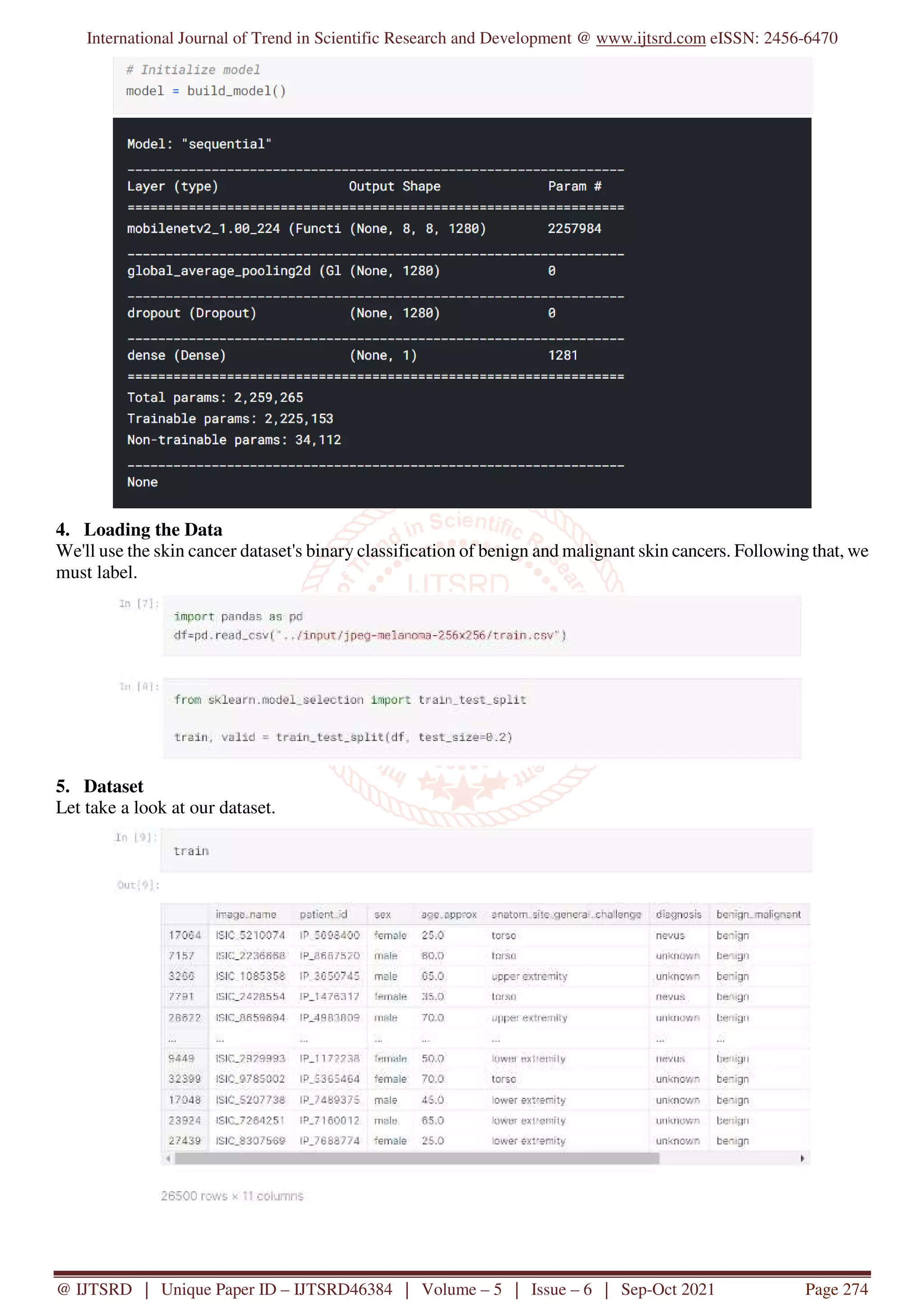
4. Loading the Data
We'll use the skin cancer dataset's binary classification of benign and malignant skin cancers. Following that, we
must label.
5. Dataset
Let take a look at our dataset.
5.
International Journal ofTrend in Scientific Research and Development @ www.ijtsrd.com eISSN: 2456-6470
@ IJTSRD | Unique Paper ID – IJTSRD46384 | Volume – 5 | Issue – 6 | Sep-Oct 2021 Page 275
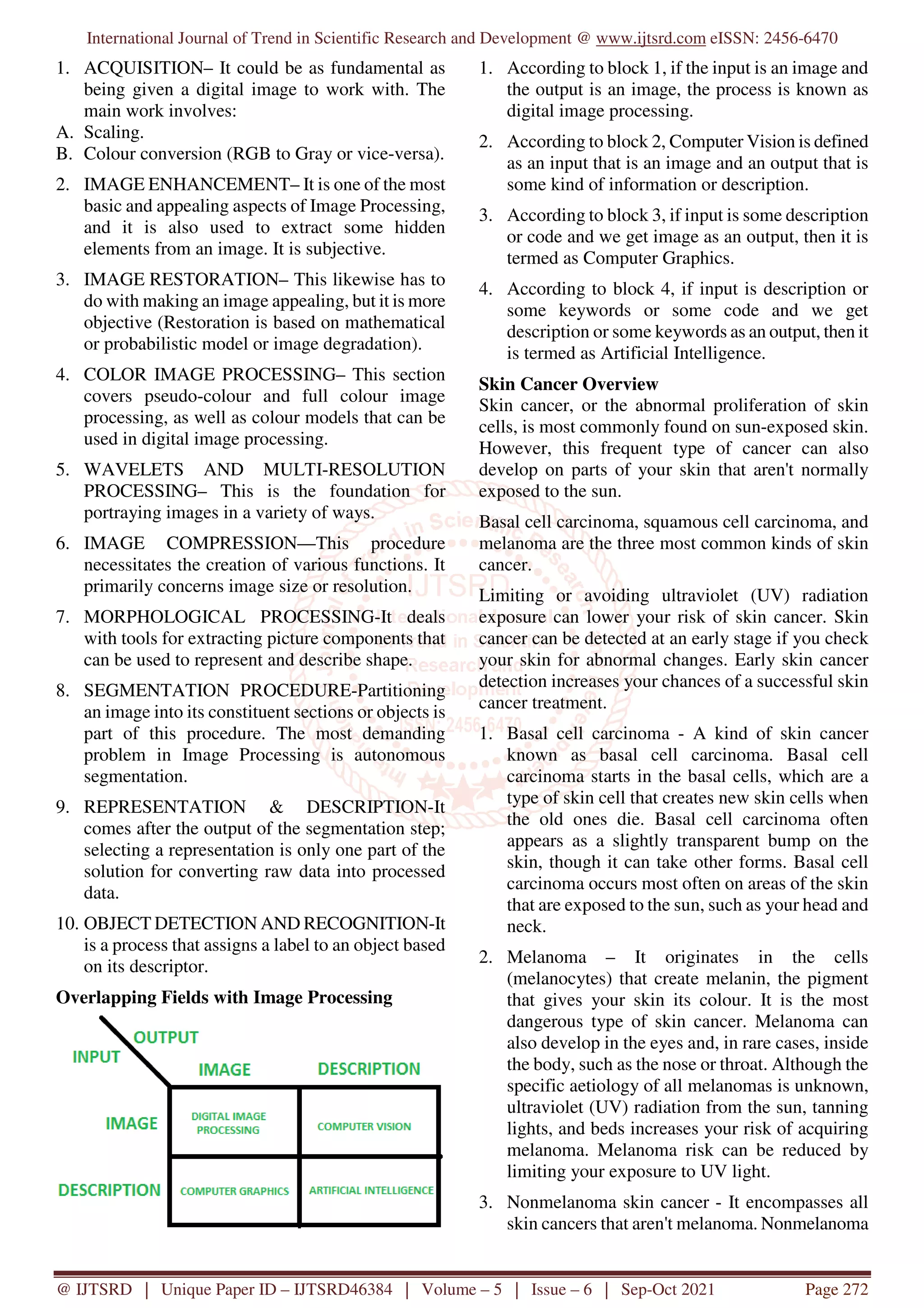
6. Data Processing
We set up generators to read images from our source folders, then we transformed them to float32 tensors, and
fed them to our network.
In our case, we'll change the pixel values to the [0, 1] range before pre-processing the images (all values are now
in the [0, 255] range). As required by the networks, the input data must be scaled to 224x224 pixels as an input.
7. Building the Model
It's as simple as layering a linear classifier on top of the feature extractor with the Hub module. For speed, we
start with a non-trainable feature extractor.
6.
International Journal ofTrend in Scientific Research and Development @ www.ijtsrd.com eISSN: 2456-6470
@ IJTSRD | Unique Paper ID – IJTSRD46384 | Volume – 5 | Issue – 6 | Sep-Oct 2021 Page 276
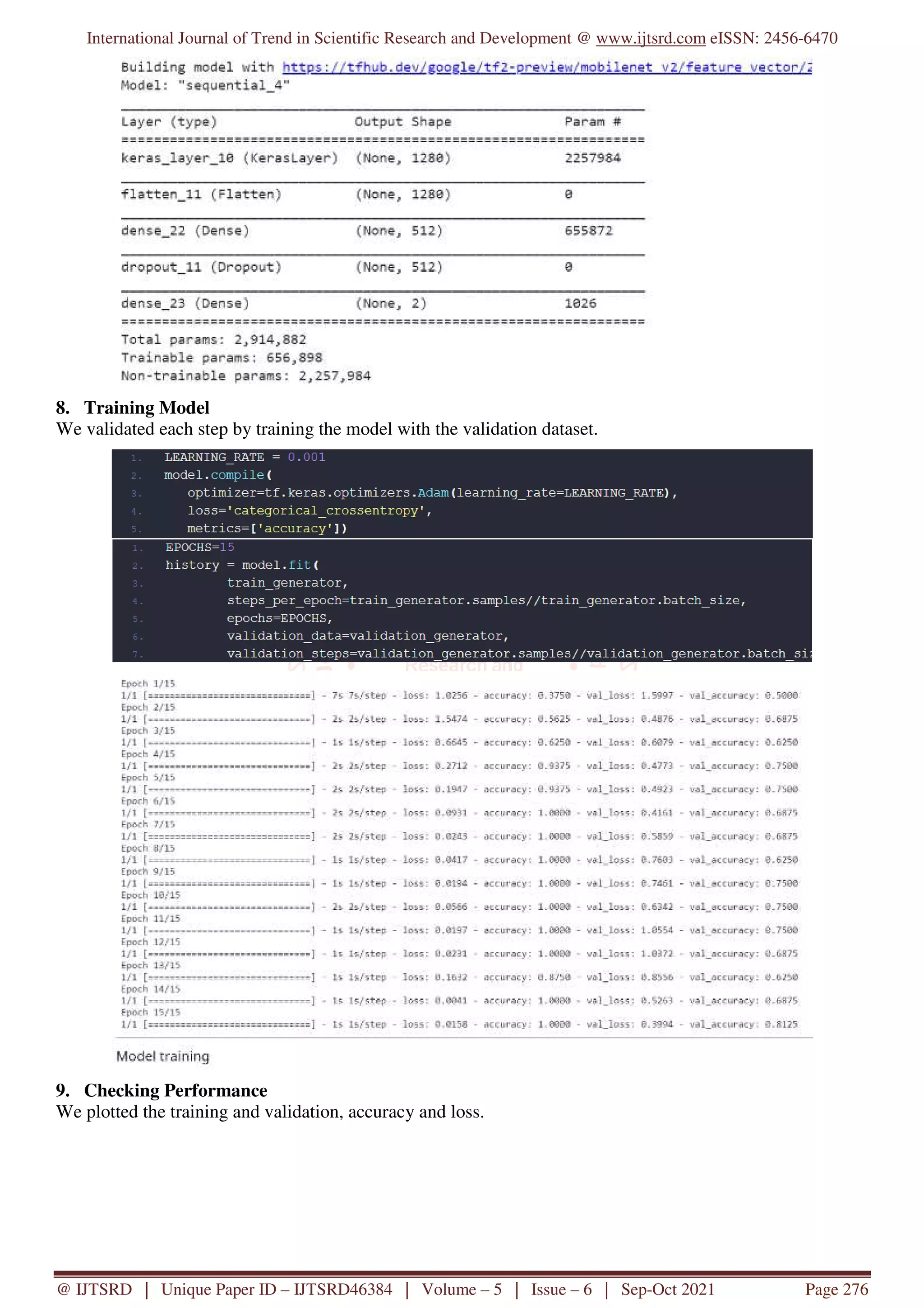
8. Training Model
We validated each step by training the model with the validation dataset.
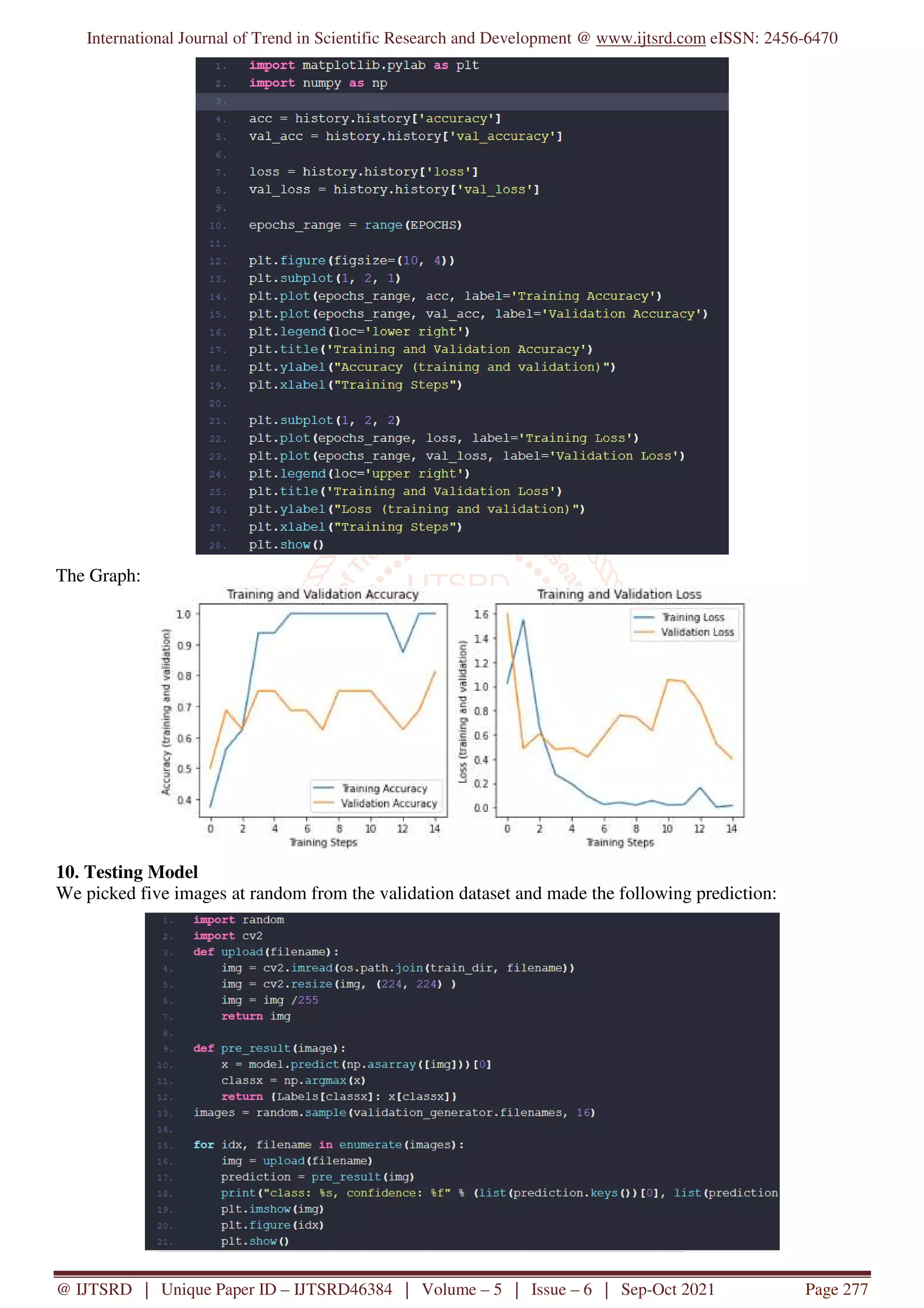
9. Checking Performance
We plotted the training and validation, accuracy and loss.
7.
International Journal ofTrend in Scientific Research and Development @ www.ijtsrd.com eISSN: 2456-6470
@ IJTSRD | Unique Paper ID – IJTSRD46384 | Volume – 5 | Issue – 6 | Sep-Oct 2021 Page 277
The Graph:
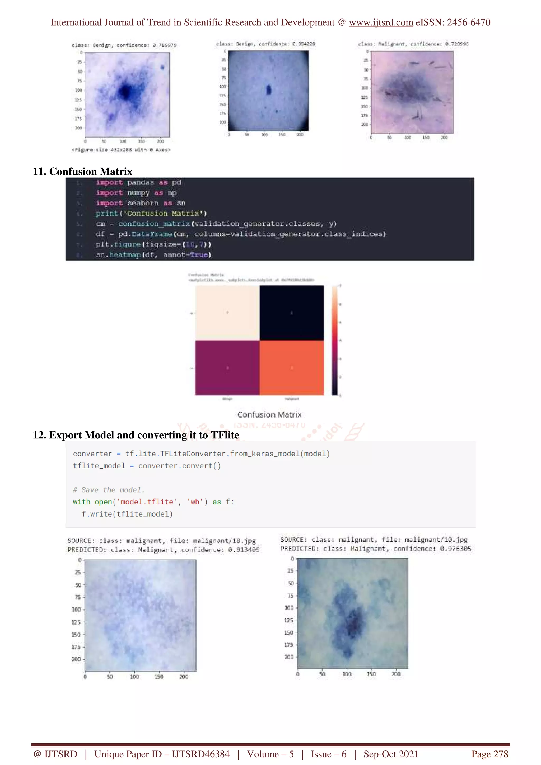
10. Testing Model
We picked five images at random from the validation dataset and made the following prediction:
8.
International Journal ofTrend in Scientific Research and Development @ www.ijtsrd.com eISSN: 2456-6470
@ IJTSRD | Unique Paper ID – IJTSRD46384 | Volume – 5 | Issue – 6 | Sep-Oct 2021 Page 278
11. Confusion Matrix
12. Export Model and converting it to TFlite
9.
International Journal ofTrend in Scientific Research and Development @ www.ijtsrd.com eISSN: 2456-6470
@ IJTSRD | Unique Paper ID – IJTSRD46384 | Volume – 5 | Issue – 6 | Sep-Oct 2021 Page 279
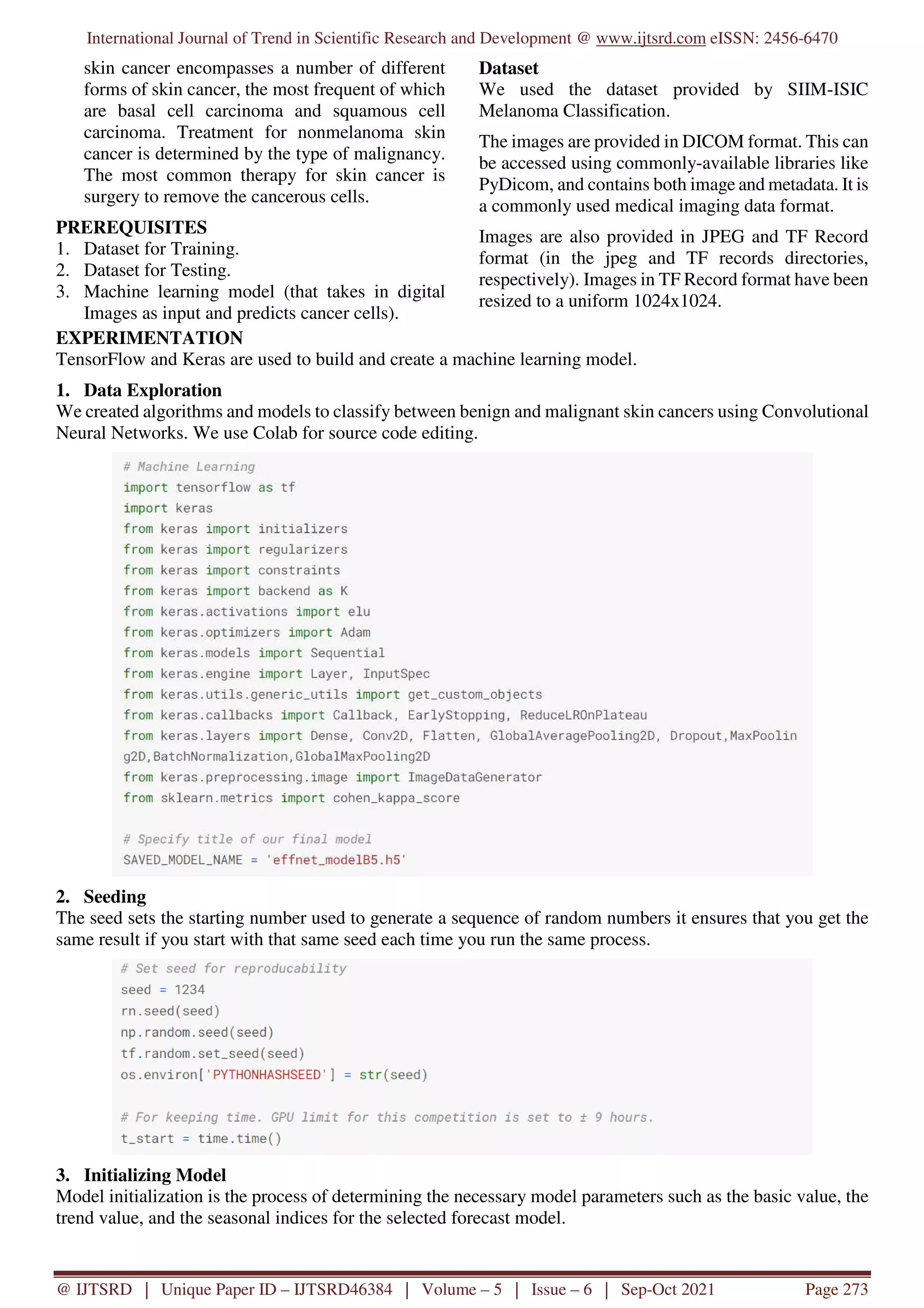
IMPLEMENTATION
We Later converted the model to TF lite format to
implement on some real-life imaging. We used the
app on an android app that we made using Java.
Out Look of Our App:
Our App while its working (Cancer Cell detected):
When Cell is benign:
The app was detecting Cancer cells at a 97%
accuracy.
CONCLUSION
Looking back on this project, the overall outcome of
results to be observed. This can be evaluated by
looking at how well our objectives were met. We
have successfully constructed our learning model that
can predict the cancer cells by image processing. The
performance at the training phase does not give the
complete picture, we have to test it on real-data. So
we later introduced it to an android application. This
passion project helped us to get the In-depth
knowledge and experience on machine learning and
image processing.
REFERENCE
[1] Jhuria, Ashwani Kumar, and Rushikesh Borse,
“Image Processing for Smart Farming:
Detection of Disease and Fruit Grading”,
Proceedings of the 2013 IEEE Second
International Conference on Image Information
Processing (ICIIP-2013)
[2] Chunxia Zhang, Xiuqing Wang, Xudong Li,
“Design of Monitoring and Control Plant
Disease System Based on DSP&FPGA”, 2010
Second International Conference on Networks
Security, Wireless Communications and
Trusted Computing.
[3] Dr. K. Thangadurai, K. Padmavathi, “Computer
Visionimage Enhancement For Plant Leaves
Disease Detection”, 2014 World Congress on
Computing and Communication Technologies.
10.
International Journal ofTrend in Scientific Research and Development @ www.ijtsrd.com eISSN: 2456-6470
@ IJTSRD | Unique Paper ID – IJTSRD46384 | Volume – 5 | Issue – 6 | Sep-Oct 2021 Page 280
[4] Peidle J., Stokes C., Hart R., Franklin M.,
Newburgh R., Pahk J., Rueckner W. & Samuel
AD, (2009) “Inexpensive microscopy for
introductory laboratory courses,” American
Journal of Physics Vol. 77 pp. 931-938.
[5] Joshi, A., Boyat, A. and Joshi, B. K. (2014)
“Impact of Wavelet Transform and Median
Filtering on removal of Salt and Pepper noise in
Digital Images,” IEEE International Conference
on Issues and Challenges in Intelligant
Computing Teachniques, Gaziabad.
[6] Salivahanan S., Vallavaraj A. &Gnanapriya C.
(2008) “Digital Signal Processing,” Tata
McgrawHill, Vol. 23, NewDelhi.
[7] Bovick A. (2000) “Handbook of Image and
Video processing,” Acedemic press, New York.
[8] Astola J. & Kuosmanen P. (1997)
“Fundamentals of nonlinear digital filtering,”
CRC Press, Boca Raton.
[9] Radenovic A., “Brownian motion and single
particle tracking,” Advanced Bioengineering
methods laboratory, Ecole polyteachenique
federal de Lausanne.
[10] Hosseini H. &Marvasti F., (2013) “Fast
restoration of natural images corrupted by high-
density impulse noise,” EURASIP Journal on
Image and Video Processing. doi:
10.1186/1687-5281-2013-15.
![International Journal of Trend in Scientific Research and Development @ www.ijtsrd.com eISSN: 2456-6470
@ IJTSRD | Unique Paper ID – IJTSRD46384 | Volume – 5 | Issue – 6 | Sep-Oct 2021 Page 275
6. Data Processing
We set up generators to read images from our source folders, then we transformed them to float32 tensors, and
fed them to our network.
In our case, we'll change the pixel values to the [0, 1] range before pre-processing the images (all values are now
in the [0, 255] range). As required by the networks, the input data must be scaled to 224x224 pixels as an input.
7. Building the Model
It's as simple as layering a linear classifier on top of the feature extractor with the Hub module. For speed, we
start with a non-trainable feature extractor.](https://image.slidesharecdn.com/41skincancerdetectionusingimage-processinginreal-time-220226121721/75/Skin-Cancer-Detection-using-Image-Processing-in-Real-Time-5-2048.jpg)
![International Journal of Trend in Scientific Research and Development @ www.ijtsrd.com eISSN: 2456-6470
@ IJTSRD | Unique Paper ID – IJTSRD46384 | Volume – 5 | Issue – 6 | Sep-Oct 2021 Page 279
IMPLEMENTATION
We Later converted the model to TF lite format to
implement on some real-life imaging. We used the
app on an android app that we made using Java.
Out Look of Our App:
Our App while its working (Cancer Cell detected):
When Cell is benign:
The app was detecting Cancer cells at a 97%
accuracy.
CONCLUSION
Looking back on this project, the overall outcome of
results to be observed. This can be evaluated by
looking at how well our objectives were met. We
have successfully constructed our learning model that
can predict the cancer cells by image processing. The
performance at the training phase does not give the
complete picture, we have to test it on real-data. So
we later introduced it to an android application. This
passion project helped us to get the In-depth
knowledge and experience on machine learning and
image processing.
REFERENCE
[1] Jhuria, Ashwani Kumar, and Rushikesh Borse,
“Image Processing for Smart Farming:
Detection of Disease and Fruit Grading”,
Proceedings of the 2013 IEEE Second
International Conference on Image Information
Processing (ICIIP-2013)
[2] Chunxia Zhang, Xiuqing Wang, Xudong Li,
“Design of Monitoring and Control Plant
Disease System Based on DSP&FPGA”, 2010
Second International Conference on Networks
Security, Wireless Communications and
Trusted Computing.
[3] Dr. K. Thangadurai, K. Padmavathi, “Computer
Visionimage Enhancement For Plant Leaves
Disease Detection”, 2014 World Congress on
Computing and Communication Technologies.](https://image.slidesharecdn.com/41skincancerdetectionusingimage-processinginreal-time-220226121721/75/Skin-Cancer-Detection-using-Image-Processing-in-Real-Time-9-2048.jpg)
![International Journal of Trend in Scientific Research and Development @ www.ijtsrd.com eISSN: 2456-6470
@ IJTSRD | Unique Paper ID – IJTSRD46384 | Volume – 5 | Issue – 6 | Sep-Oct 2021 Page 280
[4] Peidle J., Stokes C., Hart R., Franklin M.,
Newburgh R., Pahk J., Rueckner W. & Samuel
AD, (2009) “Inexpensive microscopy for
introductory laboratory courses,” American
Journal of Physics Vol. 77 pp. 931-938.
[5] Joshi, A., Boyat, A. and Joshi, B. K. (2014)
“Impact of Wavelet Transform and Median
Filtering on removal of Salt and Pepper noise in
Digital Images,” IEEE International Conference
on Issues and Challenges in Intelligant
Computing Teachniques, Gaziabad.
[6] Salivahanan S., Vallavaraj A. &Gnanapriya C.
(2008) “Digital Signal Processing,” Tata
McgrawHill, Vol. 23, NewDelhi.
[7] Bovick A. (2000) “Handbook of Image and
Video processing,” Acedemic press, New York.
[8] Astola J. & Kuosmanen P. (1997)
“Fundamentals of nonlinear digital filtering,”
CRC Press, Boca Raton.
[9] Radenovic A., “Brownian motion and single
particle tracking,” Advanced Bioengineering
methods laboratory, Ecole polyteachenique
federal de Lausanne.
[10] Hosseini H. &Marvasti F., (2013) “Fast
restoration of natural images corrupted by high-
density impulse noise,” EURASIP Journal on
Image and Video Processing. doi:
10.1186/1687-5281-2013-15.](https://image.slidesharecdn.com/41skincancerdetectionusingimage-processinginreal-time-220226121721/75/Skin-Cancer-Detection-using-Image-Processing-in-Real-Time-10-2048.jpg)