Download to read offline
![International Research Journal of Engineering and Technology (IRJET) e-ISSN: 2395-0056
Volume: 10 Issue: 05 | May 2023 www.irjet.net p-ISSN: 2395-0072
© 2023, IRJET | Impact Factor value: 8.226 | ISO 9001:2008 Certified Journal | Page 448
Detection of Skin Cancer Based on Skin Lesion Images UsingDeep
Learning
Prof. Bharath Bharadwaj B S 1, Saniya Anjum 2, Shaguftha Afreen 3 , Spoorthi T C 4,Keerthana M 5
1 Assistant professor, Dept. of computer Science and Engineering, Maharaja Institute of Technology Thandavapura
2,3,4,5Students , Dept. of Computer Science and Engineering, Maharaja Institute of Technology Thandavapura
---------------------------------------------------------------------***---------------------------------------------------------------------
Abstract - An adding number of inheritable and metabolic
anomalies have been determined to lead to cancer, generally
fatal. Cancerous cells may spread to any bodypart, wherethey
can be life- changing. Skin cancer is one of the most common
types of cancer, and its frequence is adding worldwide. The
main subtypes of skin cancer are scaled and rudimentary cell
lymphomas, and carcinoma, which is clinically aggressive and
responsible for utmost deaths. Thus, skin cancer webbing is
necessary. One of the stylish styles to directly and fleetly
identify skin cancer is using deep literacy (DL). To insure
better prognostic and death rates, early skin cancer
identification is pivotal, yet solid excrescence discovery
generally relies substantially on screening mammography
with shy perceptivity, which is also validated by clinical
samples. Cancer webbing and treatment responseevaluations
are generally not applicable uses for this approach. An adding
number of healthcare providers are using artificial
intelligence (AI) for medical diagnostics to ameliorate and
accelerate the opinion decision- making procedure
1 INTRODUCTION
The willful development of napkins in a specific
body area is known as cancer. The most snappily spreading
complaint in the world looks to be skin cancer.Skincancer is
a complaint in which abnormal skin cells develop out of
control. To determine implicit cancer rapid-fire early
discovery and accurate opinion are essential.Melanoma,the
deadliest form of skin cancer, is responsible for utmost skin
cancer-related deaths in developed countries. The skin
cancer types comprise rudimentary cell melanoma, scaled
cell melanoma, Merkel cell cancer,dermatofibroma,vascular
lesion, and benign keratosis.
1.2 PROBLEM STATEMENT
The GLOBOCAN check also points out that further
than half of the cancer deaths do in Asia about 20 of cancer
deaths are in Europe. Likewise, the areas most affected by
skin cancer are around the globe. North America reckoned
for half of the aggregate. Roughly 9,500 Americans are
diagnosed with skin cancer every day. The good news isthat
the five-time survival rate is 99 if caught and treated
beforehand.
Early discovery of skin cancer can beget by nasty lesions is
pivotal for treatment as it would increase thesurvival rate of
cases. Still, a conventional discovery system similar to
ABCDE criteria possesses colorful limitations such as
subjectivity and trip, due to the different experience
positions of dermatologists and the characteristics of nasty
skin lesions. Either, the current state-of-the-art in detecting
skin lesions using deep neural networks substantially
focuses on the skin lesions. Also, deep literacy model
infrastructures similar to ‘Resent’ used to perform these
tasks are frequently complex, heavy in size, slow, and
delicate to apply.
1.3 OBJECTIVE
The skin cancer detection project is to develop a
framework to analyze and assess the risk ofmelanoma using
dermatological photographs taken with a standard
consumer-grade camera. This step can be performed
because many features used to the risk of melanoma are
derived based on the lesion border. Our approach to finding
the lesion border is texture distinctiveness-based lesion
segmentation.
1.4 SCOPE
Skin cancer indications can be quickly and easily
diagnosed using computer-based techniques. By analyzing
images of lesions on the skin, we developed for quickly and
accurately diagnosing both benign and malignant forms of
cancer.
2 LITRATURE SURVEY
[1] Title:-Detection of Skin Cancer based on skin lesion
images
Authors:-Walaa Gouda, Noor Zaman
Publication Journal & Year:-IRJET, 2022.
Summary:-By assaying images of lesions on the skin,
we developed a fashion for snappily and directly
diagnosing both benign and nasty forms of cancer. The
suggested system uses image improvement approaches
to boost the luminance of the lesion image and reduce
noise. Resnet50, InceptionV3, and Begrudge inception
were all trained on the upper edge of the preprocessed
lesion medical images to help to over fit, as well as
meliorate the overall capabilities of the suggested DL](https://image.slidesharecdn.com/irjet-v10i570-230626065710-d1b4d8f8/75/Detection-of-Skin-Cancer-Based-on-Skin-Lesion-Images-UsingDeep-Learning-1-2048.jpg)
![International Research Journal of Engineering and Technology (IRJET) e-ISSN: 2395-0056
Volume: 10 Issue: 05 | May 2023 www.irjet.net p-ISSN: 2395-0072
© 2023, IRJET | Impact Factor value: 8.226 | ISO 9001:2008 Certified Journal | Page 449
styles. The Inception model had an overall delicacyrateo
f85.7, which is similar to that of educateddermatologists.
[2] Title:-Skin CancerClassificationUsingImageProcessing
and Machine Learning
Authors:-Arslan Javaid,MuhammadSadiq,FarazAkram
Publication Journal & Year:-IJASRT, 2021.
Summary:-In this work, a novel method of skin cancer
classification using machine learning and image
processing is implemented. In the first step, a novel
method of contrast stretching based on the mean and
standard deviation of pixels for Dermoscopy image
enhancement is proposed. Then OTSU thresholding is
performed for segmentation.
[3] Title:-Detection of Skin Cancer Lesions from Digital
Images with Image Processing Techniques
Authors:-Minakshi Waghulde, Shirish Kulkarni, Gargi
Phadke
Publication Journal & Year:-IJCDS, 2020.
Summary:-Our results are harmonious withcurrentart
on DNNs transfer knowledge is a good idea, as is fine-
tuning. We anticipated that transferring knowledge
from a related task (in our case, from Retinopathy,
another medical type task) would lead to better results,
especially in the double transfer scheme.
[4] Title:-Detection and Classification of Skin Cancer by
Using a Parallel CNN Model
Authors:-Noortaz Rezaoana, Mohammad Shahadat
Hossain, Karl Andersson
Publication Journal & Year:-IEEE, 2020.
Summary:-In this work, we propose GAN-based
methods to generate realistic synthetic skin lesion
images. Malignancy markers are present with coherent
placement and sharpness which result in visually-
appealing images.
[5] Title:- Skin Cancer Classification using Deep Learning
and Transfer Learning
Authors:- KhalidM.Hosny, MohamedA.Kassem,
Mohamed M.Foaud
Publication Journal & Year:-IJASRT-2019.
Summary:-we have proposed an improved U-Net which is
named as NABLA-N and the model is evaluated for skin
cancer segmentation tasks. Three different models are
investigated with different feature fusion between encoding
and decoding units which are evaluated on the ISIC2018
dataset. The quantitativeand qualitativeresultsdemonstrate
better performance with the model compared to the model.
3. EXISTING SYSTEM
Sample Skin cancer is on the upswing, this has been
true for the last 10 times. Because the skin is the body’s
central part, it's reasonable to assume that skin cancer isthe
most frequent complaint in humans. The first step for
identifying whether the skin lesion is nasty or benign for a
dermatologist is to do skin vivisection. In skin vivisection,
the dermatologist takes some part of the skin lesion and
examines it under a microscope. The current process takes
nearly a week or further, starting from getting a
dermatologist appointmenttogettinga vivisectionreport. At
present, to check the skin malice of a case, he needs to
witness singular webbing by a dermatologisttofetewhether
they've skin complaint or not.
4. PROPOSED SYSTEM
There is a need for accurate as well as reliable
systems that can help not only clinicians but as well persons
to descry types of lesions at early stages. This proposed skin-
cancer discovery system is developed as a clinical decision
support tool that uses computer vision which can help
croakers to diagnose skin cancer types by simply using
images with good delicacy. This design aims to dock the
current gap to just a couple of days by furnishing the
predictive model using Computer- backed opinion (CAD).
Fig. 1: Sequence Diagram](https://image.slidesharecdn.com/irjet-v10i570-230626065710-d1b4d8f8/75/Detection-of-Skin-Cancer-Based-on-Skin-Lesion-Images-UsingDeep-Learning-2-2048.jpg)
![International Research Journal of Engineering and Technology (IRJET) e-ISSN: 2395-0056
Volume: 10 Issue: 05 | May 2023 www.irjet.net p-ISSN: 2395-0072
© 2023, IRJET | Impact Factor value: 8.226 | ISO 9001:2008 Certified Journal | Page 452
Fig.8: Cancer is not found
Fig.9: Cancer is found in Keratosis Cell
Fig. 10: Cancer is not found
7. CONCLUSION
In this project, it is found that most existing skin lesion
diagnoses with deep learning technology stop at deep
learning modeling and without any further deployment or
integration with a readily available device such as a
smartphone. However, the advantage of integrating object
detection deep learning technology and smartphone in the
medical field has been discovered throughout the project.
This technology can provide a low-cost diagnosis without
requiring years of skin lesion diagnosis experience.
Moreover, users can perform diagnosis at home with a
smartphone and therefore can provide a point of care to
users from a remote area. Although the process of
development is challenging due to the immature platform of
object detection development TensorFlow Object Detection
API and requires experience for Android application
development, the success of this project has proven that the
development of this technology is feasible and should be
aware.
REFERENCES
[1] World Health Organization. GlobalHealthObservatory;
World Health Organization: Geneva, Switzerland,
2022.
[2] Han, H.S.; Choi, K.Y. Advances in nanomaterial-
mediated photo thermal cancer therapies: Toward
clinical applications. Biomedicines 2021, 9, 305.
[3] Fuzzell, L.N.; Perkins, R.B.; Christy, S.M.; Lake, P.W.;
Vadaparampil, S.T. Cervical cancer screening in the
United States: Challenges and potential solutions for
under screened groups. Prev. Med. 2021, 144,
106400.
[4] Ting, D.S.; Liu, Y.; Burlina, P.; Xu, X.; Bressler, N.M.;
Wong, T.Y. AI for medical imaging goes deep. Nat.
Med. 2018, 24, 539–540.
[5] Wolf,M.; de Boer,A.; Sharma,K.; Boor,P.; Leiner,T.;
Sunder- Plassmann,G.; Moser,E.; Caroli,A.; Jerome,N.P.
glamorous resonance imaging T1- and T2- mapping to
assess renal structure and functionAmethodical review
and statement paper. Nephron. telephone. Transplant.
2018, 33( Suppl. S2), ii41 – ii50.
BIOGRAPHIES
Bharath Bharadwaj B S
Professor,
Department of Computer
Science & Engineering,
Maharaja Institute of Technology
Thandavapura](https://image.slidesharecdn.com/irjet-v10i570-230626065710-d1b4d8f8/75/Detection-of-Skin-Cancer-Based-on-Skin-Lesion-Images-UsingDeep-Learning-5-2048.jpg)


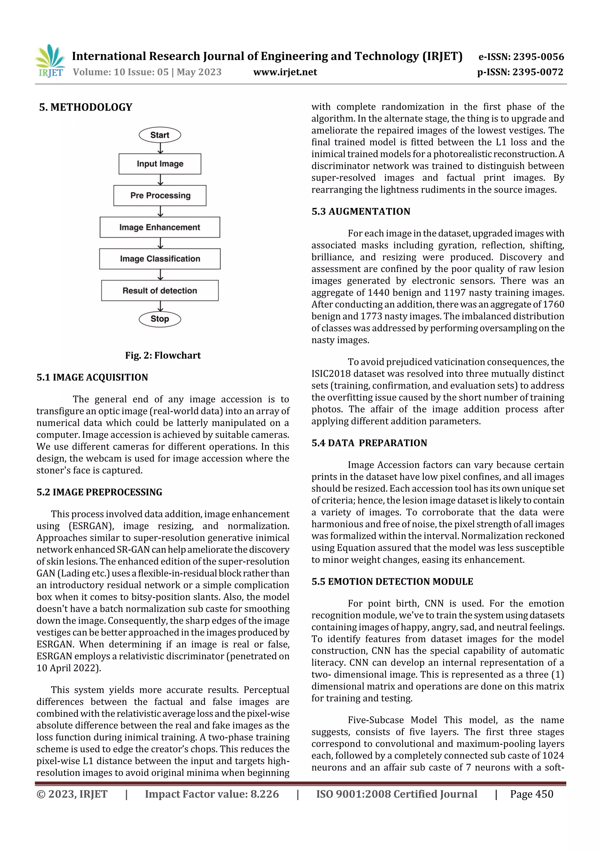
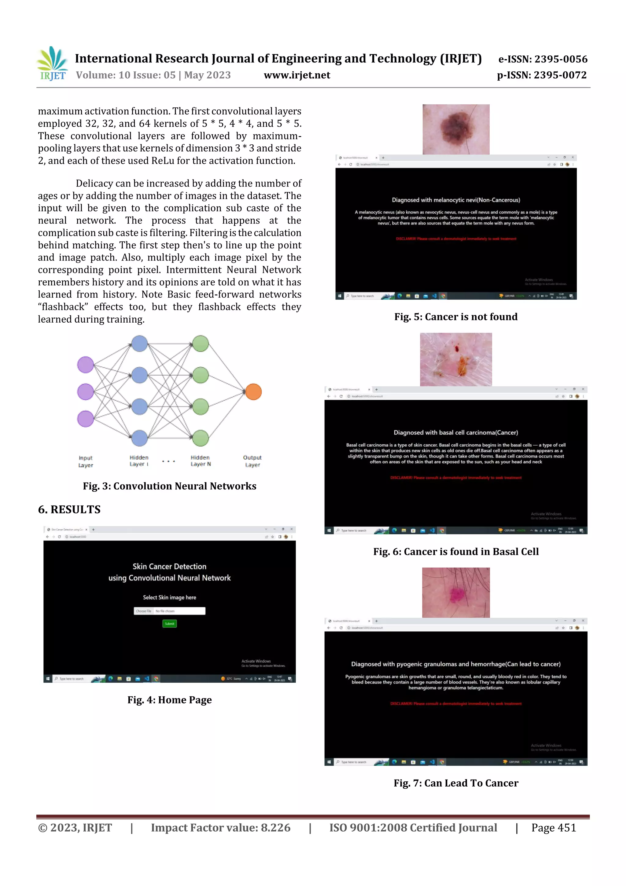
This document discusses skin cancer detection using deep learning techniques. It begins with an introduction to skin cancer and the need for early detection. It then reviews the existing methods for skin cancer detection which rely on visual examination by dermatologists. The proposed method uses a deep learning model trained on skin lesion images to classify lesions as benign or malignant. The methodology section describes the image acquisition, preprocessing including enhancement, data augmentation, and preparation steps. It then discusses training a convolutional neural network for classification. Experimental results show the system can accurately detect different types of skin cancers like basal cell carcinoma and keratosis. The conclusion discusses benefits of developing such a system for integrated use on smartphones to enable low-cost cancer screening.