The document discusses a study on melanoma skin cancer detection using automated methods involving image processing and machine learning. It details a three-phase model that includes data collection, preprocessing, and the application of AI algorithms like CNN and SVM, which achieved an accuracy of 85%. The goal is to enhance early diagnosis of skin cancer through improved analysis of lesion images, reducing diagnostic time and increasing accuracy.

![International Journal of Trend in Scientific Research and Development (IJTSRD) @ www.ijtsrd.com eISSN: 2456-6470
@ IJTSRD | Unique Paper ID - IJTSRD23936 | Volume – 3 | Issue – 4 | May-Jun 2019 Page: 781
most important and also plays a key role as it affects the
process of fore coming steps. Supervised segmentation
seems to be easy to implement by considering the
parameters like shapes, sizes, and colors along with skin
types and textures. This system-based analysis will reduce
the diagnosing time and increases the accuracy.
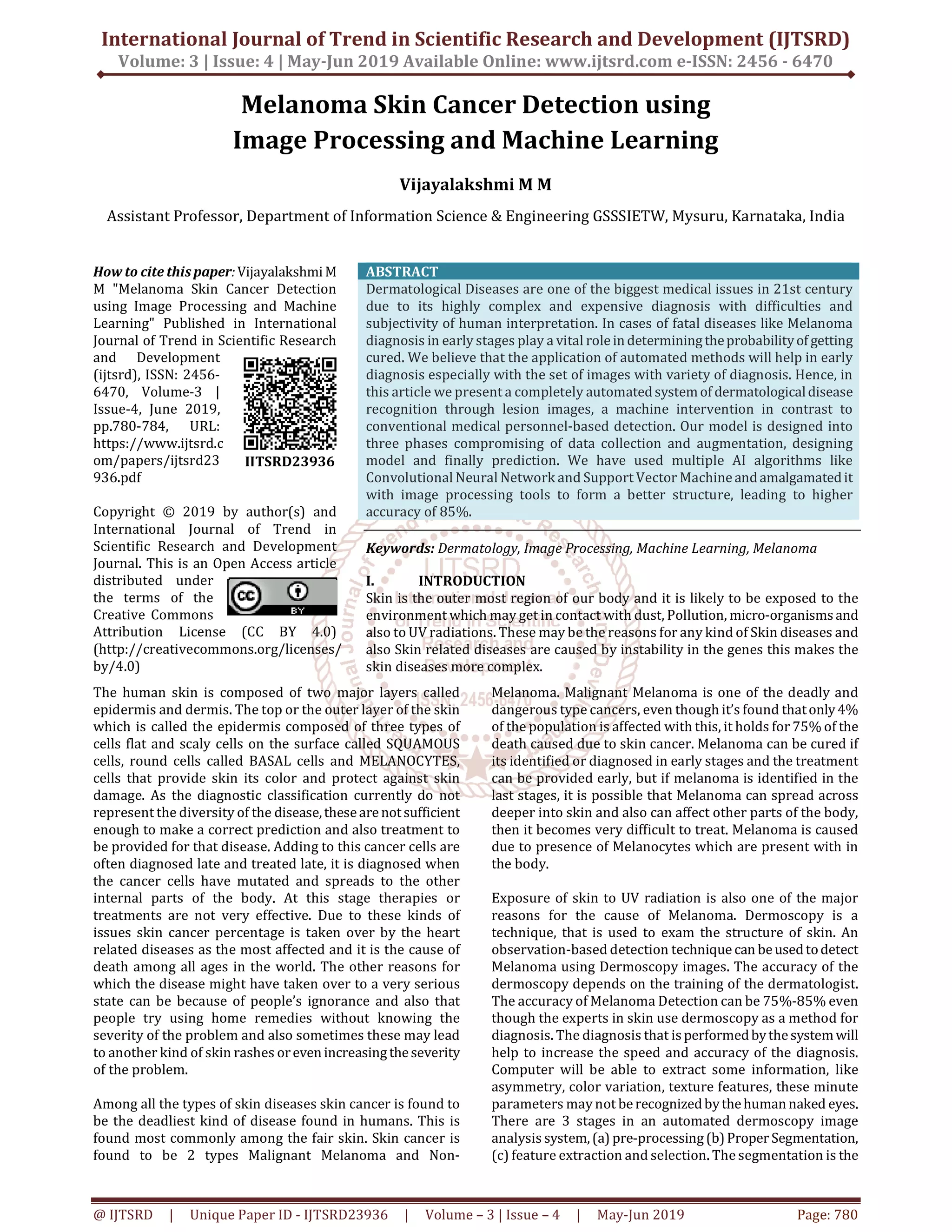
Dermatological Diseases, due to their high complexity,
variety and scarce expertise is one of the most difficult
terrains for quick, easy and accurate diagnosis especially in
developing and under-developed countries with low
healthcare budget. Also, it’s a common knowledge that the
early detection in cases on many diseases reduces the
chances of serious outcomes. The recent environmental
factors have just acted as catalyst for these skin diseases.
The general stages of these diseases are as: STAGE 1-
diseases in situ, survival 99.9%, STAGE 2- diseases in high
risk level, survival 45-79%, STAGE 3-regional metastasis,
survival 24-30%, STAGE 4- distant metastasis-survival 7-
19%
II. RELATED WORKS
The authors [1] have tried to address the same problem
using image analysis techniques. The work uses the
technique of noise removal and subsequent feature
extraction. After the noise removal, the image is fed into
classifier for further feature extraction process and finally
the prediction of the disease. Most of the earlierpublications
focused on feature extraction and then subsequent disease
prediction was done. Papers [6,3] haveused Artificial Neural
Network for dealing with thiscomplexproblemwhilepapers
[2,4,5] have used machine learning algorithms for the task.
Computer vision techniques have played a major role in
many previous literatures. As is evident, the publishers have
utilized the image processing techniques to accomplish the
pre processing task. In the similar way we also try to
implement the computer vision techniques, but out
implementation mainly focuses for dataset augmentation.
III. Methodology
Our model is designed in 3 phases as follows:
A. Phase1 – the first model involves collection of dataset,
the images are collected fromISIC dataset(International
Skin Imaging Collaboration) Phase 1 also involves the
pre-processing of the images where hair removal, glare
removal and shading removal are done
B. Removal of these parameters helps us to identify the
texture, color, size and shape like parameters in an
efficient way.
C. Phase2- this phase consists of the segmentation and
feature extraction, segmentation is explored via three
methods a. Otsu segmentation method b. Modified Otsu
segmentation method c. water shed segmentation
method. Feature are extracted for color, shape, size and
texture.
D. Phase 3- this is the most important phase of our model,
this phase involves designing of the model and training.
Our model was trained for Back Propagation Algorithm
(Neural Networks), SVM (Support Vector Machine), and
CNN (Convolutional Neural Networks) on the dataset
that was collected in the phase1,themodel aftertraining
was tested for the accurate output.
IV. COMPONENTS OF METHODOLOGY:
PRE-PROCESSING:
The pre-processing of images is an importanttaskor activity
which helps in saving time for training as well as provides
the clear enhancement forthefurtherstepsbyincreasing the
efficiency of the model. Pre-processing includes the
following:
Collection of the dataset
Hair removal
Shading removal
Glare removal
Dataset: The images were collected from the ISIC dataset;
the ISIC dataset provide the collection of images for
melanoma skin cancer. ISIC melanoma project was
undertaken to reduce the increasing deaths related to
melanoma and efficiency of melanoma early detection. This
ISIC dataset contains approximately 23,000 imagesof which
we have collected 1000-1500 images and trained and tested
over these images.
Hair Removal: for the above collected images hair removal
method was applied this method was performed using
Hough transform, Hough transform is basically used to
identify lines or elliptical or circular shapes. Performinghair
removal for the images that has hair within the tumor
provides us an clear image of tumor which also helps us to
make further more enhancements.
Shading removal: The images that is takenfromthedataset
contains shade around the region of the tumor this shadefor
few images is dark and for few is light, removal of the shade
in the region of tumor also provides us an clear vision of the
tumor which is also helpful in the further enhancements. We
have used the MATLAB filters to remove the shade for
images in the dataset.
Glare Removal: sometime the images are captured from
camera the images will contain glare this glare is not visible
to the naked eyes, we remove this glare using the MATLAB
filter, this minute noise sometimesmayaffectthe accuracyat
the end.
V. Architecture
VI. Designing The Model
In our model we have used 3 different methods i.e. Neural
Networks,SupportVectorMachineandConvolutionalNeural
Networks to find the efficient detection and classification of
the melanoma skin cancer into Malignant and benign skin
cancers. The data that is pre-processed is followed by
segmentation and feature extraction these extractedfeature
images are then passed into Neural Networks and Support](https://image.slidesharecdn.com/169melanomaskincancerdetectionusingimageprocessingandmachinelearning-190708070259/85/Melanoma-Skin-Cancer-Detection-using-Image-Processing-and-Machine-Learning-2-320.jpg)
![International Journal of Trend in Scientific Research and Development (IJTSRD) @ www.ijtsrd.com eISSN: 2456-6470
@ IJTSRD | Unique Paper ID - IJTSRD23936 | Volume – 3 | Issue – 4 | May-Jun 2019 Page: 783
demonstrated to identify faces, objects, and traffic signs
better than humans and thereforecanbefound in robotsand
self-driving cars.
CNNs are a supervised learning method and are therefore
trained using data labeled with the respective classes.
Essentially, CNNs learn the relationship between the input
objects and the class labels and comprise two components:
the hidden layers in which the features are extracted and, at
the end of the processing, the fully connected layers that are
used for the actual classification task. Unlike regular neural
networks, the hidden layers of a CNN have a specific
architecture. In regular neural networks, each layer is
formed by a set of neurons and one neuron of a layer is
connected to each neuron of the preceding layer. The
architecture of hidden layers in a CNN is slightly different.
The neurons in a layer are not connected to all neurons of
the preceding layer; rather, they are connected to only a
small number of neurons. This restriction to local
connections and additionalpoolinglayerssummarizinglocal
neuron outputs into one value results in translation-
invariant features. This results in a simpler training
procedure and a lower model complexity
VII. CONCLUTION
The aim of this project is to determine the accurate
prediction of skin cancer and also to classify the skin cancer
as malignant or non-malignant melanoma. To do so, some
pre-processing steps were carried out which followed Hair
removal, shadow removal, glare removal and also
segmentation. SVM and Deep Neural networks will be used
to classify. classifier will be trained to learn the features and
finally used to classify. The novelty of the present
methodology is that it should do the detection in very quick
time hence aiding the technicians to perfect their diagnostic
skills. The dataset used is from the available ISIC
(International Skin Image Collaboration) dataset, hence any
dataset can be used to find the efficiency.
VIII. REFERENCES
[1] abrham debasu mengistu , dagnachew melesew
alemayehu “computer vision for skin cancer diagnosis
and recognition using rbf and som “ international
journal of image processing (ijip), volume (9) : issue
(6) 2015.
[2] s.s. Mane1, s.v. Shinde “different techniques for skin
cancer detection using dermoscopy images” ,
international journal of computer sciences and
engineering vol.5(12), dec 2017, e-issn: 2347-2693.
[3] poornima m s, dr. Shailaja k “detection of skin cancer
using svm” , international research journal of
engineering and technology (irjet) volume:04issue:07
| july -2017.
[4] yuexiang li and linlin shen “skin lesion analysis
towards melanoma detection using deep learning
network”, arxiv:1904.073653v2 [cs.cv] 20 aug 2018
[5] muhammad imran razzak,saeedanazand ahmad zaib“
deep learning for medical image processing: overview,
challenges and future” arxiv:1852.3865v2 [cs.cv] 20
july 2018
[6] veronika cheplygina, marleen de bruijne, josien p. W.
Pluim, “ not-so-supervised: a survey of semi-
supervised, multi-instance, and transfer Learning in
medical image analysis” arxiv:1804.06353v2 [cs.cv]14
sep 2018
[7] salome kazeminia, christoph baur, arjan kuijper, bram
van Ginneken, nassir navab, shadi albarqouni, anirban
mukhopadhyay “gans for medical image analysis “,
arxiv:1809.06222v2 [cs.cv] 21 dec 2018
[8] andreas maier, christopher syben, tobias lasser,
christian riess “a gentle introduction to deep learning
in medical image processing”, arxiv:1810.05401v2
[cs.cv] 21 dec 2018
[9] danilo barros mendes , nilton correia da silva “skin
lesions classification using convolutional Neural
networks in clinical images”, arxiv:1812.02316v1
[cs.cv] 6 dec 2018](https://image.slidesharecdn.com/169melanomaskincancerdetectionusingimageprocessingandmachinelearning-190708070259/85/Melanoma-Skin-Cancer-Detection-using-Image-Processing-and-Machine-Learning-4-320.jpg)
![International Journal of Trend in Scientific Research and Development (IJTSRD) @ www.ijtsrd.com eISSN: 2456-6470
@ IJTSRD | Unique Paper ID - IJTSRD23936 | Volume – 3 | Issue – 4 | May-Jun 2019 Page: 784
[10] wasan kadhim saa'd “ method for detection and
diagnosis of the Area of skin disease based on color by
Wavelet transform and a rtificial neural Network” al-
qadisiya journal for engineering sciences vol. 2 no.4
year 2009
[11] li-sheng wei , quan gan, and tao ji , “skin disease
recognition method based on image color and Texture
features” hindawi computational and mathematical
methods in medicine volume 2018, article id 8145713,
10 pages
[12] rahat yasir, md. Shariful islam nibir, and nova ahmed “
a skin disease detection system for financiallyunstable
people in developing countries” global science and
technology journal vol. 3. No. 1. March 2015 issue. Pp.77
– 93
[13] t.yamunarani, “analysis of skin cancer using abcd
technique” , international research journal of
engineering and technology(irjet) volume:05 issue:04
| apr-2018
[14] rahat yasir,, md. Ashiqur rahman, and nova ahmed,
“dermatological disease detection using image
Processing and artificial neural network”,
arxiv:1012.2436v1 [cs.cv] 16 dec 2018
[15] m. Shamsul arifini, m. Golam kibria, adnan firoze, m.
Ashraful amini, hong yan, “dermatological disease
diagnosis using color-skin images”, proceedings of the
2012 international conference on machine learning
and cybernetics, xian, 15-17 july, 2012
[16] lakshay bajaj, himanshu kumar, yasha hasija,”
automated system for prediction of skin disease using
image processing and machine learning” international
journal of computer applications (0975 – 8887) volume
180 – no.19, february 2018
[17] ritesh maurya, surya kant singh,ashishk.Maurya,ajeet
kumar,” glcm and multi class support vector machine
based automated skin cancer classification” ieee
[18] prashant b. Yadav, mrs. S.s. Patil “ recognition of
dermatological disease area for identification of
disease” ijsdr may 2016 volume 1, issue 5
[19] nikita raut, aayush shah, shail vira, harmit sampat, “ a
study on different techniques for skin cancer
detection”, international research journal of
engineering and technology (irjet), volume: 05 issue:
09 | sep 2018
[20] m.yuvaraju, d.divya, a.poornima,“segmentationof skin
lesion from digital images using morphological filter”,
international research journal of engineering and
technology (irjet) volume: 03 issue: 05 | may-2016
[21] yuexiang liid and linlin shen, “skin lesion analysis
toward melanoma detection using deep learning
network” sensors mdpi 11 february 2018.
[22] mrs. S kalaiarasi, harsh kumar, sourav patra,
“dermatological disease detection using image
processing and neural networks”, s.kalaiarasi et al,
international journal of computer science and mobile
applications, vol.6 issue. , pg. 109-118 ,4 april- 2018.](https://image.slidesharecdn.com/169melanomaskincancerdetectionusingimageprocessingandmachinelearning-190708070259/85/Melanoma-Skin-Cancer-Detection-using-Image-Processing-and-Machine-Learning-5-320.jpg)