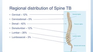
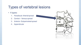
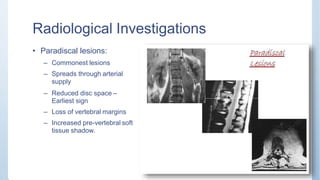
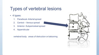
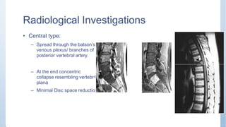
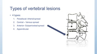
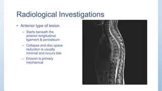
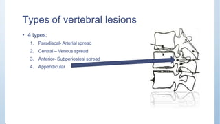
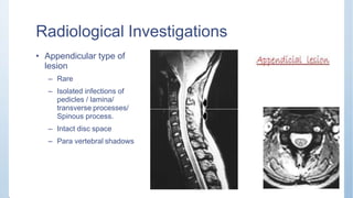
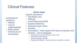
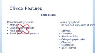
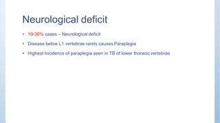
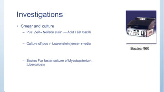
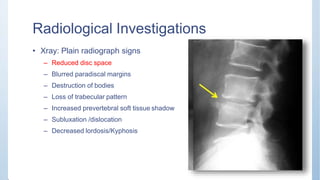
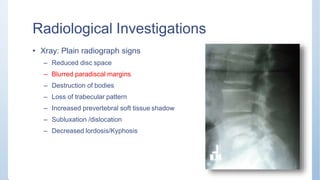
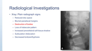
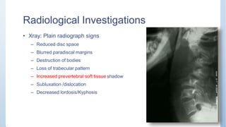
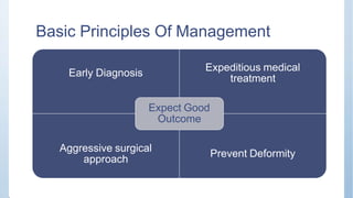
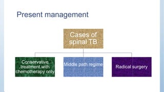
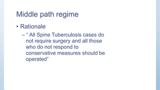
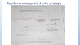
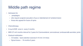
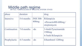
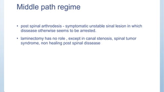
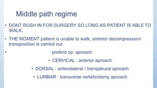
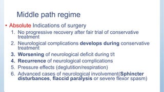
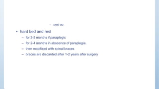
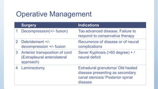
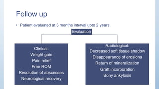
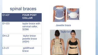
Pott’s spine & Paraplegia
PRESENTEDBY-
Dr. Ashutosh Kumar
AP Dept Of Orthopaedics
RMCH Bareilly
Epidemiology
• An estimated 2 million people have active Spinal TB
worldwide
India
• 1/5th of total TB Cases
• vertebral tuberculosis is commonest form of skeletal T.B.
Regional distribution of Spine TB
• Cervical – 12%
• Cervicodorsal – 5%
• Dorsal – 42%
• Dorsolumbar – 12%
• Lumbar – 26%
• Lumbosacral – 3%
Types of vertebral lesions
• 4 types:
1. Paradiscal- Arterial spread
2. Central – Venous spread
3. Anterior- Subperiosteal spread
4. Appendicular
Radiological Investigations
• Paradiscal lesions:
– Commonest lesions
– Spreads through arterial
supply
– Reduced disc space –
Earliest sign
– Loss of vertebral margins
– Increased pre-vertebral soft
tissue shadow.
Types of vertebral lesions
• 4 types:
1. Paradiscal- Arterial spread
2. Central – Venous spread
3. Anterior- Subperiosteal spread
4. Appendicular
vertebral body: areas of distruction or balooning
Radiological Investigations
• Central type:
– Spread through the batson’s
venous plexus/ branches of
posterior vertebral artery.
– At the end concentric
collapse resembling vertebr
plana
– Minimal Disc space reductio
a
n
Types of vertebral lesions
• 4 types:
1. Paradiscal- Arterial spread
2. Central – Venous spread
3. Anterior- Subperiosteal spread
4. Appendicular
Radiological Investigations
• Anterior type of lesion
– Starts beneath the
anterior longitudinal
ligament & periosteum
– Collapse and disc space
reduction is usually
minimal and occurs late
– Erosion is primary
mechanical
Types of vertebral lesions
• 4 types:
1. Paradiscal- Arterial spread
2. Central – Venous spread
3. Anterior- Subperiosteal spread
4. Appendicular
Radiological Investigations
• Appendicular type of
lesion
– Rare
– Isolated infections of
pedicles / lamina/
transverse processes/
Spinous process.
– Intact disc space
– Para vertebral shadows
Clinical Features
Constitutional
symptoms:
• Malaise
• Loss of
weight/appetite
• Night sweats
• Evening rise of
temperature
Active stage
Specific Symptoms:
• Pain/Night cries
• Stiffness
• painful Restricted ROM
• localised Deformity
• Enlarged lymph nodes
• Abscess
• Neurodeficit : motor appear first Sense of position and
vibration – last to disappear
• Spontaneous muscle twitching in lower limbs
• Clumsiness while walking
• Extensor plantar response
• Exagerrated reflexes – Sustained clonus of patella and ankle
Clinical Features
Healed stage
Constitutional symptoms:
• Malaise
• Loss of weight/appetite
• Night sweats
• Evening rise of temperature
Specific Symptoms:
• no pain and tenderness of spine
• Stiffness
• Deformity
• Restricted ROM
• Enlarged lymph nodes
• Abscess
• Neurodeficit
• ESR - normal
Neurological deficit
• 10-30% cases – Neurological deficit
• Disease below L1 vertebrae rarely causes Paraplegia
• Highest Incidence of paraplegia seen in TB of lower thoracic vertebrae
Classification of TB Paraplegia
Early onset paraplegia (group A)
• Appears within 2 years of onset –
during the Active phase
• Underlying pathology
– Inflammatory edema
– TB Granulation tissue
– Abscess
– Caseous tissue
– Ischaemis lesion of cord (Rare)
• Good prognosis
Late onset paraplegia (Group B)
• Appears more than 2 years of disease
in vertebral column
• Underlying pathology –due to
mechanical pressure on cord
– TB Debris
– TB Sequestra from body and disc
– Internal gibbus
– Canal stenosis / Severe deformity
• Poor prognosis
Staging of Neurological Deficit
Goel 1967, Tuli 1985, Kumar 1988, Jain 2002
Stage Severity Clinical Features
I Negligible Patient unaware of neurodeficit, physician detects plantar
extensors or ankle clonus
II Mild Patient aware of deficit but walks with support
III Moderate Non ambulatory due to spastic paralysis (in extension), sensory
deficit less than 50 %
IV Severe III + Flexor spasm / Paralysis in flexion / Flaccid/ Sensory deficit
more than 50 % / Sphincter Involved
Pathology of TB Paraplegia
Extradural mass : (mc), pus, granulation tissue. casseous material
Inflammatory edema : early cases. vascular stasis, toxins
bony disorders : sequestrum, gibbus, dislocation
meningeal changes: peridural fibrosis
infarction of spinal cord: irreversible
changes in spinal cord: myelomalacia, syringomalacia
extradural garnuloma and tuberculoma
Investigations
• CBC:
– Hb% ↓
– Lymphocytosis
• ESR:
– Raised in active stage of disease
– Normal ESR over period of 3 months suggests patient is in stage of repair
Investigations
• Mantoux test
– Erythema of more than 20 mm at 72 hours – Positive
– Negative test, in general, rules out the disease
Investigations
• Biopsy
– In case of doubt, it is mandatory to prove the diagnosis by obtaining the diseased
tissue
Investigations
• Smear and culture
– Pus: Zeill- Neilson stain → Acid Fast bacilli
– Culture of pus in Lowenstein jensen media
– Bactec For faster culture of Mycobacterium
tuberculosis
Investigation
• Serological Investigations
– ELISPOT (Enzyme- linked immunospot)
– T-cell based assay from blood
– IgM & IgG antibodies : High sensitivity, low specificity
– PCR: more sensitive
Radiological Investigations
• Xray: Plain radiograph signs
– Reduced disc space
– Blurred paradiscal margins
– Destruction of bodies
– Loss of trabecular pattern
– Increased prevertebral soft tissue shadow
– Subluxation /dislocation
– Decreased lordosis/Kyphosis
Radiological Investigations
• Xray: Plain radiograph signs
– Reduced disc space
– Blurred paradiscal margins
– Destruction of bodies
– Loss of trabecular pattern
– Increased prevertebral soft tissue shadow
– Subluxation /dislocation
– Decreased lordosis/Kyphosis
Radiological Investigations
• Xray: Plain radiograph signs
– Reduced disc space
– Blurred paradiscal margins
– Destruction of bodies
– Loss of trabecular pattern
– Increased prevertebral soft tissue shadow
– Subluxation /dislocation
– Decreased lordosis/Kyphosis
Radiological Investigations
• Xray: Plain radiograph signs
– Reduced disc space
– Blurred paradiscal margins
– Destruction of bodies
– Loss of trabecular pattern
– Increased prevertebral soft tissue shadow
– Subluxation /dislocation
– Decreased lordosis/Kyphosis
Radiological Investigations
• Xray: Plain radiograph signs
– Reduced disc space
– Blurred paradiscal margins
– Destruction of bodies
– Loss of trabecular pattern
– Increased prevertebral soft tissue shadow
– Subluxation /dislocation
– Decreased lordosis/Kyphosis
Radiological Investigations
adow
• Xray: Plain radiograph signs
– Reduced disc space
– Blurred paradiscal margins
– Destruction of bodies
– Loss of trabecular pattern
– Increased prevertebral soft tissue sh
– Subluxation /dislocation
– Decreased lordosis/Kyphosis
Radiological Investigations
• Skipped lesions:
– More than one TB
Lesion in vertebral
column with one or
more healthy vertebrae
in between the 2 lesion.
– More frequently
detected on CT/MRI
Radiological Investigations
• Lateral shift and scoliosis
– More destruction of vertebrae
on one side
• Kyphotic deformity
– Due to collapse of bone
– Forward angulations
Radiological Investigations
• Healing is indicated by
– Decreased soft tissue
shadow
– Return of normal density
– Bony ankylosis
Computed tomography (CT)
• Calcifications in abscess (pathognomic for Tb)
• Regions which are difficult to visualize on plain films, like :
1. Cranio-vertebral junction (CVJ)
2. Cervico-dorsal region,
3. Sacrum
4. Sacro-iliac joints.
5. Posterior spinal tuberculosis
because lesions less than 1.5cm are usually missed due to
overlapping of shadows on x rays.
MRI
• Detect marrow infiltration in vertebral bodies, leading to early
diagnosis.
• Changes of diskitis
• Assessment of extradural abscesses / subligamentous
spread.
• Skip lesions
• Spinal cord involvement.
• Spinal arachanoiditis.
USG
- to find out primary in abdomen
- Detect cold abscess
- Guided aspiration
Radionucleotide Scan T 99m
• Increased uptake in active tuberculosis.
• Avascular segments and abscesses show a cold spot due to
decreased uptake.
• Aid to localise the site of active disease and to detect multilevel
involvement
Clinico-radiological classification of
Typical TB Spondylitis
Basic Principles Of Management
Early Diagnosis
Expeditious medical
treatment
Aggressive surgical
approach
Prevent Deformity
Expect Good
Outcome
Present management
Cases of
spinal TB
Conservative
treatment with
chemotherapy only
Middle path regime
Radical surgery
Middle path regime
• Rationale
– ā€œ All Spine Tuberculosis cases do
not require surgery and all those
who do not respond to
conservative measures should be
operatedā€
Algorithm for management of pott’s paraplegia
Middle path regime
• Admission for
– paraplegics
– who require surgical evacuation of pus or debridement of vertebral lesion
– those who agreed for fusion of spine
• Chemotherapy
• X-ray & ESR once in every 3 months
• MRI/ CT at 6 months interval for 2 years for Craniovertebral ,cervicodorsal, lumbosacral& sacroiliac joints
• Gradual mobilization
– 3-9 weeks- back extention exercise 5-10 min 3-4 times
– Spinal brace--- 18 months-2 years
Middle path regime
phase duration drug
Intensive 5-6 months INH 300-
400mg
Rifampicin
ofloxacin400-600mg /
streptomycin
Continuation 7-8 months -do 3-4mth Pyrazinamide
1500mg
4-5mth Rifampicin
Prophylactic 4-5 months -do Ethambutol 1200mg
SM TULI, TUBERCULOSIS OF SKELETAL SYSTEM. 5TH ED.
Middle path regime
• Abcesses – aspirate near surface
– Instille 1gm Streptomycin +/- INH in sol
– all radiologicaly visible abcesses need not be drained.
– prevertebral abcess in cervical region are drained when complicated by dificulty in deglutition and
respiration.
• Neural complications if responds 3-4 weeks :- surgery unnecssary
• Excisional surgery for posterior spinal disease a/w abcess & sinus because of danger of
2' infection of meninges if dissease does not come in control in 4 wk.
• Operative debridement for patients –
– if no arrest after 3-6 months
– recurrence of disease
Middle path regime
• post spinal arthrodesis - symptomatic unstable sinal lesion in which
dissease otherwise seems to be arrested.
• laminectomy has no role , except in canal stenosis, spinal tumor
syndrome, non healing post spinal dissease
Middle path regime
• DONT RUSH IN FOR SURGERY SO LONG AS PATIENT IS ABLE TO
WALK.
• THE MOMENT patient is unable to walk, anterior decompression/
transposition is carried out.
• preferd op. aproach
• CERVICAL : anterior aproach
• DORSAL : anterolateral / transpleural aproach
• LUMBAR : transverse vertebrotomy aproach
Middle path regime
• Absolute Indications of surgery
1. No progressive recovery after fair trial of conservative
treatment
2. Neurological complications develops during conservative
treatment
3. Worsening of neurological deficit during t/t
4. Recurrence of neurological complications
5. Pressure effects (deglutition/respiration)
6. Advanced cases of neurological involvement(Sphincter
disturbances, flaccid paralysis or severe flexor spasm)
– post op:
• hard bed and rest
– for 3-5 months if paraplegic
– for 2-4 months in abscence of paraplegia.
– then mobilised with spinal braces
– braces are discarded after 1-2 years after surgery
Operative Management
Surgery Indications
1 Decompression(+/- fusion) Too advanced disease, Failure to
respond to conservative therapy
2 Debridement +/-
decompression +/- fusion
Recurrence of disease or of neural
complications
3 Anterior transposition of cord
(Extrapleural anterolateral
approach)
Sever Kyphosis (>60 degree) + /
neural deficit
4 Laminectomy Extradural granuloma/ Old healed
disease presenting as secondary
canal stenosis/ Posterior spinal
disease
Follow up
• Patient evaluated at 3 months interval upto 2 years.
Evaluation
Clinical:
Radiological:
Decreased soft tissue shadow
Disappearance of erosions
Return of mineralization
Graft incorporation
Bony ankylosis
Weight gain
Pain relief
Free ROM
Resolution of abscesses
Neurological recovery
Recovery
ļ‚§ first objective evidence of onset of recovery - 3 wks
ļ‚§ Time taken for near complete recovery varies between 3-6 months
• No significant neural recovery occurs after 12-18 months
Results
• Definition of favorable status-
– No residual neural impairment
– No sinus/ cold abscess
– No impairment of physical activity due to spinal disease / lesion
– Presence of radiographic quiescent disease
Recurrence/ Relapse
• Extradural granuloma
• Severe kyphosis
• Reactivation of lesion
– Poor nutrition
– Resistant organism
– Immuno compromised status
Recurrence/ Relapse
• Necessary surgery
• Newer anti TB drugs
• Supportive measures
spinal braces
C1-C7 FOUR POST
COLLAR
D1-D3 taylor brace with
cervical collor,
SOMI
D4-L2 taylor brsce
jawette brace
(ASH)
L3-L5 goldthwait
brace
Jawette brace
Taylor brace
Ash Brace (Hyper Extension Brace)
Four Post Cervical Brace
Sternal-Occipital-Mandibular Immobilizer (SOMI)
Pottsspine & paraplegia by dr ashutosh

Pottsspine & paraplegia by dr ashutosh

  • 1.
    Pott’s spine &Paraplegia PRESENTEDBY- Dr. Ashutosh Kumar AP Dept Of Orthopaedics RMCH Bareilly
  • 2.
    Epidemiology • An estimated2 million people have active Spinal TB worldwide India • 1/5th of total TB Cases • vertebral tuberculosis is commonest form of skeletal T.B.
  • 3.
    Regional distribution ofSpine TB • Cervical – 12% • Cervicodorsal – 5% • Dorsal – 42% • Dorsolumbar – 12% • Lumbar – 26% • Lumbosacral – 3%
  • 4.
    Types of vertebrallesions • 4 types: 1. Paradiscal- Arterial spread 2. Central – Venous spread 3. Anterior- Subperiosteal spread 4. Appendicular
  • 5.
    Radiological Investigations • Paradiscallesions: – Commonest lesions – Spreads through arterial supply – Reduced disc space – Earliest sign – Loss of vertebral margins – Increased pre-vertebral soft tissue shadow.
  • 6.
    Types of vertebrallesions • 4 types: 1. Paradiscal- Arterial spread 2. Central – Venous spread 3. Anterior- Subperiosteal spread 4. Appendicular vertebral body: areas of distruction or balooning
  • 7.
    Radiological Investigations • Centraltype: – Spread through the batson’s venous plexus/ branches of posterior vertebral artery. – At the end concentric collapse resembling vertebr plana – Minimal Disc space reductio a n
  • 8.
    Types of vertebrallesions • 4 types: 1. Paradiscal- Arterial spread 2. Central – Venous spread 3. Anterior- Subperiosteal spread 4. Appendicular
  • 9.
    Radiological Investigations • Anteriortype of lesion – Starts beneath the anterior longitudinal ligament & periosteum – Collapse and disc space reduction is usually minimal and occurs late – Erosion is primary mechanical
  • 10.
    Types of vertebrallesions • 4 types: 1. Paradiscal- Arterial spread 2. Central – Venous spread 3. Anterior- Subperiosteal spread 4. Appendicular
  • 11.
    Radiological Investigations • Appendiculartype of lesion – Rare – Isolated infections of pedicles / lamina/ transverse processes/ Spinous process. – Intact disc space – Para vertebral shadows
  • 12.
    Clinical Features Constitutional symptoms: • Malaise •Loss of weight/appetite • Night sweats • Evening rise of temperature Active stage Specific Symptoms: • Pain/Night cries • Stiffness • painful Restricted ROM • localised Deformity • Enlarged lymph nodes • Abscess • Neurodeficit : motor appear first Sense of position and vibration – last to disappear • Spontaneous muscle twitching in lower limbs • Clumsiness while walking • Extensor plantar response • Exagerrated reflexes – Sustained clonus of patella and ankle
  • 13.
    Clinical Features Healed stage Constitutionalsymptoms: • Malaise • Loss of weight/appetite • Night sweats • Evening rise of temperature Specific Symptoms: • no pain and tenderness of spine • Stiffness • Deformity • Restricted ROM • Enlarged lymph nodes • Abscess • Neurodeficit • ESR - normal
  • 14.
    Neurological deficit • 10-30%cases – Neurological deficit • Disease below L1 vertebrae rarely causes Paraplegia • Highest Incidence of paraplegia seen in TB of lower thoracic vertebrae
  • 15.
    Classification of TBParaplegia Early onset paraplegia (group A) • Appears within 2 years of onset – during the Active phase • Underlying pathology – Inflammatory edema – TB Granulation tissue – Abscess – Caseous tissue – Ischaemis lesion of cord (Rare) • Good prognosis Late onset paraplegia (Group B) • Appears more than 2 years of disease in vertebral column • Underlying pathology –due to mechanical pressure on cord – TB Debris – TB Sequestra from body and disc – Internal gibbus – Canal stenosis / Severe deformity • Poor prognosis
  • 16.
    Staging of NeurologicalDeficit Goel 1967, Tuli 1985, Kumar 1988, Jain 2002 Stage Severity Clinical Features I Negligible Patient unaware of neurodeficit, physician detects plantar extensors or ankle clonus II Mild Patient aware of deficit but walks with support III Moderate Non ambulatory due to spastic paralysis (in extension), sensory deficit less than 50 % IV Severe III + Flexor spasm / Paralysis in flexion / Flaccid/ Sensory deficit more than 50 % / Sphincter Involved
  • 17.
    Pathology of TBParaplegia Extradural mass : (mc), pus, granulation tissue. casseous material Inflammatory edema : early cases. vascular stasis, toxins bony disorders : sequestrum, gibbus, dislocation meningeal changes: peridural fibrosis infarction of spinal cord: irreversible changes in spinal cord: myelomalacia, syringomalacia extradural garnuloma and tuberculoma
  • 18.
    Investigations • CBC: – Hb%↓ – Lymphocytosis • ESR: – Raised in active stage of disease – Normal ESR over period of 3 months suggests patient is in stage of repair
  • 19.
    Investigations • Mantoux test –Erythema of more than 20 mm at 72 hours – Positive – Negative test, in general, rules out the disease
  • 20.
    Investigations • Biopsy – Incase of doubt, it is mandatory to prove the diagnosis by obtaining the diseased tissue
  • 21.
    Investigations • Smear andculture – Pus: Zeill- Neilson stain → Acid Fast bacilli – Culture of pus in Lowenstein jensen media – Bactec For faster culture of Mycobacterium tuberculosis
  • 22.
    Investigation • Serological Investigations –ELISPOT (Enzyme- linked immunospot) – T-cell based assay from blood – IgM & IgG antibodies : High sensitivity, low specificity – PCR: more sensitive
  • 23.
    Radiological Investigations • Xray:Plain radiograph signs – Reduced disc space – Blurred paradiscal margins – Destruction of bodies – Loss of trabecular pattern – Increased prevertebral soft tissue shadow – Subluxation /dislocation – Decreased lordosis/Kyphosis
  • 24.
    Radiological Investigations • Xray:Plain radiograph signs – Reduced disc space – Blurred paradiscal margins – Destruction of bodies – Loss of trabecular pattern – Increased prevertebral soft tissue shadow – Subluxation /dislocation – Decreased lordosis/Kyphosis
  • 25.
    Radiological Investigations • Xray:Plain radiograph signs – Reduced disc space – Blurred paradiscal margins – Destruction of bodies – Loss of trabecular pattern – Increased prevertebral soft tissue shadow – Subluxation /dislocation – Decreased lordosis/Kyphosis
  • 26.
    Radiological Investigations • Xray:Plain radiograph signs – Reduced disc space – Blurred paradiscal margins – Destruction of bodies – Loss of trabecular pattern – Increased prevertebral soft tissue shadow – Subluxation /dislocation – Decreased lordosis/Kyphosis
  • 27.
    Radiological Investigations • Xray:Plain radiograph signs – Reduced disc space – Blurred paradiscal margins – Destruction of bodies – Loss of trabecular pattern – Increased prevertebral soft tissue shadow – Subluxation /dislocation – Decreased lordosis/Kyphosis
  • 28.
    Radiological Investigations adow • Xray:Plain radiograph signs – Reduced disc space – Blurred paradiscal margins – Destruction of bodies – Loss of trabecular pattern – Increased prevertebral soft tissue sh – Subluxation /dislocation – Decreased lordosis/Kyphosis
  • 29.
    Radiological Investigations • Skippedlesions: – More than one TB Lesion in vertebral column with one or more healthy vertebrae in between the 2 lesion. – More frequently detected on CT/MRI
  • 30.
    Radiological Investigations • Lateralshift and scoliosis – More destruction of vertebrae on one side • Kyphotic deformity – Due to collapse of bone – Forward angulations
  • 31.
    Radiological Investigations • Healingis indicated by – Decreased soft tissue shadow – Return of normal density – Bony ankylosis
  • 32.
    Computed tomography (CT) •Calcifications in abscess (pathognomic for Tb) • Regions which are difficult to visualize on plain films, like : 1. Cranio-vertebral junction (CVJ) 2. Cervico-dorsal region, 3. Sacrum 4. Sacro-iliac joints. 5. Posterior spinal tuberculosis because lesions less than 1.5cm are usually missed due to overlapping of shadows on x rays.
  • 33.
    MRI • Detect marrowinfiltration in vertebral bodies, leading to early diagnosis. • Changes of diskitis • Assessment of extradural abscesses / subligamentous spread. • Skip lesions • Spinal cord involvement. • Spinal arachanoiditis.
  • 34.
    USG - to findout primary in abdomen - Detect cold abscess - Guided aspiration Radionucleotide Scan T 99m • Increased uptake in active tuberculosis. • Avascular segments and abscesses show a cold spot due to decreased uptake. • Aid to localise the site of active disease and to detect multilevel involvement
  • 35.
  • 36.
    Basic Principles OfManagement Early Diagnosis Expeditious medical treatment Aggressive surgical approach Prevent Deformity Expect Good Outcome
  • 37.
    Present management Cases of spinalTB Conservative treatment with chemotherapy only Middle path regime Radical surgery
  • 38.
    Middle path regime •Rationale – ā€œ All Spine Tuberculosis cases do not require surgery and all those who do not respond to conservative measures should be operatedā€
  • 39.
    Algorithm for managementof pott’s paraplegia
  • 40.
    Middle path regime •Admission for – paraplegics – who require surgical evacuation of pus or debridement of vertebral lesion – those who agreed for fusion of spine • Chemotherapy • X-ray & ESR once in every 3 months • MRI/ CT at 6 months interval for 2 years for Craniovertebral ,cervicodorsal, lumbosacral& sacroiliac joints • Gradual mobilization – 3-9 weeks- back extention exercise 5-10 min 3-4 times – Spinal brace--- 18 months-2 years
  • 41.
    Middle path regime phaseduration drug Intensive 5-6 months INH 300- 400mg Rifampicin ofloxacin400-600mg / streptomycin Continuation 7-8 months -do 3-4mth Pyrazinamide 1500mg 4-5mth Rifampicin Prophylactic 4-5 months -do Ethambutol 1200mg SM TULI, TUBERCULOSIS OF SKELETAL SYSTEM. 5TH ED.
  • 42.
    Middle path regime •Abcesses – aspirate near surface – Instille 1gm Streptomycin +/- INH in sol – all radiologicaly visible abcesses need not be drained. – prevertebral abcess in cervical region are drained when complicated by dificulty in deglutition and respiration. • Neural complications if responds 3-4 weeks :- surgery unnecssary • Excisional surgery for posterior spinal disease a/w abcess & sinus because of danger of 2' infection of meninges if dissease does not come in control in 4 wk. • Operative debridement for patients – – if no arrest after 3-6 months – recurrence of disease
  • 43.
    Middle path regime •post spinal arthrodesis - symptomatic unstable sinal lesion in which dissease otherwise seems to be arrested. • laminectomy has no role , except in canal stenosis, spinal tumor syndrome, non healing post spinal dissease
  • 44.
    Middle path regime •DONT RUSH IN FOR SURGERY SO LONG AS PATIENT IS ABLE TO WALK. • THE MOMENT patient is unable to walk, anterior decompression/ transposition is carried out. • preferd op. aproach • CERVICAL : anterior aproach • DORSAL : anterolateral / transpleural aproach • LUMBAR : transverse vertebrotomy aproach
  • 45.
    Middle path regime •Absolute Indications of surgery 1. No progressive recovery after fair trial of conservative treatment 2. Neurological complications develops during conservative treatment 3. Worsening of neurological deficit during t/t 4. Recurrence of neurological complications 5. Pressure effects (deglutition/respiration) 6. Advanced cases of neurological involvement(Sphincter disturbances, flaccid paralysis or severe flexor spasm)
  • 46.
    – post op: •hard bed and rest – for 3-5 months if paraplegic – for 2-4 months in abscence of paraplegia. – then mobilised with spinal braces – braces are discarded after 1-2 years after surgery
  • 47.
    Operative Management Surgery Indications 1Decompression(+/- fusion) Too advanced disease, Failure to respond to conservative therapy 2 Debridement +/- decompression +/- fusion Recurrence of disease or of neural complications 3 Anterior transposition of cord (Extrapleural anterolateral approach) Sever Kyphosis (>60 degree) + / neural deficit 4 Laminectomy Extradural granuloma/ Old healed disease presenting as secondary canal stenosis/ Posterior spinal disease
  • 48.
    Follow up • Patientevaluated at 3 months interval upto 2 years. Evaluation Clinical: Radiological: Decreased soft tissue shadow Disappearance of erosions Return of mineralization Graft incorporation Bony ankylosis Weight gain Pain relief Free ROM Resolution of abscesses Neurological recovery
  • 49.
    Recovery ļ‚§ first objectiveevidence of onset of recovery - 3 wks ļ‚§ Time taken for near complete recovery varies between 3-6 months • No significant neural recovery occurs after 12-18 months
  • 50.
    Results • Definition offavorable status- – No residual neural impairment – No sinus/ cold abscess – No impairment of physical activity due to spinal disease / lesion – Presence of radiographic quiescent disease
  • 51.
    Recurrence/ Relapse • Extraduralgranuloma • Severe kyphosis • Reactivation of lesion – Poor nutrition – Resistant organism – Immuno compromised status
  • 52.
    Recurrence/ Relapse • Necessarysurgery • Newer anti TB drugs • Supportive measures
  • 53.
    spinal braces C1-C7 FOURPOST COLLAR D1-D3 taylor brace with cervical collor, SOMI D4-L2 taylor brsce jawette brace (ASH) L3-L5 goldthwait brace Jawette brace Taylor brace
  • 54.
    Ash Brace (HyperExtension Brace) Four Post Cervical Brace
  • 55.