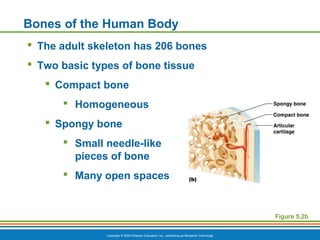
PowerPoint®
Lecture Slide Presentation
by Patty Bostwick-Taylor,
Florence-Darlington Technical College
Copyright © 2009 Pearson Education, Inc., publishing as Benjamin Cummings
PART A
5
The Skeletal
System
Copyright © 2009 Pearson Education, Inc., publishing as Benjamin Cummings
The Skeletal System
 Parts of the skeletal system
 Bones (skeleton)
 Joints
 Cartilages
 Ligaments
 Two subdivisions of the skeleton
 Axial skeleton
 Appendicular skeleton
Copyright © 2009 Pearson Education, Inc., publishing as Benjamin Cummings
Functions of Bones
 Support the body
 Protect soft organs
 Allow movement due to attached skeletal muscles
 Store minerals and fats
 Blood cell formation
Copyright © 2009 Pearson Education, Inc., publishing as Benjamin Cummings
Bones of the Human Body
 The adult skeleton has 206 bones
 Two basic types of bone tissue
 Compact bone
 Homogeneous
 Spongy bone
 Small needle-like
pieces of bone
 Many open spaces
Figure 5.2b
Copyright © 2009 Pearson Education, Inc., publishing as Benjamin Cummings
Classification of Bones on the Basis of Shape
Figure 5.1
Copyright © 2009 Pearson Education, Inc., publishing as Benjamin Cummings
Classification of Bones
 Long bones
 Typically longer than they are wide
 Have a shaft with heads at both ends
 Contain mostly compact bone
 Example:
 Femur
 Humerus
Copyright © 2009 Pearson Education, Inc., publishing as Benjamin Cummings
Classification of Bones
Figure 5.1a
Copyright © 2009 Pearson Education, Inc., publishing as Benjamin Cummings
Classification of Bones
 Short bones
 Generally cube-shape
 Contain mostly spongy bone
 Example:
 Carpals
 Tarsals
Copyright © 2009 Pearson Education, Inc., publishing as Benjamin Cummings
Classification of Bones
Figure 5.1b
Copyright © 2009 Pearson Education, Inc., publishing as Benjamin Cummings
Classification of Bones
 Flat bones
 Thin, flattened, and usually curved
 Two thin layers of compact bone surround a
layer of spongy bone
 Example:
 Skull
 Ribs
 Sternum
Copyright © 2009 Pearson Education, Inc., publishing as Benjamin Cummings
Classification of Bones
Figure 5.1c
Copyright © 2009 Pearson Education, Inc., publishing as Benjamin Cummings
Classification of Bones
 Irregular bones
 Irregular shape
 Do not fit into other bone classification
categories
 Example:
 Vertebrae
 Hip bones
Copyright © 2009 Pearson Education, Inc., publishing as Benjamin Cummings
Classification of Bones
Figure 5.1d
Copyright © 2009 Pearson Education, Inc., publishing as Benjamin Cummings
Anatomy of a Long Bone
 Diaphysis
 Shaft
 Composed of compact bone
 Epiphysis
 Ends of the bone
 Composed mostly of spongy bone
Copyright © 2009 Pearson Education, Inc., publishing as Benjamin Cummings
Anatomy of a Long Bone
Figure 5.2a
Copyright © 2009 Pearson Education, Inc., publishing as Benjamin Cummings
Anatomy of a Long Bone
 Periosteum
 Outside covering of the diaphysis
 Fibrous connective tissue membrane
 Sharpey’s fibers
 Secure periosteum to underlying bone
 Arteries
 Supply bone cells with nutrients
Copyright © 2009 Pearson Education, Inc., publishing as Benjamin Cummings
Anatomy of a Long Bone
Figure 5.2c
Copyright © 2009 Pearson Education, Inc., publishing as Benjamin Cummings
Anatomy of a Long Bone
 Articular cartilage
 Covers the external surface of the epiphyses
 Made of hyaline cartilage
 Decreases friction at joint surfaces
Copyright © 2009 Pearson Education, Inc., publishing as Benjamin Cummings
Anatomy of a Long Bone
 Epiphyseal plate
 Flat plate of hyaline cartilage seen in young,
growing bone
 Epiphyseal line
 Remnant of the epiphyseal plate
 Seen in adult bones
Copyright © 2009 Pearson Education, Inc., publishing as Benjamin Cummings
Anatomy of a Long Bone
Figure 5.2a
Copyright © 2009 Pearson Education, Inc., publishing as Benjamin Cummings
Anatomy of a Long Bone
 Medullary cavity
 Cavity inside of the shaft
 Contains yellow marrow (mostly fat) in adults
 Contains red marrow (for blood cell formation)
in infants
Copyright © 2009 Pearson Education, Inc., publishing as Benjamin Cummings
Anatomy of a Long Bone
Figure 5.2a
Copyright © 2009 Pearson Education, Inc., publishing as Benjamin Cummings
Microscopic Anatomy of Bone
 Osteon (Haversian system)
 A unit of bone containing central canal and
matrix rings
 Central (Haversian) canal
 Opening in the center of an osteon
 Carries blood vessels and nerves
 Perforating (Volkman’s) canal
 Canal perpendicular to the central canal
 Carries blood vessels and nerves
Copyright © 2009 Pearson Education, Inc., publishing as Benjamin Cummings
Microscopic Anatomy of Bone
Figure 5.3a
Copyright © 2009 Pearson Education, Inc., publishing as Benjamin Cummings
Microscopic Anatomy of Bone
 Lacunae
 Cavities containing bone cells (osteocytes)
 Arranged in concentric rings
 Lamellae
 Rings around the central canal
 Sites of lacunae
Copyright © 2009 Pearson Education, Inc., publishing as Benjamin Cummings
Microscopic Anatomy of Bone
Figure 5.3b–c
Copyright © 2009 Pearson Education, Inc., publishing as Benjamin Cummings
Formation of the Human Skeleton
 In embryos, the skeleton is primarily hyaline
cartilage
 During development, much of this cartilage is
replaced by bone
 Cartilage remains in isolated areas
 Bridge of the nose
 Parts of ribs
 Joints
Copyright © 2009 Pearson Education, Inc., publishing as Benjamin Cummings
Bone Growth (Ossification)
 Epiphyseal plates allow for lengthwise growth of
long bones during childhood
 New cartilage is continuously formed
 Older cartilage becomes ossified
 Cartilage is broken down
 Enclosed cartilage is digested away,
opening up a medullary cavity
 Bone replaces cartilage through the action
of osteoblasts
Copyright © 2009 Pearson Education, Inc., publishing as Benjamin Cummings
Bone Growth (Ossification)
 Bones are remodeled and lengthened until growth
stops
 Bones are remodeled in response to two
factors
 Blood calcium levels
 Pull of gravity and muscles on the
skeleton
 Bones grow in width (called appositional
growth)
Copyright © 2009 Pearson Education, Inc., publishing as Benjamin Cummings
Long Bone Formation and Growth
Figure 5.4a, step 1
Bone starting
to replace
cartilage
In an embryo
Bone collar
Hyaline
cartilage
model
(a)
Copyright © 2009 Pearson Education, Inc., publishing as Benjamin Cummings
Long Bone Formation and Growth
Figure 5.4a, step 2
Bone starting
to replace
cartilage
In a fetusIn an embryo
Growth
in bone
length
Blood
vessels
Hyaline
cartilage
New center of
bone growth
Medullary
cavity
Bone collar
Hyaline
cartilage
model
(a)
Copyright © 2009 Pearson Education, Inc., publishing as Benjamin Cummings
Long Bone Formation and Growth
Figure 5.4a, step 3
Bone starting
to replace
cartilage
Epiphyseal
plate
cartilage
Articular
cartilage
Spongy
bone
In a childIn a fetusIn an embryo
New bone
forming
Growth
in bone
width
Growth
in bone
length
Epiphyseal
plate cartilage
New bone
forming
Blood
vessels
Hyaline
cartilage
New center of
bone growth
Medullary
cavity
Bone collar
Hyaline
cartilage
model
(a)
Copyright © 2009 Pearson Education, Inc., publishing as Benjamin Cummings
Long Bone Formation and Growth
Figure 5.4b
Copyright © 2009 Pearson Education, Inc., publishing as Benjamin Cummings
Types of Bone Cells
 Osteocytes—mature bone cells
 Osteoblasts—bone-forming cells
 Osteoclasts—bone-destroying cells
 Break down bone matrix for remodeling and
release of calcium in response to parathyroid
hormone
 Bone remodeling is performed by both
osteoblasts and osteoclasts
Copyright © 2009 Pearson Education, Inc., publishing as Benjamin Cummings
Bone Fractures
 Fracture—break in a bone
 Types of bone fractures
 Closed (simple) fracture—break that does not
penetrate the skin
 Open (compound) fracture—broken bone
penetrates through the skin
 Bone fractures are treated by reduction and
immobilization
Copyright © 2009 Pearson Education, Inc., publishing as Benjamin Cummings
Common Types of Fractures
Table 5.2
Copyright © 2009 Pearson Education, Inc., publishing as Benjamin Cummings
Repair of Bone Fractures
 Hematoma (blood-filled swelling) is formed
 Break is splinted by fibrocartilage to form a callus
 Fibrocartilage callus is replaced by a bony callus
 Bony callus is remodeled to form a permanent
patch
Copyright © 2009 Pearson Education, Inc., publishing as Benjamin Cummings
Stages in the Healing of a Bone Fracture
Figure 5.5
Hematoma
External
callus
Bony
callus of
spongy
bone
Healed
fracture
New
blood
vessels
Internal
callus
(fibrous
tissue and
cartilage)
Spongy
bone
trabecula
Hematoma
formation
Fibrocartilage
callus formation
Bony callus
formation
Bone remodeling

CVA A&P - Chapter 5a: Standard Bone Tissue

  • 1.
    PowerPoint® Lecture Slide Presentation byPatty Bostwick-Taylor, Florence-Darlington Technical College Copyright © 2009 Pearson Education, Inc., publishing as Benjamin Cummings PART A 5 The Skeletal System
  • 2.
    Copyright © 2009Pearson Education, Inc., publishing as Benjamin Cummings The Skeletal System  Parts of the skeletal system  Bones (skeleton)  Joints  Cartilages  Ligaments  Two subdivisions of the skeleton  Axial skeleton  Appendicular skeleton
  • 3.
    Copyright © 2009Pearson Education, Inc., publishing as Benjamin Cummings Functions of Bones  Support the body  Protect soft organs  Allow movement due to attached skeletal muscles  Store minerals and fats  Blood cell formation
  • 4.
    Copyright © 2009Pearson Education, Inc., publishing as Benjamin Cummings Bones of the Human Body  The adult skeleton has 206 bones  Two basic types of bone tissue  Compact bone  Homogeneous  Spongy bone  Small needle-like pieces of bone  Many open spaces Figure 5.2b
  • 5.
    Copyright © 2009Pearson Education, Inc., publishing as Benjamin Cummings Classification of Bones on the Basis of Shape Figure 5.1
  • 6.
    Copyright © 2009Pearson Education, Inc., publishing as Benjamin Cummings Classification of Bones  Long bones  Typically longer than they are wide  Have a shaft with heads at both ends  Contain mostly compact bone  Example:  Femur  Humerus
  • 7.
    Copyright © 2009Pearson Education, Inc., publishing as Benjamin Cummings Classification of Bones Figure 5.1a
  • 8.
    Copyright © 2009Pearson Education, Inc., publishing as Benjamin Cummings Classification of Bones  Short bones  Generally cube-shape  Contain mostly spongy bone  Example:  Carpals  Tarsals
  • 9.
    Copyright © 2009Pearson Education, Inc., publishing as Benjamin Cummings Classification of Bones Figure 5.1b
  • 10.
    Copyright © 2009Pearson Education, Inc., publishing as Benjamin Cummings Classification of Bones  Flat bones  Thin, flattened, and usually curved  Two thin layers of compact bone surround a layer of spongy bone  Example:  Skull  Ribs  Sternum
  • 11.
    Copyright © 2009Pearson Education, Inc., publishing as Benjamin Cummings Classification of Bones Figure 5.1c
  • 12.
    Copyright © 2009Pearson Education, Inc., publishing as Benjamin Cummings Classification of Bones  Irregular bones  Irregular shape  Do not fit into other bone classification categories  Example:  Vertebrae  Hip bones
  • 13.
    Copyright © 2009Pearson Education, Inc., publishing as Benjamin Cummings Classification of Bones Figure 5.1d
  • 14.
    Copyright © 2009Pearson Education, Inc., publishing as Benjamin Cummings Anatomy of a Long Bone  Diaphysis  Shaft  Composed of compact bone  Epiphysis  Ends of the bone  Composed mostly of spongy bone
  • 15.
    Copyright © 2009Pearson Education, Inc., publishing as Benjamin Cummings Anatomy of a Long Bone Figure 5.2a
  • 16.
    Copyright © 2009Pearson Education, Inc., publishing as Benjamin Cummings Anatomy of a Long Bone  Periosteum  Outside covering of the diaphysis  Fibrous connective tissue membrane  Sharpey’s fibers  Secure periosteum to underlying bone  Arteries  Supply bone cells with nutrients
  • 17.
    Copyright © 2009Pearson Education, Inc., publishing as Benjamin Cummings Anatomy of a Long Bone Figure 5.2c
  • 18.
    Copyright © 2009Pearson Education, Inc., publishing as Benjamin Cummings Anatomy of a Long Bone  Articular cartilage  Covers the external surface of the epiphyses  Made of hyaline cartilage  Decreases friction at joint surfaces
  • 19.
    Copyright © 2009Pearson Education, Inc., publishing as Benjamin Cummings Anatomy of a Long Bone  Epiphyseal plate  Flat plate of hyaline cartilage seen in young, growing bone  Epiphyseal line  Remnant of the epiphyseal plate  Seen in adult bones
  • 20.
    Copyright © 2009Pearson Education, Inc., publishing as Benjamin Cummings Anatomy of a Long Bone Figure 5.2a
  • 21.
    Copyright © 2009Pearson Education, Inc., publishing as Benjamin Cummings Anatomy of a Long Bone  Medullary cavity  Cavity inside of the shaft  Contains yellow marrow (mostly fat) in adults  Contains red marrow (for blood cell formation) in infants
  • 22.
    Copyright © 2009Pearson Education, Inc., publishing as Benjamin Cummings Anatomy of a Long Bone Figure 5.2a
  • 23.
    Copyright © 2009Pearson Education, Inc., publishing as Benjamin Cummings Microscopic Anatomy of Bone  Osteon (Haversian system)  A unit of bone containing central canal and matrix rings  Central (Haversian) canal  Opening in the center of an osteon  Carries blood vessels and nerves  Perforating (Volkman’s) canal  Canal perpendicular to the central canal  Carries blood vessels and nerves
  • 24.
    Copyright © 2009Pearson Education, Inc., publishing as Benjamin Cummings Microscopic Anatomy of Bone Figure 5.3a
  • 25.
    Copyright © 2009Pearson Education, Inc., publishing as Benjamin Cummings Microscopic Anatomy of Bone  Lacunae  Cavities containing bone cells (osteocytes)  Arranged in concentric rings  Lamellae  Rings around the central canal  Sites of lacunae
  • 26.
    Copyright © 2009Pearson Education, Inc., publishing as Benjamin Cummings Microscopic Anatomy of Bone Figure 5.3b–c
  • 27.
    Copyright © 2009Pearson Education, Inc., publishing as Benjamin Cummings Formation of the Human Skeleton  In embryos, the skeleton is primarily hyaline cartilage  During development, much of this cartilage is replaced by bone  Cartilage remains in isolated areas  Bridge of the nose  Parts of ribs  Joints
  • 28.
    Copyright © 2009Pearson Education, Inc., publishing as Benjamin Cummings Bone Growth (Ossification)  Epiphyseal plates allow for lengthwise growth of long bones during childhood  New cartilage is continuously formed  Older cartilage becomes ossified  Cartilage is broken down  Enclosed cartilage is digested away, opening up a medullary cavity  Bone replaces cartilage through the action of osteoblasts
  • 29.
    Copyright © 2009Pearson Education, Inc., publishing as Benjamin Cummings Bone Growth (Ossification)  Bones are remodeled and lengthened until growth stops  Bones are remodeled in response to two factors  Blood calcium levels  Pull of gravity and muscles on the skeleton  Bones grow in width (called appositional growth)
  • 30.
    Copyright © 2009Pearson Education, Inc., publishing as Benjamin Cummings Long Bone Formation and Growth Figure 5.4a, step 1 Bone starting to replace cartilage In an embryo Bone collar Hyaline cartilage model (a)
  • 31.
    Copyright © 2009Pearson Education, Inc., publishing as Benjamin Cummings Long Bone Formation and Growth Figure 5.4a, step 2 Bone starting to replace cartilage In a fetusIn an embryo Growth in bone length Blood vessels Hyaline cartilage New center of bone growth Medullary cavity Bone collar Hyaline cartilage model (a)
  • 32.
    Copyright © 2009Pearson Education, Inc., publishing as Benjamin Cummings Long Bone Formation and Growth Figure 5.4a, step 3 Bone starting to replace cartilage Epiphyseal plate cartilage Articular cartilage Spongy bone In a childIn a fetusIn an embryo New bone forming Growth in bone width Growth in bone length Epiphyseal plate cartilage New bone forming Blood vessels Hyaline cartilage New center of bone growth Medullary cavity Bone collar Hyaline cartilage model (a)
  • 33.
    Copyright © 2009Pearson Education, Inc., publishing as Benjamin Cummings Long Bone Formation and Growth Figure 5.4b
  • 34.
    Copyright © 2009Pearson Education, Inc., publishing as Benjamin Cummings Types of Bone Cells  Osteocytes—mature bone cells  Osteoblasts—bone-forming cells  Osteoclasts—bone-destroying cells  Break down bone matrix for remodeling and release of calcium in response to parathyroid hormone  Bone remodeling is performed by both osteoblasts and osteoclasts
  • 35.
    Copyright © 2009Pearson Education, Inc., publishing as Benjamin Cummings Bone Fractures  Fracture—break in a bone  Types of bone fractures  Closed (simple) fracture—break that does not penetrate the skin  Open (compound) fracture—broken bone penetrates through the skin  Bone fractures are treated by reduction and immobilization
  • 36.
    Copyright © 2009Pearson Education, Inc., publishing as Benjamin Cummings Common Types of Fractures Table 5.2
  • 37.
    Copyright © 2009Pearson Education, Inc., publishing as Benjamin Cummings Repair of Bone Fractures  Hematoma (blood-filled swelling) is formed  Break is splinted by fibrocartilage to form a callus  Fibrocartilage callus is replaced by a bony callus  Bony callus is remodeled to form a permanent patch
  • 38.
    Copyright © 2009Pearson Education, Inc., publishing as Benjamin Cummings Stages in the Healing of a Bone Fracture Figure 5.5 Hematoma External callus Bony callus of spongy bone Healed fracture New blood vessels Internal callus (fibrous tissue and cartilage) Spongy bone trabecula Hematoma formation Fibrocartilage callus formation Bony callus formation Bone remodeling