Download to read offline

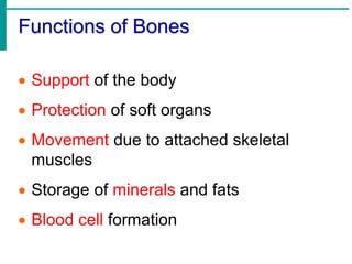
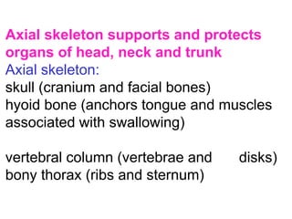
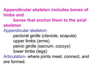
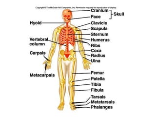
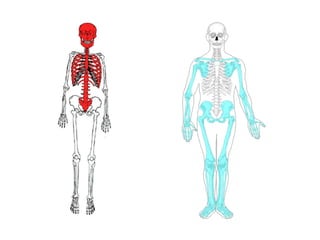
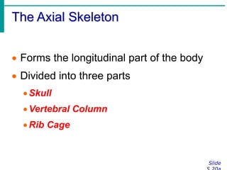
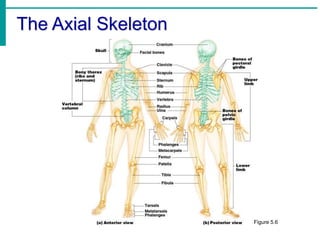
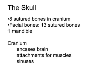
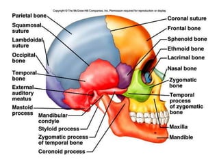


The skeletal system consists of bones, joints, cartilage, ligaments and tendons. The skeletal system is divided into the axial skeleton which includes the skull, vertebral column, and rib cage, and provides protection and support. The appendicular skeleton includes the bones of the limbs and girdles that connect the limbs to the axial skeleton. Bones are classified by their shape as long, short, flat or irregular. The skeletal system functions to support the body, protect organs, allow for movement, store minerals, and produce blood cells.