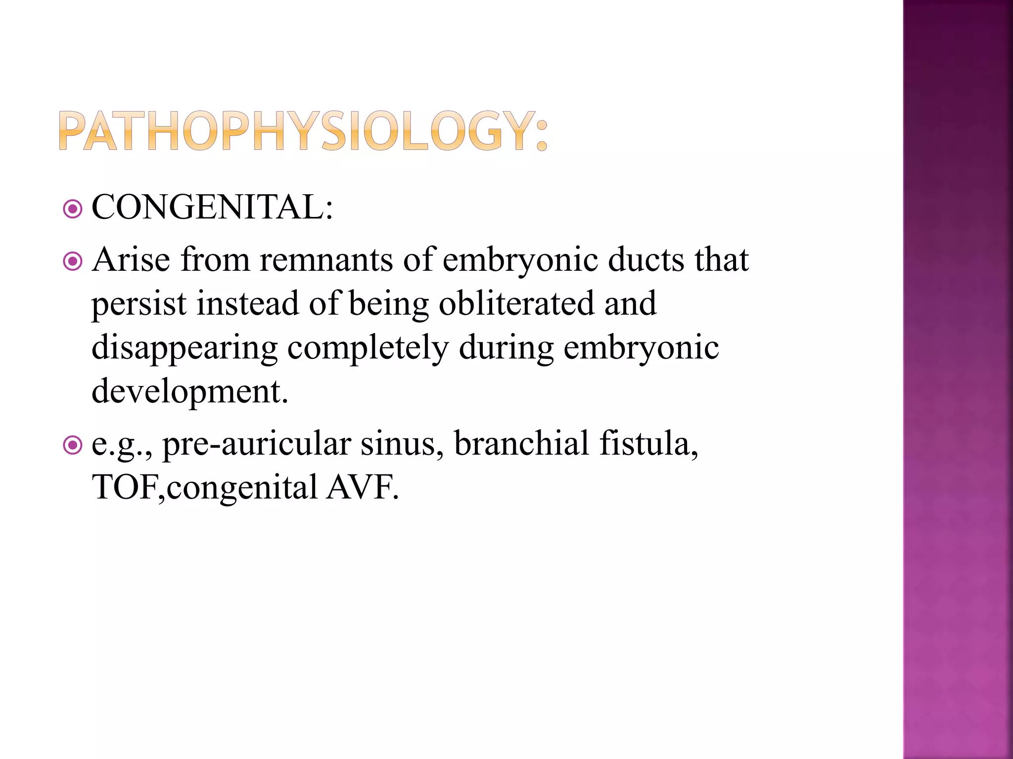
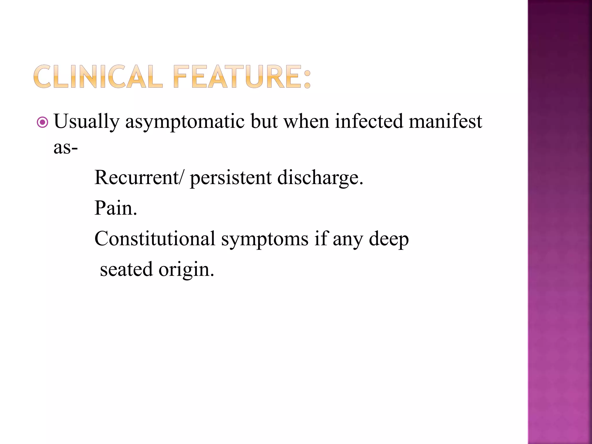
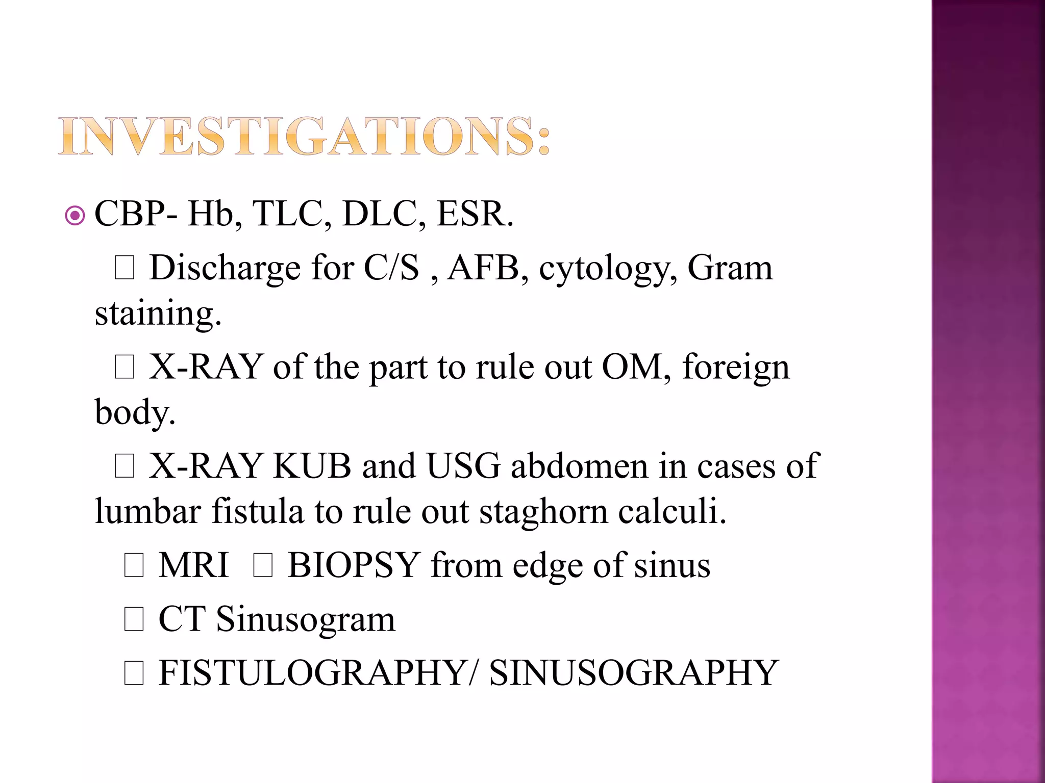
Dr.Prafulla C. Patil
Shri Chamundamata Homoeopathic
Medical College and Hospital,Jalgaon.
 It is a blind track leading from the surface down
into the tissues .
 It is lined with granulation tissue.
 Following are a few examples:
 I. Congenital sinus: Preauricular sinus, post-
amicular sinus
 2. Acquired sinus:
 -Median mental sinus : Occurs as a result of tooth
abscess.
 - Pilonidal sinus: Occurs in the mid line in the anal
region
 - Osteomyelitis: Gives rise to sinus discharging
pus with or without bony spicules.
 • Most common sinus in the neck is due to
tubercular lymphadenitis. It discharges cheesy
material. Skin surrounding the sinus shows bluish
discolouration.
 MIDLINE UPPER LIP SINUS
 PREAURICULAR SINUS
 It is an abnormal communication between the
lumen of one viscus and the lumen of another
(internal) or communicationof one hollow viscus
with the exterior, i.e. body surface(external fistula)
 Examples of internal fistula
• Tracheo-oesophageal fistula
• Colovesical fistula
 Examples of external fistula
• Orocutaneous fistula due to carcinoma of the oral
cavity infiltrating the skin
• Branchial fistula
• Thyroglossal fistula
 Presence of foreign body.
 Persistent infection.
 Distal obstruction as in enterocutaneous fistula.
 Absence of rest.
 Epithelial isation of the track.
 Malignancy.
 Nondependent drainage, inadequate drainage.
 Dense fibrosis..
 Irradiation.
 Specific causes-tuberculosis, actinomycosis.
 CONGENITAL:
 Arise from remnants of embryonic ducts that
persist instead of being obliterated and
disappearing completely during embryonic
development.
 e.g., pre-auricular sinus, branchial fistula,
TOF,congenital AVF.
 ACQUIRED :
Usually secondary to presence of foreign body,
necrotic tissue in affected area (or) microbial
infection (or) following inadequate drainage of
abscess.
e.g., perianal abscess when bursts spontaneously
into skin forming a sinus and when bursts into both
skin and anal canal forming a fistula.
 Usually asymptomatic but when infected manifest
as-
Recurrent/ persistent discharge.
Pain.
Constitutional symptoms if any deep
seated origin.
 INSPECTION:
1. Location: usually gives diagnosis in most of
the cases.
 SINUS: pre-auricular- root of helix of ear.
median mental- symphysis menti.
TB- neck.
 FISTULA: branchial- sternomastoid ant border.
parotid- parotid region
thyroglossal- midline of neck below
hyoid.
 2.Number: usually single but multiple seen in HIV
patients (or) actinomycosis.
 3. Opening:
a) sprouting with granulation tissue-foreign
body.
b) flushing with skin- TB
 4. Surrounding area:
erythematous- inflammatory
bluish- TB
excoriated- faecal
pigmented- chronic sinus/fistulae.
 5. Discharge:
White thin caseous, cheesy like- TB sinus
Faecal- faecal fistula
Yellow sulphur granules- actinomycosis
Bony granules- osteomyelitis
Yellow purulent- staph. Infections
Thin mucous like- brachial fistula
Saliva- parotid fistula
 Palpation:
a) Temperature and tenderness:
b) Discharge: after application of pressure over
the surrounding area.
c) Induration: present in chronic fistulae/sinus as
in actinomycosis.
TB Sinus induration absent.
d) Fixity:
e) Palpation at deeper plane:
lymph nodes- TB
Thickening of bone underneath- OM
 CBP- Hb, TLC, DLC, ESR.
Discharge for C/S , AFB, cytology, Gram
staining.
X-RAY of the part to rule out OM, foreign
body.
X-RAY KUB and USG abdomen in cases of
lumbar fistula to rule out staghorn calculi.
MRI BIOPSY from edge of sinus
CT Sinusogram
FISTULOGRAPHY/ SINUSOGRAPHY
 Antibiotics
 Adequate rest
 Adequate excision
 Adequate drainage.
 Hepar sulph for intense pain.
 Calcarea sulph for thick yellow discharges.
 Calcarea phos for painless fistula.

Sinus and fistula

  • 1.
    Dr.Prafulla C. Patil ShriChamundamata Homoeopathic Medical College and Hospital,Jalgaon.
  • 2.
     It isa blind track leading from the surface down into the tissues .  It is lined with granulation tissue.  Following are a few examples:  I. Congenital sinus: Preauricular sinus, post- amicular sinus  2. Acquired sinus:
  • 3.
     -Median mentalsinus : Occurs as a result of tooth abscess.  - Pilonidal sinus: Occurs in the mid line in the anal region  - Osteomyelitis: Gives rise to sinus discharging pus with or without bony spicules.  • Most common sinus in the neck is due to tubercular lymphadenitis. It discharges cheesy material. Skin surrounding the sinus shows bluish discolouration.
  • 4.
     MIDLINE UPPERLIP SINUS  PREAURICULAR SINUS
  • 5.
     It isan abnormal communication between the lumen of one viscus and the lumen of another (internal) or communicationof one hollow viscus with the exterior, i.e. body surface(external fistula)
  • 6.
     Examples ofinternal fistula • Tracheo-oesophageal fistula • Colovesical fistula  Examples of external fistula • Orocutaneous fistula due to carcinoma of the oral cavity infiltrating the skin • Branchial fistula • Thyroglossal fistula
  • 7.
     Presence offoreign body.  Persistent infection.  Distal obstruction as in enterocutaneous fistula.  Absence of rest.  Epithelial isation of the track.  Malignancy.  Nondependent drainage, inadequate drainage.  Dense fibrosis..  Irradiation.  Specific causes-tuberculosis, actinomycosis.
  • 8.
     CONGENITAL:  Arisefrom remnants of embryonic ducts that persist instead of being obliterated and disappearing completely during embryonic development.  e.g., pre-auricular sinus, branchial fistula, TOF,congenital AVF.
  • 9.
     ACQUIRED : Usuallysecondary to presence of foreign body, necrotic tissue in affected area (or) microbial infection (or) following inadequate drainage of abscess. e.g., perianal abscess when bursts spontaneously into skin forming a sinus and when bursts into both skin and anal canal forming a fistula.
  • 10.
     Usually asymptomaticbut when infected manifest as- Recurrent/ persistent discharge. Pain. Constitutional symptoms if any deep seated origin.
  • 11.
     INSPECTION: 1. Location:usually gives diagnosis in most of the cases.  SINUS: pre-auricular- root of helix of ear. median mental- symphysis menti. TB- neck.  FISTULA: branchial- sternomastoid ant border. parotid- parotid region thyroglossal- midline of neck below hyoid.
  • 12.
     2.Number: usuallysingle but multiple seen in HIV patients (or) actinomycosis.  3. Opening: a) sprouting with granulation tissue-foreign body. b) flushing with skin- TB  4. Surrounding area: erythematous- inflammatory bluish- TB excoriated- faecal pigmented- chronic sinus/fistulae.
  • 13.
     5. Discharge: Whitethin caseous, cheesy like- TB sinus Faecal- faecal fistula Yellow sulphur granules- actinomycosis Bony granules- osteomyelitis Yellow purulent- staph. Infections Thin mucous like- brachial fistula Saliva- parotid fistula
  • 14.
     Palpation: a) Temperatureand tenderness: b) Discharge: after application of pressure over the surrounding area. c) Induration: present in chronic fistulae/sinus as in actinomycosis. TB Sinus induration absent. d) Fixity: e) Palpation at deeper plane: lymph nodes- TB Thickening of bone underneath- OM
  • 15.
     CBP- Hb,TLC, DLC, ESR. Discharge for C/S , AFB, cytology, Gram staining. X-RAY of the part to rule out OM, foreign body. X-RAY KUB and USG abdomen in cases of lumbar fistula to rule out staghorn calculi. MRI BIOPSY from edge of sinus CT Sinusogram FISTULOGRAPHY/ SINUSOGRAPHY
  • 16.
     Antibiotics  Adequaterest  Adequate excision  Adequate drainage.
  • 17.
     Hepar sulphfor intense pain.  Calcarea sulph for thick yellow discharges.  Calcarea phos for painless fistula.