Download as PDF, PPTX

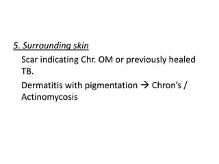
![• History
Since birth - preauricular sinus;
due to Osteomyelitis(high fever + swelling + bone pain)
TB -lymph node enlargement or TB bone or joints
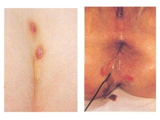
Perianal- h/o perianal/ischiorectal abscess (intermittent
contraction of anal sphincter prevent proper rest)
[Pain + inflammatory/blockage; Fever/redness of
surrounding skin inflammatory]
• Past history TB, Crohn’s, U.colitis, actinomycosis,
colloid Ca, operation complication
• Family history TB, Crohn’s, U.colitis](https://image.slidesharecdn.com/sinusfistulaedrneerajjain-200718000312/85/Sinus-fistulae-drneerajjain-6-320.jpg)


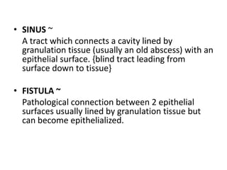
1. A sinus is a tract connecting a cavity lined by granulation tissue to the skin surface, while a fistula is an abnormal connection between two epithelial surfaces usually lined by granulation tissue. 2. Sinuses and fistulae can be congenital, traumatic, inflammatory, or neoplastic in origin. Common causes include infections from organisms like Mycobacterium tuberculosis or Actinomyces. 3. Examination of sinuses and fistulae involves inspecting their number, size, location, openings, discharge, surrounding skin, palpating for tenderness or lumps, and using probes to determine depth and connections. Relevant medical history and investigations help determine the underlying cause.


![ONFH[AVN HIP] -TRIPLE REGIME -A NOVAL SURGICAL CONCEPT .pptx](https://cdn.slidesharecdn.com/ss_thumbnails/onfhavnhip2026koaconcalicutdrgokuldevdrmashraf-260210064517-213ec005-thumbnail.jpg?width=640&height=640&fit=bounds)


