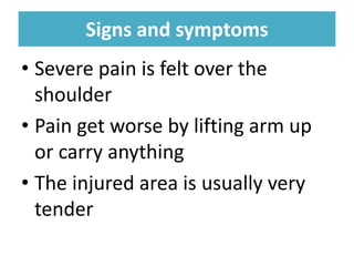
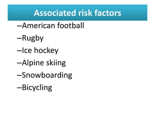
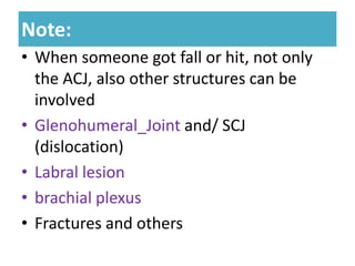
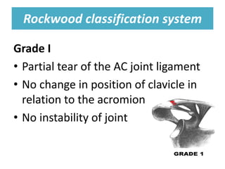
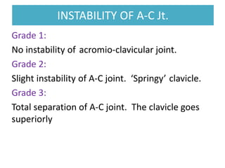
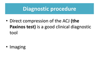
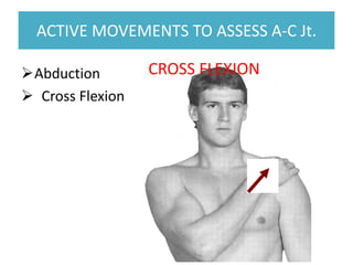
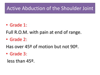
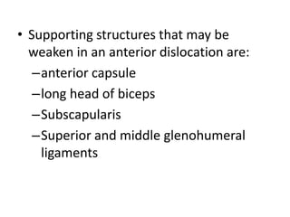
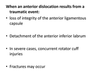
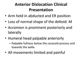
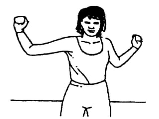
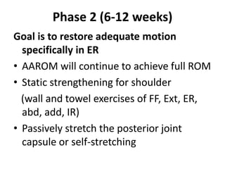
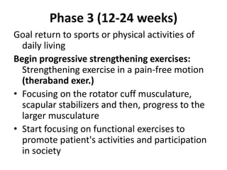
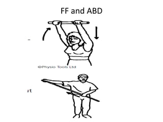
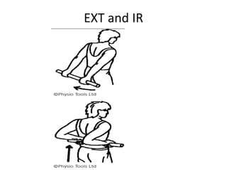
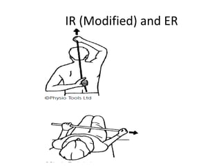
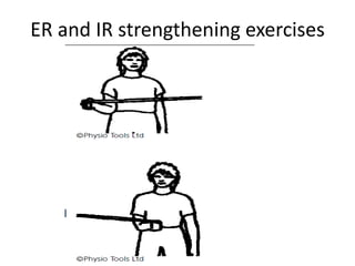
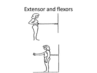
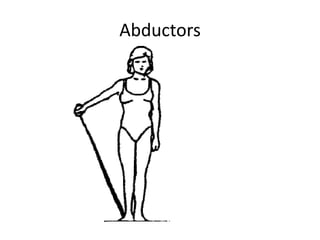
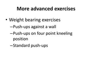
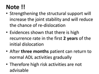
AC, SC and GH joint
disorders
Dechasa Imiru (BSc, MSc PT)
Physiotherapy Department
Jimma University
April, 2023
Acromioclavicular Joint Disorders
• Disorders is a general term to
cover a range of conditions.
• The two common conditions
affecting this joint are
–Dislocation / Subluxation
–Arthritis
Dislocation / Subluxation/separation
Mechanisms of Injury:
• Strain on the ligaments following a fall
–Onto the hand or
–Onto the Elbow or
–Onto the shoulder
• Direct hit or bump over the shoulder
(contact sports)
• Depending on the injury the joint
partially dislocates (subluxation)
Signs and symptoms
Signs and symptoms
• Severe pain is felt over the
shoulder
• Pain get worse by lifting arm up
or carry anything
• The injured area is usually very
tender
Associated risk factors
–American football
–Rugby
–Ice hockey
–Alpine skiing
–Snowboarding
–Bicycling
Note:
Note:
• When someone got fall or hit, not only
the ACJ, also other structures can be
involved
• Glenohumeral_Joint and/ SCJ
(dislocation)
• Labral lesion
• brachial plexus
• Fractures and others
Rockwood classification system
Grade I
• Partial tear of the AC joint ligament
• No change in position of clavicle in
relation to the acromion
• No instability of joint
Grade II
• Rupture/tear of the AC ligament,
Partial tear of the coracoclavicular
ligament
• Displacement of clavicle (less than
the full width of the clavicle)
Grade III
• Rupture of AC and coracoclavicular
ligament
• Displacement of clavicle (more than
the full width of the clavicle)
INSTABILITY OF A-C Jt.
Grade 1:
No instability of acromio-clavicular joint.
Grade 2:
Slight instability of A-C joint. ‘Springy’ clavicle.
Grade 3:
Total separation of A-C joint. The clavicle goes
superiorly
Diagnostic procedure
• Direct compression of the ACJ (the
Paxinos test) is a good clinical diagnostic
tool
• Imaging
ACTIVE MOVEMENTS TO ASSESS A-C Jt.
Abduction
 Cross Flexion
CROSS FLEXION
Active Abduction of the Shoulder Joint
• Grade 1:
Full R.O.M. with pain at end of range.
• Grade 2:
Has over 45º of motion but not 90º.
• Grade 3:
less than 45º.
What is the initial treatment?
• Pain modalities and anti-inflammatory
medication to alleviate pain
• Sprains and majority of dislocations require a
sling to rest the joint
• It takes about 6 weeks for the discomfort to
settle down
• Grade III injuries may require stabilizing
surgery
• Severe dislocations require surgery to put the
joint back together and to repair the torn
ligaments (arthroscopic operation)
Physiotherapy Management
• The rehab is longer for a grade II injury
but the protocol is the same for grade I
• After pain easing it is important to
start as soon as possible with
–Movement
–Active exercises
Cont …
Active exercises:
• Moving the fingers, wrist and elbow
to prevent stiffness
• Progress with more targeted
shoulder exercises
Strengthening exercises:
• As pain is reduced more mobility and
strengthening is promoted
Sterno clavicular Joint Injuries
• It is a synovial joint, gaining the majority
of its strength from surrounding ligaments
• It can become relatively unstable if
subjected to trauma
–Due to the unreserved movement it provides
and
–the small part of the joint which actually
connects
4/25/2023 20
Signs and symptoms
Signs and symptoms
• Pain will be present if the joint is only
mildly sprained
• Straight away recognizable deformity will
be observed if the SCJ has been dislocated
• Pain is worsened when moving the arms
4/25/2023 21
Two types of SCJ dislocation
• Anterior dislocations cause clavicle to poke out
of position
• Creating a readily visible bump on the chest
• Posterior dislocations can affect the throat,
limiting the ability to breath or swallow
properly
Causes
Causes
• Sporting activity, result of a falling and
accident
• The motor vehicle accident (MVA)
Mechanism of injury
• A hard strike to the shoulder
–(known as indirect force because it does not
hit the joint directly) e.g. contact sports
Treatment
Treatment
• Closed reduction or surgery (posterior
dislocation)
• Followed by immobilization for about 2 month
• Minor sprains should generally heal in a few
days on their own with suitable rest (sling)
Treatment …
Treatment …
• Cryotherapy a few times per day to reduce
pain and swelling, and anti-inflammatory pain
medication
• Avoid strenuous activities involving the arms
and shoulders
• Increasing motion of shoulder and build up
strength again using appropriate strategies
Shoulder Dislocation
anatomical predisposition to dislocation
–Glenoid Defects
– Labral Defects
– Neuromuscular Disorders
• Shoulder dislocations can occur in
two common directions:
–Anterior
–posterior
Types of dislocation
Traumatic
Atraumatic
Acquired
Anterior Shoulder Dislocation
• Accounts for 97% of recurrent or first
time dislocations
Causes;
– due to trauma from a direct posterolateral force
on the shoulder
– Due to an excessive amount of abduction and
external rotation direction force
• Supporting structures that may be
weaken in an anterior dislocation are:
–anterior capsule
–long head of biceps
–Subscapularis
–Superior and middle glenohumeral
ligaments
When an anterior dislocation results from a
traumatic event:
• loss of integrity of the anterior ligamentous
capsule
• Detachment of the anterior inferior labrum
• In severe cases, concurrent rotator cuff
injuries
• Fractures may occur
Anterior Dislocation Clinical
Presentation
• Arm held in abducted and ER position
• Loss of normal shape of the deltoid. M
• Acromion is prominent posteriorly and
laterally
• Humeral head palpable anteriorly
– Palpable fullness below the coracoid process and
towards the axilla
• All movements limited and painful
Cont …
Thorough examination is need to
check if there is;
–damage to rotator cuff musculature
–Bone fracture
–Vascular
–Nervous structures
Posterior Shoulder Dislocation
• Accounts for 3% of shoulder dislocations
• Caused by an external blow to the front of the
shoulder
• When force is applied to the humerus that
combines flexion, adduction, and internal
rotation
• Resulted from falling on an out stretched hand
(FOOSH injury)
• Traumatic mechanism of injury, posterior
dislocations may also have concurrent labral
or rotator cuff pathology or fracture
Posterior Dislocation
With acute posterior glenohumeral
dislocation:
– Arm is abducted and internal rotated (IR)
– May notice posterior prominence head of
humerus
Re-current dislocation
• Recurrence rate of dislocations in young active
individuals is as high as 92-96%
• In young patients , initial suggestions are to try
conservative rehabilitation because the risk of re-
dislocation is lower,
• Individuals whom are 40 and older also have a
low recurrence rate around less than 15%
• The recommended management is non operative
and to address associated injuries
Shoulder instability
• Instability can occur whenever the labrum is torn,
stretched or peeled back off the bone,
• This allows the head of the humerus to move
away from the glenoid
• This can occur after;
• a shoulder dislocation, shoulder trauma, or as a result of
repetitive motion
• Some patients also have a genetic predisposition
to develop shoulder instability
Continued …
• Patients with shoulder instability can suffer from
recurrent shoulder dislocations/subluxations or
shoulder pain
• Dislocations can cause fractures and rotator cuff
tears
• Osteoarthritis or wearing of the surface cartilage
of the shoulder joint can also occur as a result of
shoulder instability
Diagnostic Procedures
• Rule out a fracture if dislocation
is suspected
• Radiographs are necessary
• An MRI can be used to rule in or
rule out any soft tissue
pathologies
Management of sho. Dislocation
• Non surgical conservative management is
preferred, initially
• Surgical repair may be reasonable for fail
conservative care or
• Require extreme usage of the upper
extremity (i.e. elite level athletes)
Cont …
• Non-surgical intervention will be
a closed reduction by an
orthopedic surgeon
• Surgical intervention will be
surgical repairing by stabilization
procedures
Conservative management
Phase 1 (up to 6 weeks): Goal is to maintain
stability
Closed reduction
• Immobilization: by using sling (IR vs ER) for 2-
6 weeks
• Ice pack or pain medication (2-3 weeks)
To reduce stiffness:
• Gentle PROM of shoulder out of sling
• AROM for distal joint of the shoulder
Cont …
During the immobilization period
• Codman Exercises (pendulum exercises)
• AAROM for ER (0-30 degree) and FF (0-90
degree) & abduction
NB: Do each movt’s separately !!
• Static contraction can be incorporated for
the rotator cuff and biceps musculature
(end of this phase or the next)
Phase 2 (6-12 weeks)
Goal is to restore adequate motion
specifically in ER
• AAROM will continue to achieve full ROM
• Static strengthening for shoulder
(wall and towel exercises of FF, Ext, ER,
abd, add, IR)
• Passively stretch the posterior joint
capsule or self-stretching
Phase 3 (12-24 weeks)
Goal return to sports or physical activities of
daily living
Begin progressive strengthening exercises:
Strengthening exercise in a pain-free motion
(theraband exer.)
• Focusing on the rotator cuff musculature,
scapular stabilizers and then, progress to the
larger musculature
• Start focusing on functional exercises to
promote patient's activities and participation
in society
FF and ABD
EXT and IR
IR (Modified) and ER
ER and IR strengthening exercises
Extensor and flexors
Abductors
More advanced exercises
• Weight bearing exercises
–Push-ups against a wall
–Push-ups on four point kneeling
position
–Standard push-ups
Note !!
• Strengthening the structural support will
increase the joint stability and will reduce
the chance of re-dislocation
• Evidences shown that there is high
recurrence rate in the first 2 years of the
initial dislocation
• After three months patient can return to
normal ADL activities gradually
• Therefore high risk activities are not
advisable

0004 AC, SC and ST joints dislocation-Copy.pdf

  • 1.
    AC, SC andGH joint disorders Dechasa Imiru (BSc, MSc PT) Physiotherapy Department Jimma University April, 2023
  • 2.
    Acromioclavicular Joint Disorders AcromioclavicularJoint Disorders • Disorders is a general term to cover a range of conditions. • The two common conditions affecting this joint are –Dislocation / Subluxation –Arthritis
  • 3.
    Dislocation / Subluxation/separation Mechanismsof Injury: • Strain on the ligaments following a fall –Onto the hand or –Onto the Elbow or –Onto the shoulder • Direct hit or bump over the shoulder (contact sports) • Depending on the injury the joint partially dislocates (subluxation)
  • 4.
    Signs and symptoms Signsand symptoms • Severe pain is felt over the shoulder • Pain get worse by lifting arm up or carry anything • The injured area is usually very tender
  • 5.
    Associated risk factors Associatedrisk factors –American football –Rugby –Ice hockey –Alpine skiing –Snowboarding –Bicycling
  • 6.
    Note: Note: • When someonegot fall or hit, not only the ACJ, also other structures can be involved • Glenohumeral_Joint and/ SCJ (dislocation) • Labral lesion • brachial plexus • Fractures and others
  • 7.
    Rockwood classification system Rockwoodclassification system Grade I • Partial tear of the AC joint ligament • No change in position of clavicle in relation to the acromion • No instability of joint
  • 9.
    Grade II • Rupture/tearof the AC ligament, Partial tear of the coracoclavicular ligament • Displacement of clavicle (less than the full width of the clavicle)
  • 11.
    Grade III • Ruptureof AC and coracoclavicular ligament • Displacement of clavicle (more than the full width of the clavicle)
  • 13.
    INSTABILITY OF A-CJt. INSTABILITY OF A-C Jt. Grade 1: No instability of acromio-clavicular joint. Grade 2: Slight instability of A-C joint. ‘Springy’ clavicle. Grade 3: Total separation of A-C joint. The clavicle goes superiorly
  • 14.
    Diagnostic procedure Diagnostic procedure •Direct compression of the ACJ (the Paxinos test) is a good clinical diagnostic tool • Imaging
  • 15.
    ACTIVE MOVEMENTS TOASSESS A-C Jt. ACTIVE MOVEMENTS TO ASSESS A-C Jt. Abduction  Cross Flexion CROSS FLEXION
  • 16.
    Active Abduction ofthe Shoulder Joint • Grade 1: Full R.O.M. with pain at end of range. • Grade 2: Has over 45º of motion but not 90º. • Grade 3: less than 45º.
  • 17.
    What is theinitial treatment? What is the initial treatment? • Pain modalities and anti-inflammatory medication to alleviate pain • Sprains and majority of dislocations require a sling to rest the joint • It takes about 6 weeks for the discomfort to settle down • Grade III injuries may require stabilizing surgery • Severe dislocations require surgery to put the joint back together and to repair the torn ligaments (arthroscopic operation)
  • 18.
    Physiotherapy Management Physiotherapy Management •The rehab is longer for a grade II injury but the protocol is the same for grade I • After pain easing it is important to start as soon as possible with –Movement –Active exercises
  • 19.
    Cont … Active exercises: •Moving the fingers, wrist and elbow to prevent stiffness • Progress with more targeted shoulder exercises Strengthening exercises: • As pain is reduced more mobility and strengthening is promoted
  • 20.
    Sterno clavicular JointInjuries Sterno clavicular Joint Injuries • It is a synovial joint, gaining the majority of its strength from surrounding ligaments • It can become relatively unstable if subjected to trauma –Due to the unreserved movement it provides and –the small part of the joint which actually connects 4/25/2023 20
  • 21.
    Signs and symptoms Signsand symptoms • Pain will be present if the joint is only mildly sprained • Straight away recognizable deformity will be observed if the SCJ has been dislocated • Pain is worsened when moving the arms 4/25/2023 21
  • 22.
    Two types ofSCJ dislocation Two types of SCJ dislocation • Anterior dislocations cause clavicle to poke out of position • Creating a readily visible bump on the chest • Posterior dislocations can affect the throat, limiting the ability to breath or swallow properly
  • 23.
    Causes Causes • Sporting activity,result of a falling and accident • The motor vehicle accident (MVA) Mechanism of injury • A hard strike to the shoulder –(known as indirect force because it does not hit the joint directly) e.g. contact sports
  • 24.
    Treatment Treatment • Closed reductionor surgery (posterior dislocation) • Followed by immobilization for about 2 month • Minor sprains should generally heal in a few days on their own with suitable rest (sling)
  • 25.
    Treatment … Treatment … •Cryotherapy a few times per day to reduce pain and swelling, and anti-inflammatory pain medication • Avoid strenuous activities involving the arms and shoulders • Increasing motion of shoulder and build up strength again using appropriate strategies
  • 26.
    Shoulder Dislocation anatomical predispositionto dislocation –Glenoid Defects – Labral Defects – Neuromuscular Disorders • Shoulder dislocations can occur in two common directions: –Anterior –posterior
  • 27.
    Types of dislocation Typesof dislocation Traumatic Atraumatic Acquired
  • 28.
    Anterior Shoulder Dislocation •Accounts for 97% of recurrent or first time dislocations Causes; – due to trauma from a direct posterolateral force on the shoulder – Due to an excessive amount of abduction and external rotation direction force
  • 29.
    • Supporting structuresthat may be weaken in an anterior dislocation are: –anterior capsule –long head of biceps –Subscapularis –Superior and middle glenohumeral ligaments
  • 30.
    When an anteriordislocation results from a traumatic event: • loss of integrity of the anterior ligamentous capsule • Detachment of the anterior inferior labrum • In severe cases, concurrent rotator cuff injuries • Fractures may occur
  • 31.
    Anterior Dislocation Clinical Presentation •Arm held in abducted and ER position • Loss of normal shape of the deltoid. M • Acromion is prominent posteriorly and laterally • Humeral head palpable anteriorly – Palpable fullness below the coracoid process and towards the axilla • All movements limited and painful
  • 33.
    Cont … Thorough examinationis need to check if there is; –damage to rotator cuff musculature –Bone fracture –Vascular –Nervous structures
  • 35.
    Posterior Shoulder Dislocation •Accounts for 3% of shoulder dislocations • Caused by an external blow to the front of the shoulder • When force is applied to the humerus that combines flexion, adduction, and internal rotation • Resulted from falling on an out stretched hand (FOOSH injury) • Traumatic mechanism of injury, posterior dislocations may also have concurrent labral or rotator cuff pathology or fracture
  • 36.
    Posterior Dislocation With acuteposterior glenohumeral dislocation: – Arm is abducted and internal rotated (IR) – May notice posterior prominence head of humerus
  • 37.
    Re-current dislocation • Recurrencerate of dislocations in young active individuals is as high as 92-96% • In young patients , initial suggestions are to try conservative rehabilitation because the risk of re- dislocation is lower, • Individuals whom are 40 and older also have a low recurrence rate around less than 15% • The recommended management is non operative and to address associated injuries
  • 38.
    Shoulder instability • Instabilitycan occur whenever the labrum is torn, stretched or peeled back off the bone, • This allows the head of the humerus to move away from the glenoid • This can occur after; • a shoulder dislocation, shoulder trauma, or as a result of repetitive motion • Some patients also have a genetic predisposition to develop shoulder instability
  • 39.
    Continued … • Patientswith shoulder instability can suffer from recurrent shoulder dislocations/subluxations or shoulder pain • Dislocations can cause fractures and rotator cuff tears • Osteoarthritis or wearing of the surface cartilage of the shoulder joint can also occur as a result of shoulder instability
  • 40.
    Diagnostic Procedures • Ruleout a fracture if dislocation is suspected • Radiographs are necessary • An MRI can be used to rule in or rule out any soft tissue pathologies
  • 41.
    Management of sho.Dislocation • Non surgical conservative management is preferred, initially • Surgical repair may be reasonable for fail conservative care or • Require extreme usage of the upper extremity (i.e. elite level athletes)
  • 42.
    Cont … • Non-surgicalintervention will be a closed reduction by an orthopedic surgeon • Surgical intervention will be surgical repairing by stabilization procedures
  • 43.
    Conservative management Phase 1(up to 6 weeks): Goal is to maintain stability Closed reduction • Immobilization: by using sling (IR vs ER) for 2- 6 weeks • Ice pack or pain medication (2-3 weeks) To reduce stiffness: • Gentle PROM of shoulder out of sling • AROM for distal joint of the shoulder
  • 44.
    Cont … During theimmobilization period • Codman Exercises (pendulum exercises) • AAROM for ER (0-30 degree) and FF (0-90 degree) & abduction NB: Do each movt’s separately !! • Static contraction can be incorporated for the rotator cuff and biceps musculature (end of this phase or the next)
  • 46.
    Phase 2 (6-12weeks) Goal is to restore adequate motion specifically in ER • AAROM will continue to achieve full ROM • Static strengthening for shoulder (wall and towel exercises of FF, Ext, ER, abd, add, IR) • Passively stretch the posterior joint capsule or self-stretching
  • 47.
    Phase 3 (12-24weeks) Goal return to sports or physical activities of daily living Begin progressive strengthening exercises: Strengthening exercise in a pain-free motion (theraband exer.) • Focusing on the rotator cuff musculature, scapular stabilizers and then, progress to the larger musculature • Start focusing on functional exercises to promote patient's activities and participation in society
  • 48.
  • 49.
  • 50.
  • 51.
    ER and IRstrengthening exercises
  • 52.
  • 53.
  • 54.
    More advanced exercises •Weight bearing exercises –Push-ups against a wall –Push-ups on four point kneeling position –Standard push-ups
  • 55.
    Note !! • Strengtheningthe structural support will increase the joint stability and will reduce the chance of re-dislocation • Evidences shown that there is high recurrence rate in the first 2 years of the initial dislocation • After three months patient can return to normal ADL activities gradually • Therefore high risk activities are not advisable