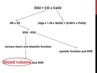
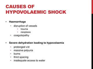
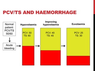
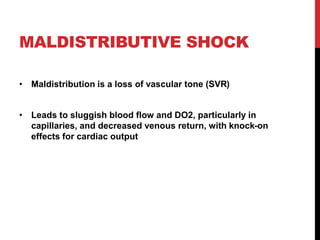
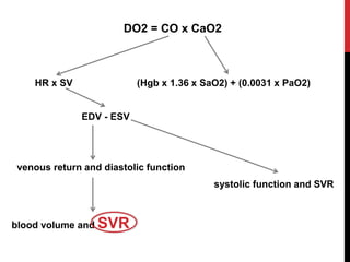
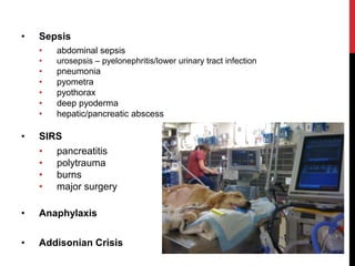
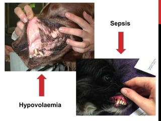
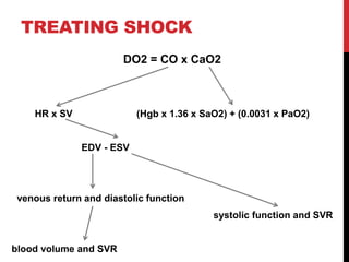
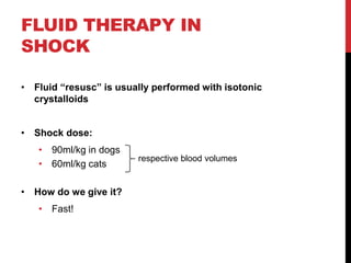
SHOCK
ERIN MOONEY, BVSC DACVECC
7TH MAY, 2014
shock is common, but under-recognised
• early recognition is important to prevent progression
of the shock state
• dramatic impact on survival
SHOCK
• Inadequate cellular energy production
• oxygen or glucose
• Usually due to inadequate oxygen delivery (DO2)
• cardiovascular dysfunction
ENERGY PRODUCTION
glycolysis
tricarboxylic
acid cycle
oxidative
phosphorylation
anaerobic metabolism
yields 2 moles ATP
aerobic metabolism
yields 36 moles ATP
WHAT’S BAD ABOUT
SHOCK?
• Preferential shunting of blood away from splanchnic
circulation, skin and muscle
• GI translocation  sepsis
• Sluggish blood flow through capillary beds
• renal
• cerebral
• myocardial
• Further anaerobic metabolism
• cellular energy deficit  MOF
OXYGEN DELIVERY (DO2)
DO2 = CO x CaO2
HR x SV (Hgb x 1.36 x SaO2) + (0.0031 x PaO2)
EDV - ESV
venous return and diastolic function
systolic function and SVR
blood volume and SVR
TYPES OF SHOCK
• Hypovolaemic
• Maldistributive
• Cardiogenic
• Obstructive
• Metabolic
• Hypoxic
HYPOVOLAEMIC SHOCK
blood volume is too low to maintain DO2
DO2 = CO x CaO2
HR x SV (Hgb x 1.36 x SaO2) + (0.0031 x PaO2)
EDV - ESV
venous return and diastolic function
systolic function and SVR
blood volume and SVR
CAUSES OF
HYPOVOLAEMIC SHOCK
• Haemorrhage
• disruption of vessels
• trauma
• neoplasia
• coagulopathy
• Severe dehydration leading to hypovolaemia
• prolonged v/d
• massive polyuria
• burns
• third spacing
• inadequate access to water
DEHYDRATION ≠ HYPOVOLAEMIA
• Dehydration is a decrease in extracellular fluid volume, i.e-
the interstitial and intravascular spaces
• Because blood volume is spared at the expense of
interstitial volume, “hydration” generally refers the
interstitial fluid volume
• Dehydrated patients may or may not be hypovolaemic
• Hypovolaemic patients may or may not be dehydrated
HYDRATION
• MM moistness
• Skin turgor
• Globe position
VOLUME
• Heart rate
• MM colour, CRT
• Pulse quality
• Temperature
PCV/TS AND HAEMORRHAGE
Hypovolaemia Euvolaemia
Acute
bleeding
PCV: 50
TS: 60
PCV: 25
TS: 30
Improving
hypovolaemia
PCV: 40
TS: 45
Normal
patient:
PCV/TS
50/60
MALDISTRIBUTIVE SHOCK
• Maldistribution is a loss of vascular tone (SVR)
• Leads to sluggish blood flow and DO2, particularly in
capillaries, and decreased venous return, with knock-on
effects for cardiac output
DO2 = CO x CaO2
HR x SV (Hgb x 1.36 x SaO2) + (0.0031 x PaO2)
EDV - ESV
venous return and diastolic function
systolic function and SVR
blood volume and SVR
• Sepsis
• abdominal sepsis
• urosepsis – pyelonephritis/lower urinary tract infection
• pneumonia
• pyometra
• pyothorax
• deep pyoderma
• hepatic/pancreatic abscess
• SIRS
• pancreatitis
• polytrauma
• burns
• major surgery
• Anaphylaxis
• Addisonian Crisis
SEPSIS
The pathogenesis of maldistributive shock in sepsis is multi-
factorial:
• increased production of nitric oxide
• over-activation of K+/ATP channels
• deficiency of vasopressin
• critical illness-related corticosteroid insufficiency
CARDIOGENIC SHOCK
Shock related to failure of the pump mechanism of the heart
• failure to fill during diastole
• failure to eject adequate stroke volume during systole
CHF adds hypoxia to the mix
DO2 = CO x CaO2
HR x SV (Hgb x 1.36 x SaO2) + (0.0031 x PaO2)
EDV - ESV
venous return and diastolic function
systolic function and SVR
blood volume and SVR
CAUSES OF
CARDIOGENIC SHOCK
• Diastolic failure
• HCM
• severe mitral insufficiency
• pericardial effusion
• Systolic failure
• DCM
• sepsis-induced myocardial dysfunction
• Tachyarrhythmias
• Bradyarrhythmias
both
OBSTRUCTIVE SHOCK
• Physical obstruction to arterial or venous blood flow
• Examples:
• GDV
• PTE
• heartworm disease
• +/- pericardial effusion
METABOLIC SHOCK
• Deranged intracellular metabolic activity
• DO2 is normal
• Examples:
• hypoglycaemia
• toxicities that cause uncoupling of ox phos:
• cyanide
• arsenic
• 1080
• bromethalin
• mitochondrial dysfunction in sepsis and SIRS
HYPOXIC SHOCK
• Decrease in arterial blood oxygen content
• i.e- not related to blood flow
• Examples:
• severe anaemia (PCV < 10%)
• CO toxicity
• methaemoglobinaemia
• severe pulmonary disease
IDENTIFYING SHOCK
Mixture of history, physical exam and laboratory evidence
Physical exam is your most important tool
IDENTIFYING SHOCK
Perfusion parameters:
• MM colour*
• CRT
• HR
• pulse quality
• temperature (particularly of extremities)
• mentation
• urine output
• blood pressure
* may be injected with maldistribution
LABORATORY
EVIDENCE OF SHOCK
Less fancy:
• metabolic acidosis
• decreased SBE
• decreased HCO3
• +/- acidaemia (depends on compensation)
• hyperlactataemia
• pre-renal azotaemia
• oliguria/anuria
• estimate volume status and CO via echocardiography
LACTATE
• Produced from pyruvate, a waste product of glycolysis
• During anaerobic metabolism, there is increased
production of pyruvate  increased lactate production
• The most common cause of hyperlactataemia is anaerobic
metabolism during shock (“Type A hyperlactataemia”)
• Production also increases with SIRS/sepsis,
administration of steroids and some types of neoplasia
(“Type B hyperlactataemia”)
LABORATORY
EVIDENCE OF SHOCK
Fancy:
• direct CO monitoring via PAC (gold standard)
• mixed venous oxygen via PAC
• combined CVP and direct BP
• central venous oxygen via CVC
CLASSIFYING SHOCK
compensated
shock
early,
decompensated
shock
late,
decompensated
shock
COMPENSATED SHOCK
• Catecholamine surge to maintain perfusion/DO2 causes
tachycardia and vasoconstriction
• Can be hard to identify; tachycardia in a patient that’s not
boisterous or stressed
• HR 130 - 160
• tall, narrow pulses
• pink/pale pink MM (+/- “injected” in sepsis/SIRS)
• CRT 0.5 – 1.5 sec
• normal body temperature (+/- elevated in sepsis/SIRS)
• extremities may be cool
• quiet mentation
• normal BP
• normal UOP
• normal  mild hyperlactataemia
Hypovolaemia
Sepsis
EARLY DECOMPENSATED
SHOCK
Compensatory mechanisms become overwhelmed and DO2
starts to fall
• HR 160 – 200
• reduced femoral pulse quality, absent metatarsal pulses
• pale pink MM (+/- injected in SIRS/sepsis)
• CRT (1.5 – 2 sec)
• mild hypothermia with cool extremities
• quiet mentation
• mild  moderate hypotension
• decreased UOP
• mild moderate hyperlactataemia
LATE DECOMPENSATED
SHOCK
• During this phase, DO2 is inadequate to maintain organ
function.
• Death is imminent
• HR >200
• ** bradycardia may develop shortly before death.
• weak to absent femoral pulses
• grey/patchy MM
• CRT > 2 sec or undetectable
• hypothermia and cold extremities
• obtundation
• severe hypotension
• decreased  absent urine output
• severe hyperlactataemia
CATS ARE NOT SMALL DOGS
• Cats tend to become bradycardic and hypothermic earlier
in shock, particularly in sepsis
• A sick bradycardic cat is a very sick cat indeed
TREATING SHOCK
DO2 = CO x CaO2
HR x SV (Hgb x 1.36 x SaO2) + (0.0031 x PaO2)
EDV - ESV
venous return and diastolic function
systolic function and SVR
blood volume and SVR
FLUID THERAPY IN
SHOCK
• Fluid “resusc” is usually performed with isotonic
crystalloids
• Shock dose:
• 90ml/kg in dogs
• 60ml/kg cats
• How do we give it?
• Fast!
respective blood volumes
• Administer ¼ shock dose over 10 – 15 min then re-assess
your patient.
• If your goals of resusc have not been met, keep going
• Goals of resusc:
• HR 80 – 120
• pink MM, CRT 1 – 2 s
• normal pulse quality
• warm extremities, normothermia
• normal mentation
• SBP 100 – 140
• normal UOP
FAST?
http://www.impactednurse.com/pics5/notstatH.jpg
OTHER FLUID TYPES
• Hypertonic saline (7%)
• hypertonic crystalloid
• dose: 3 – 5ml/kg once
• Artificial colloids
• use in shock is controversial
• renal failure, coagulopathies
• NEVER bolus hypotonic fluids
• 0.45% saline
• 0.45% saline + 2.5% glucose
• 5% glucose in water
• plasmalyte-56
• normosol-M
I’VE GIVEN SHOCK FLUIDS …
NOW WHAT?
• In cases of uncomplicated hypovolaemia, your work is
done
• In most cases, shock will continue unless you address the
the underlying cause, because only 25% of your
crystalloids will still be in IV space 30 min after infusion
• Must diagnose and address the underlying cause ASAP
• Sepsis
• volume first
• vasopressors if needed, once resusc
• treat the septic focus
• Active haemorrhage
• need to stop the haemorrhage
• large amounts of fluids can exacerbate bleeding
• patients may benefit from a low-volume resusc strategy
until haemostasis can be achieved
• blood products early
• GDV
• hypovolaemia plays a role
• need to also relieve the obstruction
• Concurrent shock and head trauma
• treat the CV system first!
• essential for adequate cerebral DO2
• once perfusion is restored, administer hyperosmolar
agents
OTHER TYPES OF
SHOCK
• Cardiogenic shock
• NO FLUIDS
• pulmonary oedema
• oxygen, furosemide
• nitroprusside
• pleural effusion
• thoracocentesis
• dobutamine/pimobendan for systolic failure
• anti-arrhythmics for tachycardias
• pacemaker for bradycardias
• pericardial effusion
• pericardiocentesis STAT!
• Severe anaemia
• red cells!
• usually pRBC
STEROIDS IN SHOCK
Don’t use them
…except in anaphylaxis and addisonian crisis
IN SUMMARY…
• Shock is common! Particularly after trauma
• Treating shock is relatively straight-forward
• Treating shock is a life-saving move!

SASH : Shock by Dr Erin Mooney

  • 1.
    SHOCK ERIN MOONEY, BVSCDACVECC 7TH MAY, 2014
  • 2.
    shock is common,but under-recognised • early recognition is important to prevent progression of the shock state • dramatic impact on survival
  • 3.
    SHOCK • Inadequate cellularenergy production • oxygen or glucose • Usually due to inadequate oxygen delivery (DO2) • cardiovascular dysfunction
  • 4.
    ENERGY PRODUCTION glycolysis tricarboxylic acid cycle oxidative phosphorylation anaerobicmetabolism yields 2 moles ATP aerobic metabolism yields 36 moles ATP
  • 5.
    WHAT’S BAD ABOUT SHOCK? •Preferential shunting of blood away from splanchnic circulation, skin and muscle • GI translocation  sepsis • Sluggish blood flow through capillary beds • renal • cerebral • myocardial • Further anaerobic metabolism • cellular energy deficit  MOF
  • 6.
    OXYGEN DELIVERY (DO2) DO2= CO x CaO2 HR x SV (Hgb x 1.36 x SaO2) + (0.0031 x PaO2) EDV - ESV venous return and diastolic function systolic function and SVR blood volume and SVR
  • 7.
    TYPES OF SHOCK •Hypovolaemic • Maldistributive • Cardiogenic • Obstructive • Metabolic • Hypoxic
  • 8.
    HYPOVOLAEMIC SHOCK blood volumeis too low to maintain DO2
  • 9.
    DO2 = COx CaO2 HR x SV (Hgb x 1.36 x SaO2) + (0.0031 x PaO2) EDV - ESV venous return and diastolic function systolic function and SVR blood volume and SVR
  • 10.
    CAUSES OF HYPOVOLAEMIC SHOCK •Haemorrhage • disruption of vessels • trauma • neoplasia • coagulopathy • Severe dehydration leading to hypovolaemia • prolonged v/d • massive polyuria • burns • third spacing • inadequate access to water
  • 11.
    DEHYDRATION ≠ HYPOVOLAEMIA •Dehydration is a decrease in extracellular fluid volume, i.e- the interstitial and intravascular spaces • Because blood volume is spared at the expense of interstitial volume, “hydration” generally refers the interstitial fluid volume • Dehydrated patients may or may not be hypovolaemic • Hypovolaemic patients may or may not be dehydrated
  • 12.
    HYDRATION • MM moistness •Skin turgor • Globe position VOLUME • Heart rate • MM colour, CRT • Pulse quality • Temperature
  • 13.
    PCV/TS AND HAEMORRHAGE HypovolaemiaEuvolaemia Acute bleeding PCV: 50 TS: 60 PCV: 25 TS: 30 Improving hypovolaemia PCV: 40 TS: 45 Normal patient: PCV/TS 50/60
  • 14.
    MALDISTRIBUTIVE SHOCK • Maldistributionis a loss of vascular tone (SVR) • Leads to sluggish blood flow and DO2, particularly in capillaries, and decreased venous return, with knock-on effects for cardiac output
  • 15.
    DO2 = COx CaO2 HR x SV (Hgb x 1.36 x SaO2) + (0.0031 x PaO2) EDV - ESV venous return and diastolic function systolic function and SVR blood volume and SVR
  • 16.
    • Sepsis • abdominalsepsis • urosepsis – pyelonephritis/lower urinary tract infection • pneumonia • pyometra • pyothorax • deep pyoderma • hepatic/pancreatic abscess • SIRS • pancreatitis • polytrauma • burns • major surgery • Anaphylaxis • Addisonian Crisis
  • 17.
    SEPSIS The pathogenesis ofmaldistributive shock in sepsis is multi- factorial: • increased production of nitric oxide • over-activation of K+/ATP channels • deficiency of vasopressin • critical illness-related corticosteroid insufficiency
  • 18.
    CARDIOGENIC SHOCK Shock relatedto failure of the pump mechanism of the heart • failure to fill during diastole • failure to eject adequate stroke volume during systole CHF adds hypoxia to the mix
  • 19.
    DO2 = COx CaO2 HR x SV (Hgb x 1.36 x SaO2) + (0.0031 x PaO2) EDV - ESV venous return and diastolic function systolic function and SVR blood volume and SVR
  • 20.
    CAUSES OF CARDIOGENIC SHOCK •Diastolic failure • HCM • severe mitral insufficiency • pericardial effusion • Systolic failure • DCM • sepsis-induced myocardial dysfunction • Tachyarrhythmias • Bradyarrhythmias both
  • 21.
    OBSTRUCTIVE SHOCK • Physicalobstruction to arterial or venous blood flow • Examples: • GDV • PTE • heartworm disease • +/- pericardial effusion
  • 22.
    METABOLIC SHOCK • Derangedintracellular metabolic activity • DO2 is normal • Examples: • hypoglycaemia • toxicities that cause uncoupling of ox phos: • cyanide • arsenic • 1080 • bromethalin • mitochondrial dysfunction in sepsis and SIRS
  • 23.
    HYPOXIC SHOCK • Decreasein arterial blood oxygen content • i.e- not related to blood flow • Examples: • severe anaemia (PCV < 10%) • CO toxicity • methaemoglobinaemia • severe pulmonary disease
  • 25.
    IDENTIFYING SHOCK Mixture ofhistory, physical exam and laboratory evidence Physical exam is your most important tool
  • 26.
    IDENTIFYING SHOCK Perfusion parameters: •MM colour* • CRT • HR • pulse quality • temperature (particularly of extremities) • mentation • urine output • blood pressure * may be injected with maldistribution
  • 27.
    LABORATORY EVIDENCE OF SHOCK Lessfancy: • metabolic acidosis • decreased SBE • decreased HCO3 • +/- acidaemia (depends on compensation) • hyperlactataemia • pre-renal azotaemia • oliguria/anuria • estimate volume status and CO via echocardiography
  • 28.
    LACTATE • Produced frompyruvate, a waste product of glycolysis • During anaerobic metabolism, there is increased production of pyruvate  increased lactate production • The most common cause of hyperlactataemia is anaerobic metabolism during shock (“Type A hyperlactataemia”) • Production also increases with SIRS/sepsis, administration of steroids and some types of neoplasia (“Type B hyperlactataemia”)
  • 29.
    LABORATORY EVIDENCE OF SHOCK Fancy: •direct CO monitoring via PAC (gold standard) • mixed venous oxygen via PAC • combined CVP and direct BP • central venous oxygen via CVC
  • 30.
  • 31.
    COMPENSATED SHOCK • Catecholaminesurge to maintain perfusion/DO2 causes tachycardia and vasoconstriction • Can be hard to identify; tachycardia in a patient that’s not boisterous or stressed
  • 32.
    • HR 130- 160 • tall, narrow pulses • pink/pale pink MM (+/- “injected” in sepsis/SIRS) • CRT 0.5 – 1.5 sec • normal body temperature (+/- elevated in sepsis/SIRS) • extremities may be cool • quiet mentation • normal BP • normal UOP • normal  mild hyperlactataemia
  • 33.
  • 34.
    EARLY DECOMPENSATED SHOCK Compensatory mechanismsbecome overwhelmed and DO2 starts to fall
  • 35.
    • HR 160– 200 • reduced femoral pulse quality, absent metatarsal pulses • pale pink MM (+/- injected in SIRS/sepsis) • CRT (1.5 – 2 sec) • mild hypothermia with cool extremities • quiet mentation • mild  moderate hypotension • decreased UOP • mild moderate hyperlactataemia
  • 36.
    LATE DECOMPENSATED SHOCK • Duringthis phase, DO2 is inadequate to maintain organ function. • Death is imminent
  • 37.
    • HR >200 •** bradycardia may develop shortly before death. • weak to absent femoral pulses • grey/patchy MM • CRT > 2 sec or undetectable • hypothermia and cold extremities • obtundation • severe hypotension • decreased  absent urine output • severe hyperlactataemia
  • 38.
    CATS ARE NOTSMALL DOGS • Cats tend to become bradycardic and hypothermic earlier in shock, particularly in sepsis • A sick bradycardic cat is a very sick cat indeed
  • 39.
    TREATING SHOCK DO2 =CO x CaO2 HR x SV (Hgb x 1.36 x SaO2) + (0.0031 x PaO2) EDV - ESV venous return and diastolic function systolic function and SVR blood volume and SVR
  • 40.
    FLUID THERAPY IN SHOCK •Fluid “resusc” is usually performed with isotonic crystalloids • Shock dose: • 90ml/kg in dogs • 60ml/kg cats • How do we give it? • Fast! respective blood volumes
  • 41.
    • Administer ¼shock dose over 10 – 15 min then re-assess your patient. • If your goals of resusc have not been met, keep going • Goals of resusc: • HR 80 – 120 • pink MM, CRT 1 – 2 s • normal pulse quality • warm extremities, normothermia • normal mentation • SBP 100 – 140 • normal UOP FAST?
  • 43.
  • 44.
    OTHER FLUID TYPES •Hypertonic saline (7%) • hypertonic crystalloid • dose: 3 – 5ml/kg once • Artificial colloids • use in shock is controversial • renal failure, coagulopathies • NEVER bolus hypotonic fluids • 0.45% saline • 0.45% saline + 2.5% glucose • 5% glucose in water • plasmalyte-56 • normosol-M
  • 45.
    I’VE GIVEN SHOCKFLUIDS … NOW WHAT? • In cases of uncomplicated hypovolaemia, your work is done • In most cases, shock will continue unless you address the the underlying cause, because only 25% of your crystalloids will still be in IV space 30 min after infusion • Must diagnose and address the underlying cause ASAP
  • 46.
    • Sepsis • volumefirst • vasopressors if needed, once resusc • treat the septic focus • Active haemorrhage • need to stop the haemorrhage • large amounts of fluids can exacerbate bleeding • patients may benefit from a low-volume resusc strategy until haemostasis can be achieved • blood products early
  • 47.
    • GDV • hypovolaemiaplays a role • need to also relieve the obstruction • Concurrent shock and head trauma • treat the CV system first! • essential for adequate cerebral DO2 • once perfusion is restored, administer hyperosmolar agents
  • 48.
    OTHER TYPES OF SHOCK •Cardiogenic shock • NO FLUIDS • pulmonary oedema • oxygen, furosemide • nitroprusside • pleural effusion • thoracocentesis • dobutamine/pimobendan for systolic failure • anti-arrhythmics for tachycardias • pacemaker for bradycardias • pericardial effusion • pericardiocentesis STAT! • Severe anaemia • red cells! • usually pRBC
  • 49.
    STEROIDS IN SHOCK Don’tuse them …except in anaphylaxis and addisonian crisis
  • 50.
    IN SUMMARY… • Shockis common! Particularly after trauma • Treating shock is relatively straight-forward • Treating shock is a life-saving move!