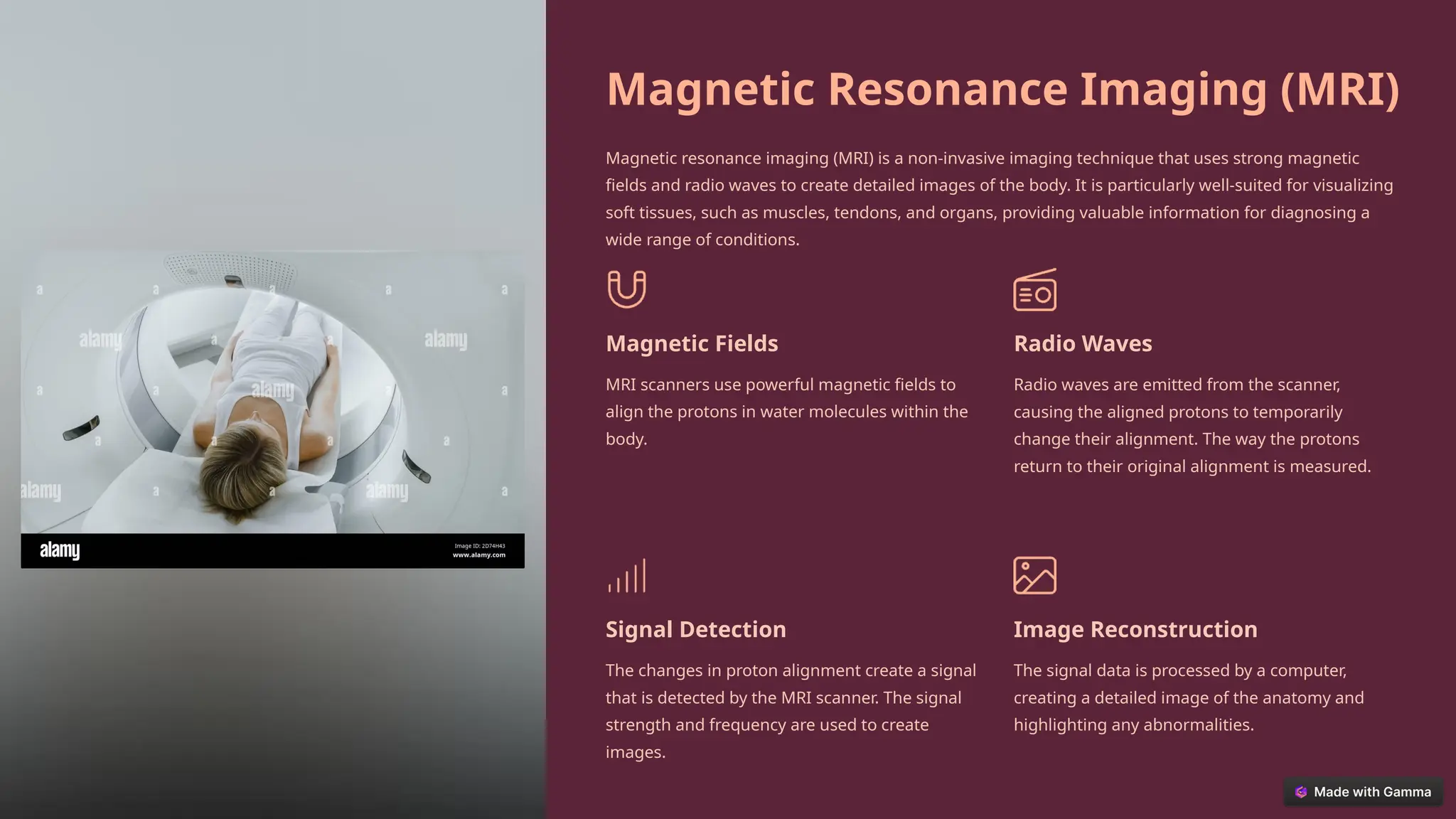
Radiology
presentation – Saurabh Sharma
Group - 418
Introduction to
Radiology and
Radiation
Diagnostics
Radiology is a branch of medicine that uses imaging techniques to diagnose and
treat diseases. Radiation diagnostics is a key component of radiology,
encompassing the use of ionizing radiation to produce images of the human body.
This field utilizes various imaging modalities, including X-rays, computed
tomography (CT), magnetic resonance imaging (MRI), and nuclear medicine
techniques, to provide valuable insights into the internal structures and functions
of the body.
SF
Principles of Radiation Physics
Radiation physics is the study of the physical properties and behaviors of radiation, encompassing both ionizing and
non-ionizing radiation. It forms the foundation of radiology and radiation diagnostics, providing the scientific
framework for understanding how radiation interacts with matter and how it can be harnessed for medical imaging
and treatment. Key principles include the nature of electromagnetic radiation, the energy levels of photons, and the
interactions of radiation with biological tissues.
1 Electromagnetic Spectrum
Understanding the different types of radiation
within the electromagnetic spectrum is essential
for radiology. This includes understanding the
energy levels of photons and their interactions
with matter.
2 Radiation Energy
The energy of radiation is measured in electron
volts (eV). Higher energy radiation, like X-rays, has
more penetrating power and is better suited for
imaging dense structures. Lower energy radiation,
like visible light, interacts primarily with the
surface of materials.
3 Radiation Doses
Radiologists must understand the different units
used to measure radiation dose, such as the gray
(Gy) and the sievert (Sv). This knowledge is
essential for ensuring patient safety and
minimizing radiation exposure during diagnostic
procedures.
4 Interaction with Matter
The way radiation interacts with matter depends
on its energy level and the composition of the
material. Understanding these interactions is
crucial for interpreting radiological images and
designing appropriate imaging techniques.
Ionizing and Non-Ionizing Radiation
Radiation is classified into two main types: ionizing and non-ionizing radiation. The distinction lies in the energy level of the radiation and its ability
to interact with matter, particularly with electrons in atoms. Ionizing radiation carries enough energy to remove electrons from atoms, creating ions,
while non-ionizing radiation does not possess sufficient energy to cause ionization.
Ionizing Radiation
Examples include X-rays, gamma rays, and
alpha and beta particles. It is used in medical
imaging, radiation therapy, and industrial
applications.
• X-rays
• Gamma rays
• Alpha particles
• Beta particles
Non-Ionizing Radiation
Examples include radio waves, microwaves,
infrared radiation, visible light, and ultraviolet
radiation. It is used in various technologies
and applications, including communication,
heating, and light.
• Radio waves
• Microwaves
• Infrared radiation
• Visible light
• Ultraviolet radiation
Applications
Radiology primarily utilizes ionizing radiation
to produce images of the human body.
However, non-ionizing radiation also plays a
role in specific imaging techniques like
magnetic resonance imaging (MRI) and
ultrasound.
Interaction of Radiation with
Matter
The interaction of radiation with matter is a complex process that depends on the energy of the
radiation and the composition of the material. Different types of interactions occur, leading to the
absorption or scattering of radiation. These interactions are fundamental to understanding how
radiation is used for imaging and treatment.
Photoelectric Effect
A photon interacts with an inner shell electron, transferring all its energy to the
electron, causing it to be ejected from the atom.
Compton Scattering
A photon interacts with an outer shell electron, losing some of its energy and
changing direction. The electron is also ejected from the atom.
Pair Production
A high-energy photon interacts with the nucleus of an atom, creating an electron and
a positron (anti-electron) pair. This occurs at energies greater than 1.022 MeV.
Imaging Modalities in Radiology
Radiology encompasses a variety of imaging modalities, each utilizing different physical principles and
techniques to produce images of the body. These modalities provide different types of information and are
chosen based on the specific clinical needs and the anatomical region of interest.
X-ray Imaging
X-ray imaging is the most basic and widely used
modality in radiology. It uses high-energy X-rays
to create images of bones, teeth, and other
dense tissues.
Computed Tomography (CT)
CT imaging uses X-rays to create detailed cross-
sectional images of the body. It is a powerful
tool for diagnosing a wide range of conditions,
including cancer, trauma, and heart disease.
Magnetic Resonance Imaging
(MRI)
MRI imaging uses strong magnetic fields and
radio waves to create detailed images of soft
tissues, including muscles, tendons, and organs.
It is particularly valuable for diagnosing brain
disorders, spinal cord injuries, and joint
problems.
Nuclear Medicine
Nuclear medicine utilizes radioactive substances
to create images of specific organs and tissues.
It is used to diagnose a variety of conditions,
including cancer, heart disease, and thyroid
disorders.
Computed Tomography (CT) Imaging
Computed tomography (CT) imaging is a powerful diagnostic tool that uses X-rays to create detailed cross-sectional images of the body. This allows for the visualization of internal
structures in three dimensions, providing valuable information about anatomy, pathology, and physiology.
1
X-ray Beam Rotation
A thin X-ray beam rotates around the patient, acquiring data from
multiple angles.
2
Data Acquisition
The X-ray detectors capture the transmitted radiation, generating
signals that are processed by a computer.
3
Image Reconstruction
The computer uses algorithms to reconstruct the data into a
series of cross-sectional images, creating a detailed 3D model of
the anatomy.
4
Image Visualization
The reconstructed images are displayed on a monitor, allowing
radiologists to analyze the anatomy, identify abnormalities, and
make diagnoses.
Magnetic Resonance Imaging (MRI)
Magnetic resonance imaging (MRI) is a non-invasive imaging technique that uses strong magnetic
fields and radio waves to create detailed images of the body. It is particularly well-suited for visualizing
soft tissues, such as muscles, tendons, and organs, providing valuable information for diagnosing a
wide range of conditions.
Magnetic Fields
MRI scanners use powerful magnetic fields to
align the protons in water molecules within the
body.
Radio Waves
Radio waves are emitted from the scanner,
causing the aligned protons to temporarily
change their alignment. The way the protons
return to their original alignment is measured.
Signal Detection
The changes in proton alignment create a signal
that is detected by the MRI scanner. The signal
strength and frequency are used to create
images.
Image Reconstruction
The signal data is processed by a computer,
creating a detailed image of the anatomy and
highlighting any abnormalities.
Positron Emission Tomography (PET)
Positron emission tomography (PET) is a nuclear medicine imaging technique that uses a radioactive tracer
to create images of metabolic activity within the body. This technique provides valuable information about
organ function and can be used to diagnose a variety of conditions, including cancer, heart disease, and
neurological disorders.
Radioactive Tracer A radioactive substance is injected into the body.
Metabolic Activity The tracer accumulates in areas of high
metabolic activity, such as tumors or areas of
inflammation.
Positron Emission The tracer emits positrons, which interact with
electrons in the body, releasing gamma rays.
Gamma Ray Detection The gamma rays are detected by the PET
scanner, which creates an image of the tracer
distribution.
Image Interpretation Radiologists analyze the image, interpreting the
areas of high tracer accumulation to identify
metabolic abnormalities.
Radiation Exposure and Safety
Considerations
Radiation exposure is a serious concern in radiology and other medical imaging procedures. Ionizing
radiation can damage cells and tissues, potentially leading to health risks. However, modern medical
imaging techniques and strict safety protocols minimize radiation exposure to patients and healthcare
professionals.
1 ALARA Principle
The ALARA principle (As Low As Reasonably
Achievable) is a fundamental principle in
radiation protection, emphasizing the need
to minimize radiation exposure to the lowest
practical level.
2 Radiation Shielding
Radiation shielding, such as lead aprons and
gloves, is used to protect individuals from
radiation exposure during medical
procedures.
3 Exposure Optimization
Medical imaging techniques are optimized to
minimize the radiation dose while providing
diagnostic quality images. This involves
adjusting exposure parameters and using
specialized imaging techniques.
4 Dose Monitoring
Radiation dose monitoring devices, such as
dosimeters, are used to track the radiation
exposure received by healthcare
professionals. This helps ensure that
exposure levels remain within acceptable
limits.
Conclusion and Future Trends
Radiology is a dynamic field that continues to evolve with technological advancements. New imaging techniques, such as advanced
MRI sequences and AI-powered image analysis, are being developed to enhance diagnostic accuracy and improve patient care. The
future of radiology is promising, with the potential for personalized medicine, more precise diagnoses, and less invasive treatment
options.

Saurabh - 418.pptxftd5d5dtdydydyftfydyfyfyf

  • 1.
  • 2.
    Introduction to Radiology and Radiation Diagnostics Radiologyis a branch of medicine that uses imaging techniques to diagnose and treat diseases. Radiation diagnostics is a key component of radiology, encompassing the use of ionizing radiation to produce images of the human body. This field utilizes various imaging modalities, including X-rays, computed tomography (CT), magnetic resonance imaging (MRI), and nuclear medicine techniques, to provide valuable insights into the internal structures and functions of the body. SF
  • 3.
    Principles of RadiationPhysics Radiation physics is the study of the physical properties and behaviors of radiation, encompassing both ionizing and non-ionizing radiation. It forms the foundation of radiology and radiation diagnostics, providing the scientific framework for understanding how radiation interacts with matter and how it can be harnessed for medical imaging and treatment. Key principles include the nature of electromagnetic radiation, the energy levels of photons, and the interactions of radiation with biological tissues. 1 Electromagnetic Spectrum Understanding the different types of radiation within the electromagnetic spectrum is essential for radiology. This includes understanding the energy levels of photons and their interactions with matter. 2 Radiation Energy The energy of radiation is measured in electron volts (eV). Higher energy radiation, like X-rays, has more penetrating power and is better suited for imaging dense structures. Lower energy radiation, like visible light, interacts primarily with the surface of materials. 3 Radiation Doses Radiologists must understand the different units used to measure radiation dose, such as the gray (Gy) and the sievert (Sv). This knowledge is essential for ensuring patient safety and minimizing radiation exposure during diagnostic procedures. 4 Interaction with Matter The way radiation interacts with matter depends on its energy level and the composition of the material. Understanding these interactions is crucial for interpreting radiological images and designing appropriate imaging techniques.
  • 4.
    Ionizing and Non-IonizingRadiation Radiation is classified into two main types: ionizing and non-ionizing radiation. The distinction lies in the energy level of the radiation and its ability to interact with matter, particularly with electrons in atoms. Ionizing radiation carries enough energy to remove electrons from atoms, creating ions, while non-ionizing radiation does not possess sufficient energy to cause ionization. Ionizing Radiation Examples include X-rays, gamma rays, and alpha and beta particles. It is used in medical imaging, radiation therapy, and industrial applications. • X-rays • Gamma rays • Alpha particles • Beta particles Non-Ionizing Radiation Examples include radio waves, microwaves, infrared radiation, visible light, and ultraviolet radiation. It is used in various technologies and applications, including communication, heating, and light. • Radio waves • Microwaves • Infrared radiation • Visible light • Ultraviolet radiation Applications Radiology primarily utilizes ionizing radiation to produce images of the human body. However, non-ionizing radiation also plays a role in specific imaging techniques like magnetic resonance imaging (MRI) and ultrasound.
  • 5.
    Interaction of Radiationwith Matter The interaction of radiation with matter is a complex process that depends on the energy of the radiation and the composition of the material. Different types of interactions occur, leading to the absorption or scattering of radiation. These interactions are fundamental to understanding how radiation is used for imaging and treatment. Photoelectric Effect A photon interacts with an inner shell electron, transferring all its energy to the electron, causing it to be ejected from the atom. Compton Scattering A photon interacts with an outer shell electron, losing some of its energy and changing direction. The electron is also ejected from the atom. Pair Production A high-energy photon interacts with the nucleus of an atom, creating an electron and a positron (anti-electron) pair. This occurs at energies greater than 1.022 MeV.
  • 6.
    Imaging Modalities inRadiology Radiology encompasses a variety of imaging modalities, each utilizing different physical principles and techniques to produce images of the body. These modalities provide different types of information and are chosen based on the specific clinical needs and the anatomical region of interest. X-ray Imaging X-ray imaging is the most basic and widely used modality in radiology. It uses high-energy X-rays to create images of bones, teeth, and other dense tissues. Computed Tomography (CT) CT imaging uses X-rays to create detailed cross- sectional images of the body. It is a powerful tool for diagnosing a wide range of conditions, including cancer, trauma, and heart disease. Magnetic Resonance Imaging (MRI) MRI imaging uses strong magnetic fields and radio waves to create detailed images of soft tissues, including muscles, tendons, and organs. It is particularly valuable for diagnosing brain disorders, spinal cord injuries, and joint problems. Nuclear Medicine Nuclear medicine utilizes radioactive substances to create images of specific organs and tissues. It is used to diagnose a variety of conditions, including cancer, heart disease, and thyroid disorders.
  • 7.
    Computed Tomography (CT)Imaging Computed tomography (CT) imaging is a powerful diagnostic tool that uses X-rays to create detailed cross-sectional images of the body. This allows for the visualization of internal structures in three dimensions, providing valuable information about anatomy, pathology, and physiology. 1 X-ray Beam Rotation A thin X-ray beam rotates around the patient, acquiring data from multiple angles. 2 Data Acquisition The X-ray detectors capture the transmitted radiation, generating signals that are processed by a computer. 3 Image Reconstruction The computer uses algorithms to reconstruct the data into a series of cross-sectional images, creating a detailed 3D model of the anatomy. 4 Image Visualization The reconstructed images are displayed on a monitor, allowing radiologists to analyze the anatomy, identify abnormalities, and make diagnoses.
  • 8.
    Magnetic Resonance Imaging(MRI) Magnetic resonance imaging (MRI) is a non-invasive imaging technique that uses strong magnetic fields and radio waves to create detailed images of the body. It is particularly well-suited for visualizing soft tissues, such as muscles, tendons, and organs, providing valuable information for diagnosing a wide range of conditions. Magnetic Fields MRI scanners use powerful magnetic fields to align the protons in water molecules within the body. Radio Waves Radio waves are emitted from the scanner, causing the aligned protons to temporarily change their alignment. The way the protons return to their original alignment is measured. Signal Detection The changes in proton alignment create a signal that is detected by the MRI scanner. The signal strength and frequency are used to create images. Image Reconstruction The signal data is processed by a computer, creating a detailed image of the anatomy and highlighting any abnormalities.
  • 9.
    Positron Emission Tomography(PET) Positron emission tomography (PET) is a nuclear medicine imaging technique that uses a radioactive tracer to create images of metabolic activity within the body. This technique provides valuable information about organ function and can be used to diagnose a variety of conditions, including cancer, heart disease, and neurological disorders. Radioactive Tracer A radioactive substance is injected into the body. Metabolic Activity The tracer accumulates in areas of high metabolic activity, such as tumors or areas of inflammation. Positron Emission The tracer emits positrons, which interact with electrons in the body, releasing gamma rays. Gamma Ray Detection The gamma rays are detected by the PET scanner, which creates an image of the tracer distribution. Image Interpretation Radiologists analyze the image, interpreting the areas of high tracer accumulation to identify metabolic abnormalities.
  • 10.
    Radiation Exposure andSafety Considerations Radiation exposure is a serious concern in radiology and other medical imaging procedures. Ionizing radiation can damage cells and tissues, potentially leading to health risks. However, modern medical imaging techniques and strict safety protocols minimize radiation exposure to patients and healthcare professionals. 1 ALARA Principle The ALARA principle (As Low As Reasonably Achievable) is a fundamental principle in radiation protection, emphasizing the need to minimize radiation exposure to the lowest practical level. 2 Radiation Shielding Radiation shielding, such as lead aprons and gloves, is used to protect individuals from radiation exposure during medical procedures. 3 Exposure Optimization Medical imaging techniques are optimized to minimize the radiation dose while providing diagnostic quality images. This involves adjusting exposure parameters and using specialized imaging techniques. 4 Dose Monitoring Radiation dose monitoring devices, such as dosimeters, are used to track the radiation exposure received by healthcare professionals. This helps ensure that exposure levels remain within acceptable limits.
  • 11.
    Conclusion and FutureTrends Radiology is a dynamic field that continues to evolve with technological advancements. New imaging techniques, such as advanced MRI sequences and AI-powered image analysis, are being developed to enhance diagnostic accuracy and improve patient care. The future of radiology is promising, with the potential for personalized medicine, more precise diagnoses, and less invasive treatment options.