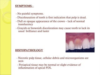
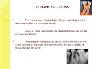
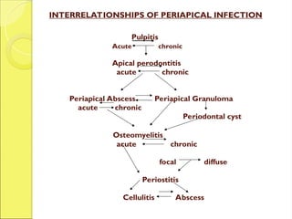
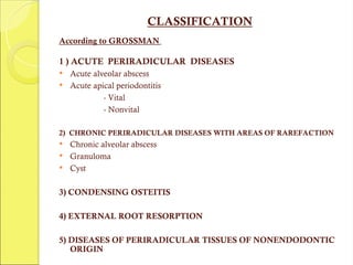
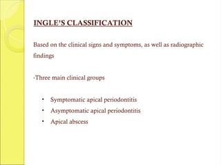
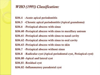
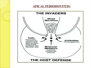
SYMPTOMS :
- No painful symptoms.
- Discolouration of tooth is first indication that pulp is dead.
- Dull or opaque appearance of the crown – lack of normal
translucency
- Grayish or brownish discoloration may cause tooth to lack its
usual brilliance and luster
HISTOPATHOLOGY
- Necrotic pulp tissue, cellular debris and microorganisms are
seen
- Periapical tissue may be normal or slight evidence of
inflammation of apical PDL
POISONOUS INTERMEDIATE & END PRODUCTS
FOUND IN NECROSIS
1. Intermediate proteolytic products that emit foul odour
a. Indole and Skatole
b. Putriscine and Cadaverine
c. Indican
2. End products
Hydrogen sulphide, ammonia, water, carbon dioxide &
fatty acids
3. Exotoxins
4. Endotoxins
5. Foreign bacterial protein
DIAGNOSIS
- Pain is absent with total necrosis
 Swelling - negative
 Mobility - negative
 Tenderness to percusion - negative
 Radiographic findings are normal except if there is apical
periodontitis.
 No response to vitality tests.
 Sometimes positive electric test as result of liquefaction necrosis.
 Discoloration - as result of haemolysis of RBC s or
decomposition of pulp tissue (grey/ brown)
TREATMENT - Root Canal Therapy
PERIAPICAL LESIONS
 As a consequence of pathologic changes in dental pulp, the
root canal can harbor numerous irritants.
 Egress of these irritants into the periapical tissues can initiate
periradicular lesions.
 Depending on the nature and quality of these irritants as well
as the duration of exposure of the periradicular tissues, a variety of
tissue changes can occur.
Pulpitis
Acute chronic
Apical perodontitis
acute chronic
Periapical Abscess Periapical Granuloma
acute chronic
Periodontal cyst
Osteomyelitis
acute chronic
focal diffuse
Periostitis
Cellulitis Abscess
INTERRELATIONSHIPS OF PERIAPICAL INFECTION
CLASSIFICATION
According to GROSSMAN
1 ) ACUTE PERIRADICULAR DISEASES
 Acute alveolar abscess
 Acute apical periodontitis
- Vital
- Nonvital
2) CHRONIC PERIRADICULAR DISEASES WITH AREAS OF RAREFACTION
 Chronic alveolar abscess
 Granuloma
 Cyst
3) CONDENSING OSTEITIS
4) EXTERNAL ROOT RESORPTION
5) DISEASES OF PERIRADICULAR TISSUES OF NONENDODONTIC
ORIGIN
INGLE’S CLASSIFICATION
Based on the clinical signs and symptoms, as well as radiographic
findings
-Three main clinical groups
• Symptomatic apical periodontitis
• Asymptomatic apical periodontitis
• Apical abscess
WEINE’S CLASSIFICATION
PULPOPERIAPICAL DISEASE:
1] PAINFUL PULPOPERIAPICAL PATHOSES
a) Incipient acute periapical periodontitis
b) Advanced acute periapical periodontitis
i. Acute periapical abscess
ii. Recrudescent abscess
iii. Subacute periapical abscess
2] NON PAINFUL PULPOPERIAPICAL PATHOSES
a) Pulpoperiapical osteosclerosis
b) Incipient chronic periapical periodontitis
c) Advanced chronic periapical periodontitis
i. Periapical granuloma
ii. Chronic periapical abscess
iii. Periapical cyst
WHO (1995) Classification:
K04.4 -Acute apical periodontitis
K04.5 -Chronic apical periodontitis (Apical granuloma)
K04.6 -Periapical abscess with sinus
K04.60 -Periapical abscess with sinus to maxillary antrum
K04.61 -Periapical abscess with sinus to nasal cavity
K04.62 -Periapical abscess with sinus to oral cavity
K04.63 -Periapical abscess with sinus to skin
K04.7 -Periapical abscess without sinus
K04.8 -Radicular cyst (Apical periodontal cyst, Periapical cyst)
K04.80 -Apical and lateral cyst
K04.81 -Residual cyst
K04.82 -Inflammatory paradental cyst
ACUTE ALVEOLAR ABSCESS
(Synonyms: Acute periapical abscess, Acute dentoalveolar abscess )
Definition
Localised collection of pus in the alveolar bone at the root apex of
tooth following death of pulp, with extension of infection through
apical foramen into periradicular tissues.
Etiology
Bacterial invasion , trauma, chemical or mechanical irritation.
HISTOPATHOLOGY
HISTOPATHOLOGY
PMN’S infiltration
Accumulation of inflammatory exudates
Distention of PDL
Prosses continues, PDL separates
Mobile tooth
Bone resorption at apex
Liquefaction necrosis containing PMN’S ,debris,
cell remnants & purulent exudates
SYMPTOMS
 First symptom - tenderness
 Later - patient has severe throbbing pain with swelling of the
overlying soft tissue
 As the infection progresses, swelling becomes more pronounced
and extends beyond the original site
 Tooth becomes more painful, elongated and mobile
 If untreated - progresses to osteitis, periostitis, cellulitis
or osteomyelitis
 Sinus tract - opens to buccal mucosa
 When Maxillary anterior teeth - swelling of upper lip
and chin (extend both eyelids)
 When Maxillary posterior teeth - the Cheek may
swell.
 Mandibular posterior teeth- swelling extends around
border of jaw into submaxillary region or ear.
Gutta-percha is placed in the sinus tract - points to involved tooth:
SINUS TRACT TRACING
SINUS TRACT TRACING
 General Systemic reaction is seen ( Septic products)
 Patient appears - Pale, Irritable & Weakened from pain and
loss of sleep
DIAGNOSIS
 Early stage - difficult to locate tooth.
 Once infection progresses to process of periodontitis & extrusion, a
radiographic evaluation shows thickening of periodontal ligament
space & breakdown of bone
 Electric test & thermal tests: No response
 Tooth is tender on percussion
 Apical mucosa is tender on palpation
 Tooth may be mobile & extruded.
DIFFERENTIAL DIAGNOSIS
Periodontal abscess & Irreversible pulpitis
Treatment:
Establish drainage & control systemic infection.
ON FIRST VISIT : Tooth is left open for drainage.
Thorough instrumentation & irrigation
before medicating and sealing.
Once the swelling and pain subsides
endodontic treatment is done.
If diffuse swelling : Antibiotic coverage is
prescribed along with hot mouth rinse, Once area
is localised, incision and drainage is instituted
ACUTE APICAL PERIODONTITIS
A painful inflammation of periodontium as a result of trauma, irritation
or infection through root canal regardless of pulp is vital or non-vital.
HISTOPATHOLOGIC CLASSIFICATION: (P.N.R Nair)
1) Acute apical periodontitis ( PMN’s) : Primary & secondary
2) Chronic apical periodontitis ( Lymphocytes, macrophages, plasma cells)
3) Cystic apical periodontitis - True cyst, Pocket cyst (bay cyst)
APICAL PERIODONTITIS:
ETIOLOGY
 Occlusal trauma
 Wedging of foreign object between teeth
 Non-vital tooth as a sequelae to pulpal dieseases.
 Iatrogenic: during over instrumentation & extrusion of irritating
medicaments
 Perforation of root
SYMPTOMS
- Pain and tenderness
- Tooth may be slightly sore, when percussed.
HISTOPATHOLOGY
 Inflammatory reaction in apical periodontal ligament
 Blood vessels are dilated, PMNL’s are present
 Cholestrol clefts are the commom finding.
 Accumulation of serous exudate distends the periodontal
ligament
 Osteoclasts are seen.
DIAGNOSIS
 Tooth is tender to percussion
 Symptoms are due to Overinstrumentation, Irritating medicament or
Overfilling
 Radiographically: thickened periodontal ligament or small area of
rarefaction
TREATMENT
 Endodontic therapy
 Postoperative pain is controlled analgesics & antibiotics
 Hyper-occlusion relieve the occlusion.
ACUTE EXACERBATION OF A CHRONIC LESION
(PHOENIX ABSCESS)
An acute inflammatory reaction superimposed on an existing chronic
lesion, such as a cyst or granuloma
ETIOLOGY
1. Noxious stimuli from a diseased pulp with chronic periradicular
disease.
2. Because of influx of necrotic products or bacteria and their toxins, the
dormant lesions may become reactive & cause an acute inflammatory
response.
3. Lowering of the body's defenses in the presence of bacteria - may also
trigger an acute inflammatory response.
4. Mechanical irritation during root canal instrumentation
SYMPTOMS
 Tooth - tender to touch & elevated in its socket
 Mucosa - sensitive to palpation & appears red & swollen
HISTOPATHOLOGY :
Liquefaction necrosis with disintegrating PMNL & cellular debris
(pus), surrounded by infiltration of macrophages, lymphocytes &
plasma cells
DIAGNOSIS
History of patient
vitality tests Lack of response
RADIOGRAPHICALLY :- Large area of radiolucency at the apex.
DIFFERENTIAL DIAGNOSIS :
Acute alveolar abscess, Acute irreversible pulpitis
TREATMENT
 Establishment of drainage
 Once symptoms subside - RCT
CHRONIC ALVEOLAR ABSCESS
(Chronic Suppurative Apical Periodontitis)
DEFINITION
A chronic alveolar abscess is a long-standing, low-
grade infection of the periradicular alveolar bone.
ETIOLOGY
1. Death of the pulp with extension of the infective process
periapically
2. A pre-existing acute abscess
SIGNS & SYMPTOMS
- Asymptomatic
- Detected either by the presence
of sinus tract or on routine
radiograph.
- If the sinus tract drainage becomes blocked – pain & swelling
- Range of sensitivity to percussion & palpation – depends on
the sinus tract is open, draining or closed .
A radiograph with a gutta-percha cone into the sinus tract
often shows involved tooth by tracing the sinus tract to its
origin.

Pulp and periapical pathology presentation part 3

  • 1.
    SYMPTOMS : - Nopainful symptoms. - Discolouration of tooth is first indication that pulp is dead. - Dull or opaque appearance of the crown – lack of normal translucency - Grayish or brownish discoloration may cause tooth to lack its usual brilliance and luster HISTOPATHOLOGY - Necrotic pulp tissue, cellular debris and microorganisms are seen - Periapical tissue may be normal or slight evidence of inflammation of apical PDL
  • 2.
    POISONOUS INTERMEDIATE &END PRODUCTS FOUND IN NECROSIS 1. Intermediate proteolytic products that emit foul odour a. Indole and Skatole b. Putriscine and Cadaverine c. Indican 2. End products Hydrogen sulphide, ammonia, water, carbon dioxide & fatty acids 3. Exotoxins 4. Endotoxins 5. Foreign bacterial protein
  • 3.
    DIAGNOSIS - Pain isabsent with total necrosis  Swelling - negative  Mobility - negative  Tenderness to percusion - negative  Radiographic findings are normal except if there is apical periodontitis.  No response to vitality tests.  Sometimes positive electric test as result of liquefaction necrosis.  Discoloration - as result of haemolysis of RBC s or decomposition of pulp tissue (grey/ brown) TREATMENT - Root Canal Therapy
  • 4.
    PERIAPICAL LESIONS  Asa consequence of pathologic changes in dental pulp, the root canal can harbor numerous irritants.  Egress of these irritants into the periapical tissues can initiate periradicular lesions.  Depending on the nature and quality of these irritants as well as the duration of exposure of the periradicular tissues, a variety of tissue changes can occur.
  • 5.
    Pulpitis Acute chronic Apical perodontitis acutechronic Periapical Abscess Periapical Granuloma acute chronic Periodontal cyst Osteomyelitis acute chronic focal diffuse Periostitis Cellulitis Abscess INTERRELATIONSHIPS OF PERIAPICAL INFECTION
  • 6.
    CLASSIFICATION According to GROSSMAN 1) ACUTE PERIRADICULAR DISEASES  Acute alveolar abscess  Acute apical periodontitis - Vital - Nonvital 2) CHRONIC PERIRADICULAR DISEASES WITH AREAS OF RAREFACTION  Chronic alveolar abscess  Granuloma  Cyst 3) CONDENSING OSTEITIS 4) EXTERNAL ROOT RESORPTION 5) DISEASES OF PERIRADICULAR TISSUES OF NONENDODONTIC ORIGIN
  • 7.
    INGLE’S CLASSIFICATION Based onthe clinical signs and symptoms, as well as radiographic findings -Three main clinical groups • Symptomatic apical periodontitis • Asymptomatic apical periodontitis • Apical abscess
  • 8.
    WEINE’S CLASSIFICATION PULPOPERIAPICAL DISEASE: 1]PAINFUL PULPOPERIAPICAL PATHOSES a) Incipient acute periapical periodontitis b) Advanced acute periapical periodontitis i. Acute periapical abscess ii. Recrudescent abscess iii. Subacute periapical abscess 2] NON PAINFUL PULPOPERIAPICAL PATHOSES a) Pulpoperiapical osteosclerosis b) Incipient chronic periapical periodontitis c) Advanced chronic periapical periodontitis i. Periapical granuloma ii. Chronic periapical abscess iii. Periapical cyst
  • 9.
    WHO (1995) Classification: K04.4-Acute apical periodontitis K04.5 -Chronic apical periodontitis (Apical granuloma) K04.6 -Periapical abscess with sinus K04.60 -Periapical abscess with sinus to maxillary antrum K04.61 -Periapical abscess with sinus to nasal cavity K04.62 -Periapical abscess with sinus to oral cavity K04.63 -Periapical abscess with sinus to skin K04.7 -Periapical abscess without sinus K04.8 -Radicular cyst (Apical periodontal cyst, Periapical cyst) K04.80 -Apical and lateral cyst K04.81 -Residual cyst K04.82 -Inflammatory paradental cyst
  • 10.
    ACUTE ALVEOLAR ABSCESS (Synonyms:Acute periapical abscess, Acute dentoalveolar abscess ) Definition Localised collection of pus in the alveolar bone at the root apex of tooth following death of pulp, with extension of infection through apical foramen into periradicular tissues. Etiology Bacterial invasion , trauma, chemical or mechanical irritation.
  • 11.
    HISTOPATHOLOGY HISTOPATHOLOGY PMN’S infiltration Accumulation ofinflammatory exudates Distention of PDL Prosses continues, PDL separates Mobile tooth Bone resorption at apex Liquefaction necrosis containing PMN’S ,debris, cell remnants & purulent exudates
  • 12.
    SYMPTOMS  First symptom- tenderness  Later - patient has severe throbbing pain with swelling of the overlying soft tissue  As the infection progresses, swelling becomes more pronounced and extends beyond the original site  Tooth becomes more painful, elongated and mobile  If untreated - progresses to osteitis, periostitis, cellulitis or osteomyelitis  Sinus tract - opens to buccal mucosa
  • 13.
     When Maxillaryanterior teeth - swelling of upper lip and chin (extend both eyelids)  When Maxillary posterior teeth - the Cheek may swell.  Mandibular posterior teeth- swelling extends around border of jaw into submaxillary region or ear.
  • 14.
    Gutta-percha is placedin the sinus tract - points to involved tooth: SINUS TRACT TRACING SINUS TRACT TRACING  General Systemic reaction is seen ( Septic products)  Patient appears - Pale, Irritable & Weakened from pain and loss of sleep
  • 15.
    DIAGNOSIS  Early stage- difficult to locate tooth.  Once infection progresses to process of periodontitis & extrusion, a radiographic evaluation shows thickening of periodontal ligament space & breakdown of bone  Electric test & thermal tests: No response  Tooth is tender on percussion  Apical mucosa is tender on palpation  Tooth may be mobile & extruded. DIFFERENTIAL DIAGNOSIS Periodontal abscess & Irreversible pulpitis
  • 16.
    Treatment: Establish drainage &control systemic infection. ON FIRST VISIT : Tooth is left open for drainage. Thorough instrumentation & irrigation before medicating and sealing. Once the swelling and pain subsides endodontic treatment is done. If diffuse swelling : Antibiotic coverage is prescribed along with hot mouth rinse, Once area is localised, incision and drainage is instituted
  • 17.
    ACUTE APICAL PERIODONTITIS Apainful inflammation of periodontium as a result of trauma, irritation or infection through root canal regardless of pulp is vital or non-vital. HISTOPATHOLOGIC CLASSIFICATION: (P.N.R Nair) 1) Acute apical periodontitis ( PMN’s) : Primary & secondary 2) Chronic apical periodontitis ( Lymphocytes, macrophages, plasma cells) 3) Cystic apical periodontitis - True cyst, Pocket cyst (bay cyst)
  • 18.
  • 19.
    ETIOLOGY  Occlusal trauma Wedging of foreign object between teeth  Non-vital tooth as a sequelae to pulpal dieseases.  Iatrogenic: during over instrumentation & extrusion of irritating medicaments  Perforation of root SYMPTOMS - Pain and tenderness - Tooth may be slightly sore, when percussed. HISTOPATHOLOGY  Inflammatory reaction in apical periodontal ligament  Blood vessels are dilated, PMNL’s are present  Cholestrol clefts are the commom finding.  Accumulation of serous exudate distends the periodontal ligament  Osteoclasts are seen.
  • 20.
    DIAGNOSIS  Tooth istender to percussion  Symptoms are due to Overinstrumentation, Irritating medicament or Overfilling  Radiographically: thickened periodontal ligament or small area of rarefaction TREATMENT  Endodontic therapy  Postoperative pain is controlled analgesics & antibiotics  Hyper-occlusion relieve the occlusion.
  • 21.
    ACUTE EXACERBATION OFA CHRONIC LESION (PHOENIX ABSCESS) An acute inflammatory reaction superimposed on an existing chronic lesion, such as a cyst or granuloma ETIOLOGY 1. Noxious stimuli from a diseased pulp with chronic periradicular disease. 2. Because of influx of necrotic products or bacteria and their toxins, the dormant lesions may become reactive & cause an acute inflammatory response. 3. Lowering of the body's defenses in the presence of bacteria - may also trigger an acute inflammatory response. 4. Mechanical irritation during root canal instrumentation SYMPTOMS  Tooth - tender to touch & elevated in its socket  Mucosa - sensitive to palpation & appears red & swollen
  • 22.
    HISTOPATHOLOGY : Liquefaction necrosiswith disintegrating PMNL & cellular debris (pus), surrounded by infiltration of macrophages, lymphocytes & plasma cells DIAGNOSIS History of patient vitality tests Lack of response RADIOGRAPHICALLY :- Large area of radiolucency at the apex.
  • 23.
    DIFFERENTIAL DIAGNOSIS : Acutealveolar abscess, Acute irreversible pulpitis TREATMENT  Establishment of drainage  Once symptoms subside - RCT
  • 24.
    CHRONIC ALVEOLAR ABSCESS CHRONICALVEOLAR ABSCESS (Chronic Suppurative Apical Periodontitis) (Chronic Suppurative Apical Periodontitis) DEFINITION A chronic alveolar abscess is a long-standing, low- grade infection of the periradicular alveolar bone. ETIOLOGY 1. Death of the pulp with extension of the infective process periapically 2. A pre-existing acute abscess
  • 25.
    SIGNS & SYMPTOMS -Asymptomatic - Detected either by the presence of sinus tract or on routine radiograph. - If the sinus tract drainage becomes blocked – pain & swelling - Range of sensitivity to percussion & palpation – depends on the sinus tract is open, draining or closed . A radiograph with a gutta-percha cone into the sinus tract often shows involved tooth by tracing the sinus tract to its origin.