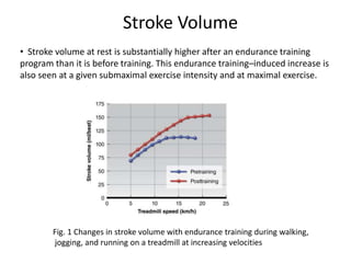
CENTRE FOR PHYSIOTHERAPY AND REHABILITATION SCIENCES
JAMIA MILLIA ISLAMIA
Presented By-
Purnima kushwaha
MPT-Cardiopulmonary (3rd semester)
Roll no. -18MPC003
Topic- Potential Mechanism of exercise benefits during
Cardiac Rehabilitation
Exercise Benefits By Following
Adaptation
• Cardiovascular Adaptation
• Respiratory Adaptation
• Muscle Adaptation
• Metabolic Adaptation
Cardiovascular Adaptation
• Multiple cardiovascular adaptations occurs in response to
exercise training, including changes in the following:
 Heart size
 Stroke volume
 Heart rate
 Cardiac output
 Blood flow
 Blood pressure
 Blood volume
Oxygen Transport System
• The ability of the cardiovascular and respiratory systems to deliver oxygen
to active tissues is defined by the Fick Equation, which states that whole-
body oxygen consumption is determined by both the delivery of oxygen
via blood flow (cardiac output) and the amount of oxygen extracted by
the tissues, the (a-v) O2 difference. The product of cardiac output and the
(a-v¯)O2 difference determines the rate at which oxygen is being
consumed:
V.O2 = stroke volume x heart rate x(a-v¯)O2 diff
and
V.O2 max = maximal stroke volume x maximal heart rate x maximal (a-
v¯)O2 diff.
• Because HRmax either stays the same or decreases slightly with training,
increases in V. O2 max depend on adaptations in maximal stroke volume
and maximal (a-v¯)O2 difference.
Heart size
• As an adaptation to the increased work demand, cardiac muscle mass and
ventricular volume increase with training. Cardiac muscle, like skeletal
muscle, undergoes morphological adaptations as a result of chronic
endurance training (Fagard, R.H. 1996, Milliken et al., 1988)
• The type of ventricular adaptation depends on the type of exercise training
performed.
• With endurance training, left ventricular chamber size increases. This
allows for increased left ventricular filling and consequently an increase in
stroke volume.
• The increases in plasma volume and diastolic filling time increase left
ventricular chamber size at the end of diastole. This effect of endurance
training on the left ventricle is often called a volume loading effect.
Stroke Volume
• Stroke volume at rest is substantially higher after an endurance training
program than it is before training. This endurance training–induced increase is
also seen at a given submaximal exercise intensity and at maximal exercise.
Fig. 1 Changes in stroke volume with endurance training during walking,
jogging, and running on a treadmill at increasing velocities
Table 1 Stroke Volumes at Rest (SV rest) and During Maximal Exercise (SV
max) for Different States of Training
• After aerobic training, the left ventricle fills more completely during
diastole. Plasma volume expands with training, which allows for more
blood to enter the ventricle during diastole, increasing end-diastolic
volume (EDV). The heart rate of a trained heart is also lower at rest and at
the same absolute exercise intensity than that of an untrained heart,
allowing more time for the increased diastolic filling. More blood entering
the ventricle increases the stretch on the ventricular walls; by the Frank-
Starling mechanism, this results in an increased force of contraction.
Subjects SV rest (ml/beat) SV max (ml/beat)
Untrained 50-70 80-110
Trained 70-90 110-150
Highly trained 90- 110 150-220+
• The thickness of the posterior and septal walls of the left ventricle also
increases slightly with endurance training. Increased ventricular muscle
mass results in increased contractile force, in turn causing a lower end-
systolic volume.
• The decrease in end-systolic volume is facilitated by the decrease in
peripheral resistance that occurs with training. Increased contractility
resulting from an increase in left ventricular thickness and greater diastolic
filling (Frank-Starling mechanism), coupled with the reduction in systemic
peripheral resistance, increases the ejection fraction [equal to (EDV – ESV)/
EDV] in the trained heart. More blood enters the left ventricle, and a greater
percentage of what enters is forced out with each contraction, resulting in
an increase in stroke volume. (Ehsani et al., 1991)
Heart rate
• Aerobic training has a major impact on heart rate at rest, during
submaximal exercise, and during the post exercise recovery period.
The effect of aerobic training on maximal heart rate is rather
negligible.
• Resting Heart Rate: Resting heart rate decreases markedly as a
result of endurance training. The actual mechanisms responsible for
this decrease are not entirely understood, but training appears to
increase parasympathetic activity in the heart while decreasing
sympathetic activity.
• Submaximal Heart Rate: During submaximal exercise, aerobic
training results in a lower heart rate at any given absolute exercise
intensity. The training-induced decrease in heart rate is typically
greater at higher intensities.
• Maximum Heart Rate : A person’s maximal heart rate (HRmax) tends to be stable
and typically remains relatively unchanged after endurance training.
• Heart Rate Recovery : When the exercise bout is finished, heart rate does not
instantly return to its resting level. Instead, it remains elevated for a while,
slowly returning to its resting rate. The time it takes for heart rate to return to its
resting rate is called the heart rate recovery period.
• After endurance training, as shown in figure 2,heart rate returns to its resting
level much more quickly after an exercise bout than it does before training.
This is true after both submaximal and maximal exercise.
Figure 2 Changes in heart rate during recovery after a 4 min, all-out bout
of exercise before and after endurance training.
Cardiac output
• Cardiac output at rest and during submaximal exercise at a given exercise intensity
does not change much following endurance training. In fact, cardiac output can
decrease slightly. This is likely the result of an increase in the (a-v¯)O2 difference
(reflecting greater oxygen extraction by the tissues) or a decrease in the rate of
oxygen consumption (reflecting an increased mechanical efficiency). Generally,
cardiac output matches the oxygen consumption required for any given intensity of
effort.
• Maximal cardiac output, however, increases considerably in response to aerobic
training, as seen in figure 3, and is largely responsible for the increase in V.O2max.
This increase in cardiac output must result from an increase in maximal stroke
volume, because HRmax changes little, if any.
Figure 3 Changes in cardiac output with
endurance training during walking then
jogging, and finally running on a
treadmill as velocity increases
Blood flow
• With endurance training, the cardiovascular system adapts to increase
blood flow to exercising muscles to meet their higher demand for oxygen
and metabolic substrates. Four factors account for this enhanced blood
flow to muscle following training:
 Increased capillarization (Hermansen, L., & Wachtlova, M. 1971).
 Greater recruitment of existing capillaries
 More effective blood flow redistribution from inactive regions
(Armstrong, R.B., & Laughlin, M.H. 1984).
 Increased total blood volume
Blood Pressure
• Resting blood pressure does not change significantly in healthy subjects in
response to endurance training, but some studies have shown modest
reductions after training in borderline or moderately hypertensive
individuals. Reductions in both systolic and diastolic blood pressure of
approximately 6 to 7 mmHg may result in hypertensive subjects.
• The mechanisms underlying this reduction are unknown. Following
endurance training, blood pressure is reduced at a given submaximal
exercise intensity; but at maximal exercise capacity, systolic blood
pressure is increased and diastolic pressure is decreased.
Blood volume
• Endurance training increases total blood volume, and this effect is larger at
higher training intensities. Furthermore, the effect occurs rapidly. This
increased blood volume results primarily from an increase in plasma
volume, but there is also an increase in the volume of red blood cells. The
time course and mechanism for the increase of each of these components
of blood are quite different( Sawka et al., 2000)
• Plasma Volume The increase in plasma volume with training is thought to
result from two mechanisms. The first mechanism, which has two phases,
results in increases in plasma proteins, particularly albumin. As plasma
protein concentration increases, so does oncotic pressure, and fluid is
reabsorbed from the interstitial fluid into the blood vessels.
• During an intense bout of exercise, proteins leave the vascular space and
move into the interstitial space. They are then returned in greater
amounts through the lymph system. It is likely that the first phase of rapid
plasma volume increase is the result of the increased plasma albumin,
which is noted within the first hour of recovery from the first training bout.
In the second phase, protein synthesis is turned on (upregulated) by
repeated exercise, and new proteins are formed .
• With the second mechanism, exercise increases the release of antidiuretic
hormone and aldosterone, hormones that cause reabsorption of water and
sodium in the kidneys, which increases blood plasma. That increased fluid is
kept in the vascular space by the oncotic pressure exerted by the proteins.
Nearly all of the increase in blood volume during the first two weeks of
training can be explained by the increase in plasma volume.
• Red Blood Cells An increase in red blood cell volume with endurance
training also contributes to the overall increase in blood volume, but this is
an inconsistent finding. Although the actual number of red blood cells may
increase, the hematocrit—the ratio of the red blood cell volume to the total
blood volume—may actually decrease.
• The increased ratio of plasma to cells resulting from a greater increase in
the fluid portion reduces the blood’s viscosity, or thickness. Reduced
viscosity may aid the smooth flow of blood through the blood vessels,
particularly through the smaller vessels such as the capillaries. One of the
physiological benefits of decreasing blood viscosity is that it enhances
oxygen delivery to the active muscle mass.
Respiratory Adaptation
• As with the cardiovascular system, the respiratory system undergoes
specific adaptations to endurance training to maximize its efficiency.
• Pulmonary Ventilation After training, pulmonary ventilation is essentially
unchanged at rest. Although endurance training does not change the
structure or basic physiology of the lung, it does decrease ventilation
during submaximal exercise by as much as 20% to 30% at a given
submaximal intensity.
• Maximal pulmonary ventilation is substantially increased from a rate of
about 100 to 120 L/min in untrained sedentary individuals to about 130 to
150 L/ min or more following endurance training. Pulmonary ventilation
rates typically increase to about 180 L/ min in highly trained athletes and
can exceed 200 L/ min in very large, highly trained endurance athletes.
• Two factors can account for the increase in maximal pulmonary
ventilation following training: increased tidal volume and increased
respiratory frequency at maximal exercise.
• Pulmonary Diffusion Pulmonary diffusion, or gas exchange occurring in the
alveoli, is unaltered at rest and during submaximal exercise following
training. However, it increases at maximal exercise intensity.
• Pulmonary blood flow (blood coming from the right side of the heart to the
lungs) increases following training, particularly flow to the upper regions of
the lungs when a person is sitting or standing. This increases lung perfusion.
More blood is brought into the lungs for gas exchange, and at the same time
ventilation increases so that more air is brought into the lungs. This means
that more alveoli will be involved in pulmonary diffusion.
• The net result is that pulmonary diffusion increases.
• Arterial–Venous Oxygen Difference The oxygen content of arterial blood
changes very little with endurance training. Even though total hemoglobin
is increased, the amount of hemoglobin per unit of blood is the same or
even slightly reduced. The (a-v¯)O2 difference, however, does increase
with training, particularly at maximal exercise intensity.
• This increase results from a lower mixed venous oxygen content, which
means that the blood returning to the heart (which is a mixture of venous
blood from all body parts, not just the active tissues) contains less oxygen
than it would in an untrained person. This reflects both greater oxygen
extraction by active tissues and a more effective distribution of blood flow
to active tissues.
Muscle Adaptation
• Repeated excitation and contraction of muscle fibers during endurance
training stimulate changes in their structure and function.
• Muscle Fiber Type : low- to moderate-intensity aerobic activities rely
extensively on type I (slow twitch) fibers. In response to aerobic training,
type I fibers become larger. More specifically, they develop a larger cross-
sectional area, although the magnitude of change depends on the
intensity and duration of each training bout and the length of the training
program. Increases in cross-sectional area of up to 25% have been
reported.
• Fast-twitch (type II) fibers, because they are not being recruited to the
same extent during endurance exercise, generally do not increase cross
sectional area.
• Capillary Supply: One of the most important adaptations to aerobic
training is an increase in the number of capillaries surrounding each
muscle fiber.
• With long periods of aerobic training, the number of capillaries may
increase by more than 15%(Rico-Sanz et al., 2003). Having more
capillaries allows for greater exchange of gases, heat, nutrients, and
metabolic by-products between the blood and contracting muscle fibers.
• In fact, the increase in capillary density (i.e., increase in capillaries per
muscle fiber) is potentially one of the most important alterations in
response to training that causes the increase in V. O2max.
• Myoglobin Content: When oxygen enters the muscle fiber, it binds to
myoglobin, a molecule similar to hemoglobin. This iron containing
molecule shuttles the oxygen molecules from the cell membrane to the
mitochondria.
• Myoglobin transports oxygen and releases it to the mitochondria when
oxygen becomes limited during muscle action.
• Endurance training has been shown to increase muscle myoglobin
content by 75% to 80%. This adaptation clearly supports a muscle’s
increased capacity for oxidative metabolism after training.
• Mitochondrial Function: oxidative energy production takes place in the
mitochondria. Not surprisingly, then, aerobic training also induces changes
in mitochondrial function that improve the muscle fibers’ capacity to
produce ATP.
• The ability to use oxygen and produce ATP via oxidation depends on the
number and size of the muscle mitochondria. Both increase with aerobic
training(Holloszy et al., 1971)
• Oxidative Enzymes: These changes are further enhanced by an increase in
mitochondrial capacity. The oxidative breakdown of fuels and the ultimate
production of ATP depend on the action of mitochondrial oxidative
enzymes, the specialized proteins that catalyze (i.e., speed up) the
breakdown of nutrients to form ATP. Aerobic training increases the activity
of these important enzymes. The activities of muscle enzymes such as SDH
and citrate synthase are dramatically influenced by aerobic training.
Metabolic Adaptation
• Changes in three important physiological variables related to metabolism:
• Lactate threshold
• Respiratory exchange ratio
• Oxygen consumption
• Lactate Threshold: Lactate threshold, is a physiological marker that is closely
associated with endurance performance—the higher the lactate threshold,
the better the performance capacity.
• Respiratory Exchange Ratio :The respiratory exchange ratio (RER) is the ratio
of carbon dioxide released to oxygen consumed during metabolism. The RER
reflects the composition of the mixture of substrates being used as an energy
source, with a lower RER reflecting an increased reliance on fats for energy
production and a higher RER reflecting a higher contribution of
carbohydrates.
• After training, the RER decreases at both absolute and relative submaximal
exercise intensities. These changes are attributable to a greater utilization of
free fatty acids instead of carbohydrate at these work rates following training.
• Resting and Submaximal Oxygen Consumption : Oxygen consumption (V.
O2 ) at rest is unchanged following endurance training(Wilmore et al.,
1998)
• During submaximal exercise at a given intensity, V.O2 is either unchanged
or slightly reduced following training.
• Maximal Oxygen Consumption: V.O2max is the best indicator of
cardiorespiratory endurance capacity and increases substantially in
response to endurance training.
• While small and very large increases have been reported, an increase of
15% to 20% is typical for a previously sedentary person who trains at 50%
to 85% of his or her V. O2max three to five times per week, 20 to 60 min
per day, for six months.
(Kenney, Wilmore & Costill,2012)
JOURNAL/AUTHOR/
YEAR/IMPACT
FACTOR
TITLE METHODOLOGY RESULT CONCLUSION
•The Journal of
Tehran University
Heart Center
•Mahdavi Anari L
et al.,
•2015
•IF: 0.22
Effect of Cardiac
Rehabilitation
Program on
Heart Rate
Recovery in
Coronary Heart
Disease
•Patients with a previous
diagnosis of coronary artery
disease were enrolled.
• All the patients
participated in
rehabilitation sessions 3
times a week for 12 weeks.
•Heart rate recovery (HRR)
was measured as an
indicator of the autonomic
system balance. In order to
calculate HRR, the
maximum heart rate during
the exercise test was
recorded.
• At the end of the exercise
test, the patients were
asked to sit down without
having a cool down period
and their heart rate was
recorded again after 1
minute. The difference
between these 2
measurements was
considered as HRR.
•A total of 108
patients, including
86 (79.6%) men and
22 (20.4%) women,
with mean age of
58.25 ± 9.83 years.
• A statistically
significant
improvement was
observed in HRR (p
value = 0.040).
•Significant declines
were also observed
in the patients'
waist circumference
(p value < 0.001)
and systolic and
diastolic blood
pressures (p value =
0.018 and 0.003,
respectively).
The cardiac
rehabilitation
program may
help to improve
HRR and several
components of
the metabolic
syndrome in
patients with
coronary heart
disease.
JOURNAL/AU
THOR/YEAR/
IMPACT
FACTOR
TITLE METHODOLOGY RESULTS CONCLUSION
•Journal of
Cardiology
• M.
Nishitani et
al.,
•2013
•IF: 2.57
Effect of cardiac
rehabilitation on
muscle mass,
muscle strength,
and exercise
tolerance in
diabetic patients
after coronary
artery bypass
grafting
•Enrolled 78
consecutive patients
who completed a
supervised CR for 6
months after CABG (DM
group, n = 37; non-DM
group, n = 41).
• Measured mid-upper
arm muscle area
(MAMA), handgrip
power (HGP), muscle
strength of the knee
extensor (Ext) and
flexor (Flex), and
exercise tolerance at
the beginning and end
of CR.
•At the end of CR,
significant
improvement in the
levels of muscle
strength, HGP, and
exercise tolerance
was observed in
both groups.
• However,the
levels of Ext muscle
strength, HGP, peak
VO2,thigh
circumference, and
MAMA were
significantly lower
in the DM group
than in the non-DM
group
• In addition, no
significant
improvement in
thigh circumference
and MAMA was
observed in the DM
group.
These data suggest that
improvement in muscle
strength may be influenced
by changes in muscle mass
and high glucose levels in
DM patients undergoing CR
after CABG. A CR program,
including muscle mass
intervention and blood
glucose control, may
improve deterioration in
exercise tolerance in DM
patients after CABG.
References
1.Armstrong, R.B., & Laughlin, M.H. (1984). Exercise blood flow patterns within
and among rat muscles after training. American Journal of Physiology, 246, H59-
H68.
2. Ehsani, A.A., Ogawa, T., Miller, T.R., Spina, R.J., & Jilka, S.M. (1991). Exercise
training improves left ventricular systolic function in older men. Circulation, 83,
96-103.
3. Fagard, R.H. (1996). Athlete’s heart: A meta-analysis of the echocardiographic
experience. International Journal of Sports Medicine, 17, S140-S144.
4.Hermansen, L., & Wachtlova, M. (1971). Capillary density of skeletal muscle in
well-trained and untrained men. Journal of Applied Physiology, 30, 860-863.
5. Holloszy, J.O., Oscai, L.B., Mole, P.A., & Don, I.J. (1971).Biochemical
adaptations to endurance exercise in skeletal muscle. In B. Pernow & B. Saltin
(Eds.), Muscle
metabolism during exercise (pp. 51-61). New York: Plenum Press.
6. Milliken, M.C., Stray-Gundersen, J., Peshock, R.M., Katz, J., & Mitchell, J.H.
(1988). Left ventricular mass as determined by magnetic resonance imaging in
male endurance athletes. American Journal of Cardiology, 62, 301-305.
7.Rico-Sanz, J., Rankinen, T., Joanisse, D.R., Leon, A.S., Skinner, J.S., Wilmore,
J.H., Rao, D.C., & Bouchard, C. (2003). Familial resemblance for muscle
phenotypes in The Heritage Family Study. Medicine and Science in Sports
and Exercise, 35(8): 1360-1366.
8. Sawka, M.N., Convertino, V.A., Eichner, E.R., Schnieder, S.M., & Young, A.J.
(2000). Blood volume: Importance and adaptations to exercise training,
environmental stresses, and trauma/sickness. Medicine and Science in
Sports and Exercise, 32, 332-348.
9. Wilmore, J.H., Stanforth, P.R., Hudspeth, L.A., Gagnon, J., Daw, E.W., Leon,
A.S., Rao, D.C., Skinner, J.S., & Bouchard, C. (1998). Alterations in resting
metabolic rate as a consequence of 20 wk of endurance training: The
HERITAGE Family Study. American Journal of Clinical Nutrition, 68, 66-71.
10.W. Larry Kenny, Jack H. Wilmore & David L. Costill (2012) Physiology of
sport and exercise (5th ed) United states: Human Kinetics.
THANK YOU

Seminar presentation 1

  • 1.
    CENTRE FOR PHYSIOTHERAPYAND REHABILITATION SCIENCES JAMIA MILLIA ISLAMIA Presented By- Purnima kushwaha MPT-Cardiopulmonary (3rd semester) Roll no. -18MPC003 Topic- Potential Mechanism of exercise benefits during Cardiac Rehabilitation
  • 2.
    Exercise Benefits ByFollowing Adaptation • Cardiovascular Adaptation • Respiratory Adaptation • Muscle Adaptation • Metabolic Adaptation
  • 3.
    Cardiovascular Adaptation • Multiplecardiovascular adaptations occurs in response to exercise training, including changes in the following:  Heart size  Stroke volume  Heart rate  Cardiac output  Blood flow  Blood pressure  Blood volume
  • 4.
    Oxygen Transport System •The ability of the cardiovascular and respiratory systems to deliver oxygen to active tissues is defined by the Fick Equation, which states that whole- body oxygen consumption is determined by both the delivery of oxygen via blood flow (cardiac output) and the amount of oxygen extracted by the tissues, the (a-v) O2 difference. The product of cardiac output and the (a-v¯)O2 difference determines the rate at which oxygen is being consumed: V.O2 = stroke volume x heart rate x(a-v¯)O2 diff and V.O2 max = maximal stroke volume x maximal heart rate x maximal (a- v¯)O2 diff. • Because HRmax either stays the same or decreases slightly with training, increases in V. O2 max depend on adaptations in maximal stroke volume and maximal (a-v¯)O2 difference.
  • 5.
    Heart size • Asan adaptation to the increased work demand, cardiac muscle mass and ventricular volume increase with training. Cardiac muscle, like skeletal muscle, undergoes morphological adaptations as a result of chronic endurance training (Fagard, R.H. 1996, Milliken et al., 1988) • The type of ventricular adaptation depends on the type of exercise training performed. • With endurance training, left ventricular chamber size increases. This allows for increased left ventricular filling and consequently an increase in stroke volume. • The increases in plasma volume and diastolic filling time increase left ventricular chamber size at the end of diastole. This effect of endurance training on the left ventricle is often called a volume loading effect.
  • 6.
    Stroke Volume • Strokevolume at rest is substantially higher after an endurance training program than it is before training. This endurance training–induced increase is also seen at a given submaximal exercise intensity and at maximal exercise. Fig. 1 Changes in stroke volume with endurance training during walking, jogging, and running on a treadmill at increasing velocities
  • 7.
    Table 1 StrokeVolumes at Rest (SV rest) and During Maximal Exercise (SV max) for Different States of Training • After aerobic training, the left ventricle fills more completely during diastole. Plasma volume expands with training, which allows for more blood to enter the ventricle during diastole, increasing end-diastolic volume (EDV). The heart rate of a trained heart is also lower at rest and at the same absolute exercise intensity than that of an untrained heart, allowing more time for the increased diastolic filling. More blood entering the ventricle increases the stretch on the ventricular walls; by the Frank- Starling mechanism, this results in an increased force of contraction. Subjects SV rest (ml/beat) SV max (ml/beat) Untrained 50-70 80-110 Trained 70-90 110-150 Highly trained 90- 110 150-220+
  • 8.
    • The thicknessof the posterior and septal walls of the left ventricle also increases slightly with endurance training. Increased ventricular muscle mass results in increased contractile force, in turn causing a lower end- systolic volume. • The decrease in end-systolic volume is facilitated by the decrease in peripheral resistance that occurs with training. Increased contractility resulting from an increase in left ventricular thickness and greater diastolic filling (Frank-Starling mechanism), coupled with the reduction in systemic peripheral resistance, increases the ejection fraction [equal to (EDV – ESV)/ EDV] in the trained heart. More blood enters the left ventricle, and a greater percentage of what enters is forced out with each contraction, resulting in an increase in stroke volume. (Ehsani et al., 1991)
  • 9.
    Heart rate • Aerobictraining has a major impact on heart rate at rest, during submaximal exercise, and during the post exercise recovery period. The effect of aerobic training on maximal heart rate is rather negligible. • Resting Heart Rate: Resting heart rate decreases markedly as a result of endurance training. The actual mechanisms responsible for this decrease are not entirely understood, but training appears to increase parasympathetic activity in the heart while decreasing sympathetic activity. • Submaximal Heart Rate: During submaximal exercise, aerobic training results in a lower heart rate at any given absolute exercise intensity. The training-induced decrease in heart rate is typically greater at higher intensities.
  • 10.
    • Maximum HeartRate : A person’s maximal heart rate (HRmax) tends to be stable and typically remains relatively unchanged after endurance training. • Heart Rate Recovery : When the exercise bout is finished, heart rate does not instantly return to its resting level. Instead, it remains elevated for a while, slowly returning to its resting rate. The time it takes for heart rate to return to its resting rate is called the heart rate recovery period. • After endurance training, as shown in figure 2,heart rate returns to its resting level much more quickly after an exercise bout than it does before training. This is true after both submaximal and maximal exercise. Figure 2 Changes in heart rate during recovery after a 4 min, all-out bout of exercise before and after endurance training.
  • 11.
    Cardiac output • Cardiacoutput at rest and during submaximal exercise at a given exercise intensity does not change much following endurance training. In fact, cardiac output can decrease slightly. This is likely the result of an increase in the (a-v¯)O2 difference (reflecting greater oxygen extraction by the tissues) or a decrease in the rate of oxygen consumption (reflecting an increased mechanical efficiency). Generally, cardiac output matches the oxygen consumption required for any given intensity of effort. • Maximal cardiac output, however, increases considerably in response to aerobic training, as seen in figure 3, and is largely responsible for the increase in V.O2max. This increase in cardiac output must result from an increase in maximal stroke volume, because HRmax changes little, if any. Figure 3 Changes in cardiac output with endurance training during walking then jogging, and finally running on a treadmill as velocity increases
  • 12.
    Blood flow • Withendurance training, the cardiovascular system adapts to increase blood flow to exercising muscles to meet their higher demand for oxygen and metabolic substrates. Four factors account for this enhanced blood flow to muscle following training:  Increased capillarization (Hermansen, L., & Wachtlova, M. 1971).  Greater recruitment of existing capillaries  More effective blood flow redistribution from inactive regions (Armstrong, R.B., & Laughlin, M.H. 1984).  Increased total blood volume
  • 13.
    Blood Pressure • Restingblood pressure does not change significantly in healthy subjects in response to endurance training, but some studies have shown modest reductions after training in borderline or moderately hypertensive individuals. Reductions in both systolic and diastolic blood pressure of approximately 6 to 7 mmHg may result in hypertensive subjects. • The mechanisms underlying this reduction are unknown. Following endurance training, blood pressure is reduced at a given submaximal exercise intensity; but at maximal exercise capacity, systolic blood pressure is increased and diastolic pressure is decreased.
  • 14.
    Blood volume • Endurancetraining increases total blood volume, and this effect is larger at higher training intensities. Furthermore, the effect occurs rapidly. This increased blood volume results primarily from an increase in plasma volume, but there is also an increase in the volume of red blood cells. The time course and mechanism for the increase of each of these components of blood are quite different( Sawka et al., 2000) • Plasma Volume The increase in plasma volume with training is thought to result from two mechanisms. The first mechanism, which has two phases, results in increases in plasma proteins, particularly albumin. As plasma protein concentration increases, so does oncotic pressure, and fluid is reabsorbed from the interstitial fluid into the blood vessels. • During an intense bout of exercise, proteins leave the vascular space and move into the interstitial space. They are then returned in greater amounts through the lymph system. It is likely that the first phase of rapid plasma volume increase is the result of the increased plasma albumin, which is noted within the first hour of recovery from the first training bout. In the second phase, protein synthesis is turned on (upregulated) by repeated exercise, and new proteins are formed .
  • 15.
    • With thesecond mechanism, exercise increases the release of antidiuretic hormone and aldosterone, hormones that cause reabsorption of water and sodium in the kidneys, which increases blood plasma. That increased fluid is kept in the vascular space by the oncotic pressure exerted by the proteins. Nearly all of the increase in blood volume during the first two weeks of training can be explained by the increase in plasma volume. • Red Blood Cells An increase in red blood cell volume with endurance training also contributes to the overall increase in blood volume, but this is an inconsistent finding. Although the actual number of red blood cells may increase, the hematocrit—the ratio of the red blood cell volume to the total blood volume—may actually decrease. • The increased ratio of plasma to cells resulting from a greater increase in the fluid portion reduces the blood’s viscosity, or thickness. Reduced viscosity may aid the smooth flow of blood through the blood vessels, particularly through the smaller vessels such as the capillaries. One of the physiological benefits of decreasing blood viscosity is that it enhances oxygen delivery to the active muscle mass.
  • 16.
    Respiratory Adaptation • Aswith the cardiovascular system, the respiratory system undergoes specific adaptations to endurance training to maximize its efficiency. • Pulmonary Ventilation After training, pulmonary ventilation is essentially unchanged at rest. Although endurance training does not change the structure or basic physiology of the lung, it does decrease ventilation during submaximal exercise by as much as 20% to 30% at a given submaximal intensity. • Maximal pulmonary ventilation is substantially increased from a rate of about 100 to 120 L/min in untrained sedentary individuals to about 130 to 150 L/ min or more following endurance training. Pulmonary ventilation rates typically increase to about 180 L/ min in highly trained athletes and can exceed 200 L/ min in very large, highly trained endurance athletes. • Two factors can account for the increase in maximal pulmonary ventilation following training: increased tidal volume and increased respiratory frequency at maximal exercise.
  • 17.
    • Pulmonary DiffusionPulmonary diffusion, or gas exchange occurring in the alveoli, is unaltered at rest and during submaximal exercise following training. However, it increases at maximal exercise intensity. • Pulmonary blood flow (blood coming from the right side of the heart to the lungs) increases following training, particularly flow to the upper regions of the lungs when a person is sitting or standing. This increases lung perfusion. More blood is brought into the lungs for gas exchange, and at the same time ventilation increases so that more air is brought into the lungs. This means that more alveoli will be involved in pulmonary diffusion. • The net result is that pulmonary diffusion increases.
  • 18.
    • Arterial–Venous OxygenDifference The oxygen content of arterial blood changes very little with endurance training. Even though total hemoglobin is increased, the amount of hemoglobin per unit of blood is the same or even slightly reduced. The (a-v¯)O2 difference, however, does increase with training, particularly at maximal exercise intensity. • This increase results from a lower mixed venous oxygen content, which means that the blood returning to the heart (which is a mixture of venous blood from all body parts, not just the active tissues) contains less oxygen than it would in an untrained person. This reflects both greater oxygen extraction by active tissues and a more effective distribution of blood flow to active tissues.
  • 19.
    Muscle Adaptation • Repeatedexcitation and contraction of muscle fibers during endurance training stimulate changes in their structure and function. • Muscle Fiber Type : low- to moderate-intensity aerobic activities rely extensively on type I (slow twitch) fibers. In response to aerobic training, type I fibers become larger. More specifically, they develop a larger cross- sectional area, although the magnitude of change depends on the intensity and duration of each training bout and the length of the training program. Increases in cross-sectional area of up to 25% have been reported. • Fast-twitch (type II) fibers, because they are not being recruited to the same extent during endurance exercise, generally do not increase cross sectional area. • Capillary Supply: One of the most important adaptations to aerobic training is an increase in the number of capillaries surrounding each muscle fiber.
  • 20.
    • With longperiods of aerobic training, the number of capillaries may increase by more than 15%(Rico-Sanz et al., 2003). Having more capillaries allows for greater exchange of gases, heat, nutrients, and metabolic by-products between the blood and contracting muscle fibers. • In fact, the increase in capillary density (i.e., increase in capillaries per muscle fiber) is potentially one of the most important alterations in response to training that causes the increase in V. O2max. • Myoglobin Content: When oxygen enters the muscle fiber, it binds to myoglobin, a molecule similar to hemoglobin. This iron containing molecule shuttles the oxygen molecules from the cell membrane to the mitochondria. • Myoglobin transports oxygen and releases it to the mitochondria when oxygen becomes limited during muscle action. • Endurance training has been shown to increase muscle myoglobin content by 75% to 80%. This adaptation clearly supports a muscle’s increased capacity for oxidative metabolism after training.
  • 21.
    • Mitochondrial Function:oxidative energy production takes place in the mitochondria. Not surprisingly, then, aerobic training also induces changes in mitochondrial function that improve the muscle fibers’ capacity to produce ATP. • The ability to use oxygen and produce ATP via oxidation depends on the number and size of the muscle mitochondria. Both increase with aerobic training(Holloszy et al., 1971) • Oxidative Enzymes: These changes are further enhanced by an increase in mitochondrial capacity. The oxidative breakdown of fuels and the ultimate production of ATP depend on the action of mitochondrial oxidative enzymes, the specialized proteins that catalyze (i.e., speed up) the breakdown of nutrients to form ATP. Aerobic training increases the activity of these important enzymes. The activities of muscle enzymes such as SDH and citrate synthase are dramatically influenced by aerobic training.
  • 22.
    Metabolic Adaptation • Changesin three important physiological variables related to metabolism: • Lactate threshold • Respiratory exchange ratio • Oxygen consumption • Lactate Threshold: Lactate threshold, is a physiological marker that is closely associated with endurance performance—the higher the lactate threshold, the better the performance capacity. • Respiratory Exchange Ratio :The respiratory exchange ratio (RER) is the ratio of carbon dioxide released to oxygen consumed during metabolism. The RER reflects the composition of the mixture of substrates being used as an energy source, with a lower RER reflecting an increased reliance on fats for energy production and a higher RER reflecting a higher contribution of carbohydrates. • After training, the RER decreases at both absolute and relative submaximal exercise intensities. These changes are attributable to a greater utilization of free fatty acids instead of carbohydrate at these work rates following training.
  • 23.
    • Resting andSubmaximal Oxygen Consumption : Oxygen consumption (V. O2 ) at rest is unchanged following endurance training(Wilmore et al., 1998) • During submaximal exercise at a given intensity, V.O2 is either unchanged or slightly reduced following training. • Maximal Oxygen Consumption: V.O2max is the best indicator of cardiorespiratory endurance capacity and increases substantially in response to endurance training. • While small and very large increases have been reported, an increase of 15% to 20% is typical for a previously sedentary person who trains at 50% to 85% of his or her V. O2max three to five times per week, 20 to 60 min per day, for six months. (Kenney, Wilmore & Costill,2012)
  • 24.
    JOURNAL/AUTHOR/ YEAR/IMPACT FACTOR TITLE METHODOLOGY RESULTCONCLUSION •The Journal of Tehran University Heart Center •Mahdavi Anari L et al., •2015 •IF: 0.22 Effect of Cardiac Rehabilitation Program on Heart Rate Recovery in Coronary Heart Disease •Patients with a previous diagnosis of coronary artery disease were enrolled. • All the patients participated in rehabilitation sessions 3 times a week for 12 weeks. •Heart rate recovery (HRR) was measured as an indicator of the autonomic system balance. In order to calculate HRR, the maximum heart rate during the exercise test was recorded. • At the end of the exercise test, the patients were asked to sit down without having a cool down period and their heart rate was recorded again after 1 minute. The difference between these 2 measurements was considered as HRR. •A total of 108 patients, including 86 (79.6%) men and 22 (20.4%) women, with mean age of 58.25 ± 9.83 years. • A statistically significant improvement was observed in HRR (p value = 0.040). •Significant declines were also observed in the patients' waist circumference (p value < 0.001) and systolic and diastolic blood pressures (p value = 0.018 and 0.003, respectively). The cardiac rehabilitation program may help to improve HRR and several components of the metabolic syndrome in patients with coronary heart disease.
  • 25.
    JOURNAL/AU THOR/YEAR/ IMPACT FACTOR TITLE METHODOLOGY RESULTSCONCLUSION •Journal of Cardiology • M. Nishitani et al., •2013 •IF: 2.57 Effect of cardiac rehabilitation on muscle mass, muscle strength, and exercise tolerance in diabetic patients after coronary artery bypass grafting •Enrolled 78 consecutive patients who completed a supervised CR for 6 months after CABG (DM group, n = 37; non-DM group, n = 41). • Measured mid-upper arm muscle area (MAMA), handgrip power (HGP), muscle strength of the knee extensor (Ext) and flexor (Flex), and exercise tolerance at the beginning and end of CR. •At the end of CR, significant improvement in the levels of muscle strength, HGP, and exercise tolerance was observed in both groups. • However,the levels of Ext muscle strength, HGP, peak VO2,thigh circumference, and MAMA were significantly lower in the DM group than in the non-DM group • In addition, no significant improvement in thigh circumference and MAMA was observed in the DM group. These data suggest that improvement in muscle strength may be influenced by changes in muscle mass and high glucose levels in DM patients undergoing CR after CABG. A CR program, including muscle mass intervention and blood glucose control, may improve deterioration in exercise tolerance in DM patients after CABG.
  • 26.
    References 1.Armstrong, R.B., &Laughlin, M.H. (1984). Exercise blood flow patterns within and among rat muscles after training. American Journal of Physiology, 246, H59- H68. 2. Ehsani, A.A., Ogawa, T., Miller, T.R., Spina, R.J., & Jilka, S.M. (1991). Exercise training improves left ventricular systolic function in older men. Circulation, 83, 96-103. 3. Fagard, R.H. (1996). Athlete’s heart: A meta-analysis of the echocardiographic experience. International Journal of Sports Medicine, 17, S140-S144. 4.Hermansen, L., & Wachtlova, M. (1971). Capillary density of skeletal muscle in well-trained and untrained men. Journal of Applied Physiology, 30, 860-863. 5. Holloszy, J.O., Oscai, L.B., Mole, P.A., & Don, I.J. (1971).Biochemical adaptations to endurance exercise in skeletal muscle. In B. Pernow & B. Saltin (Eds.), Muscle metabolism during exercise (pp. 51-61). New York: Plenum Press. 6. Milliken, M.C., Stray-Gundersen, J., Peshock, R.M., Katz, J., & Mitchell, J.H. (1988). Left ventricular mass as determined by magnetic resonance imaging in male endurance athletes. American Journal of Cardiology, 62, 301-305.
  • 27.
    7.Rico-Sanz, J., Rankinen,T., Joanisse, D.R., Leon, A.S., Skinner, J.S., Wilmore, J.H., Rao, D.C., & Bouchard, C. (2003). Familial resemblance for muscle phenotypes in The Heritage Family Study. Medicine and Science in Sports and Exercise, 35(8): 1360-1366. 8. Sawka, M.N., Convertino, V.A., Eichner, E.R., Schnieder, S.M., & Young, A.J. (2000). Blood volume: Importance and adaptations to exercise training, environmental stresses, and trauma/sickness. Medicine and Science in Sports and Exercise, 32, 332-348. 9. Wilmore, J.H., Stanforth, P.R., Hudspeth, L.A., Gagnon, J., Daw, E.W., Leon, A.S., Rao, D.C., Skinner, J.S., & Bouchard, C. (1998). Alterations in resting metabolic rate as a consequence of 20 wk of endurance training: The HERITAGE Family Study. American Journal of Clinical Nutrition, 68, 66-71. 10.W. Larry Kenny, Jack H. Wilmore & David L. Costill (2012) Physiology of sport and exercise (5th ed) United states: Human Kinetics.
  • 29.