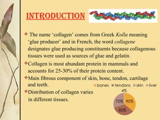
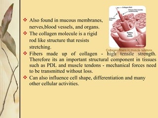
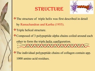
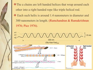
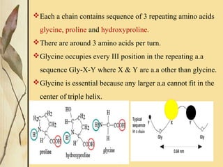
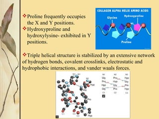
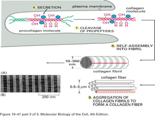
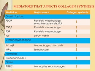
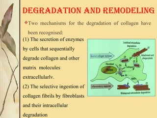
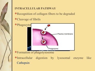
COLLAGEN
GUIDED BY :
DR. RUPINDER KAUR
PRESENTED BY:
DR. MALVIKA THAKUR
PG I YEAR
α 1
α 1
α 2
( G ly - X a a - Y a a ) 3 3 8
CONTENTS
1. Introduction
2. Structure of collagen
3. Types and tissue distribution of collagen
4. Synthesis of collagen
- Regulation of collagen synthesis
5. Degradation and remodeling
6. Age changes in collagen
7. Pathological alterations of collagen in periodontium
8. Uses of collagen in Periodontics
9. Conclusion
INTRODUCTION
 The name ‘collagen’ comes from Greek Kolla meaning
‘glue producer’ and in French, the word collagene
designates glue producing constituents because collagenous
tissues were used as sources of glue and gelatin.
Collagen is most abundant protein in mammals and
accounts for 25-30% of their protein content.
Main fibrous component of skin, bone, tendon, cartilage
and teeth.
Distribution of collagen varies
in different tissues.
 Also found in mucous membranes,
nerves,blood vessels, and organs.
 The collagen molecule is a rigid
rod like structure that resists
stretching.
 Fibers made up of collagen - high tensile strength.
Therefore its an important structural component in tissues
such as PDL and muscle tendons - mechanical forces need
to be transmitted without loss.
 Can also influence cell shape, differentiation and many
other cellular activities.
Collagen fibers in muscle tendons
STRUCTURE
The structure of triple helix was first described in detail
by Ramachandran and Kartha (1955).
Triple helical structure.
Composed of 3 polypeptide alpha chains coiled around each
other to form the triple helix configuration.
The individual polypeptide chains of collagen contain app.
1000 amino acid residues.
α1
α1
α2
( G l y - X a a - Y a a ) 3 3 8
The a chains are left handed helices that wrap around each
other into a right handed rope like triple helical rod.
 Each such helix is around 1.4 nanometers in diameter and
300 nanometers in length. (Ramchandran & Ramakrishnan
1976; Piez 1976).
α1
α1
α2
300 nm
(Gly-Xaa-Yaa)338
1.5 nm
Each a chain contains sequence of 3 repeating amino acids
glycine, proline and hydroxyproline.
There are around 3 amino acids per turn.
Glycine occupies every III position in the repeating a.a
sequence Gly-X-Y where X & Y are a.a other than glycine.
Glycine is essential because any larger a.a cannot fit in the
center of triple helix.
Proline frequently occupies
the X and Y positions.
Hydroxyproline and
hydroxylysine- exhibited in Y
positions.
Triple helical structure is stabilized by an extensive network
of hydrogen bonds, covalent crosslinks, electrostatic and
hydrophobic interactions, and vander waals forces.
The fibril formation occurs by a quarter staggered alignment
i.e.each helix is displaced longitudinally from its neighbour
by about one-quarter of is length (67nm)
The triple helical molecules of collagen assemble and form
elongated fibrils, and then rod like fibers in the tissues.
TYPES OF COLLAGEN
19 different types of collagen have been described by
Prockkop and Kivirrikko 1995 ; van der Rest and
Garrone 1991.
It ranges from type I to type XIX.
However, 90% of the collagen in the body are of type I,
II, III, and IV.
They are divided into three groups depending on their
ability to form fibrils.
1.Fibril forming collagens.
2.FACIT collagens.
3.Non fibrillar collagens
MAJOR PRODUCERS OF
COLLAGEN
Fibroblasts
Chondroblasts
Osteoblasts
Odontoblasts
Cementoblasts
LIMITED AMOUNT OF
COLLAGEN IS PRODUCED
Epithelial cells
Endothelial cells
Muscle cells
Schwann cells
SYNTHESIS OF COLLAGEN
The entire process of collagen biosynthesis can be best
understood under the following stages
Intracellular steps in collagen synthesis
Extracellular collagen biosynthetic events
Regulation of synthesis
Mediators Major source Collagen synthesis
Growth factors
PDGF Platelets, macrophage,
smooth muscle cells, Epi.
TGF β Platelets, macrophage
FGF Platelets, macrophage
IGF Serum matrix
Cytokine/Lymphokine
IL-1 α,β Macrophages, most cells
INF-γ Lymphocytes
Hormones
Glucocorticoides
Others
PGE-2 Monocytes, macrophages
MEDIATORS THAT AFFECTS COLLAGEN SYNTHESIS
Two mechanisms for the degradation of collagen have
been recognised:
DEGRADATION AND REMODELING
(1) The secretion of enzymes
by cells that sequentially
degrade collagen and other
matrix molecules
extracellularlv.
(2) The selective ingestion of
collagen fibrils by fibroblasts
and their intracellular
degradation
INTRACELLULAR PATHWAY
Recognition of collagen fibers to be degraded
Cleavage of fibrils
Phagocytosis of cleaved fibrils
Formation of phagolysosome
Intracellular digestion by lysosomal enzyme like
Cathepsin
EXTRACELLULAR DEGRADATION
It is achieved by MMP secreted by fibroblasts and other
cells
MMP – Matrix Metallo-Proteinases
These enzymes degrade collagen and other matrix
molecules into smaller peptides
Protoglycanase and Stromelysin remove the proteoglycans
around the collagen fibres and expose them to collagenase
Inactive collagenase (bound with inhibitor)
is secreted
Activating enzyme removes the inhibitor
Activated collagenase binds to fibrillar collagen
Collagen is split into 2 fragments (TCA & TCB)
Increased collagenase
activity
a. acute inflammation
b. immune mediated cell
injury
c. mast cell degranulation
d. bacterial infection
e. tumor invasion
Increased susceptibility of
collagen
a. denaturation of collagen
i. tissue hyperthermia
ii.Under hydroxylation
of proline
b. deficient cross-links
Deficient degradation
Decreased collagenase
activity
a. cirrhosis
b. scleroderma
c. osteopetrosis
Decreased susceptibility
of collagen
a. diabetes mellitus
b. hypertrophic scar
CONTROL MECHAMISMS
Birkedal- Hansen et al 1993 have studied three
mechanisms in vivo which keep the destructive properties
of MMPs’ under control.They are:
1)They are present in tissues as inactive precursors and
require activation by plasmin , trypsin or other proteinases.
2)These activating proteinases are themselves regulated by
other proteins such as tissue plasminogen activator.
3)Regulation of MMP synthesis by factors such as interleukin
–1 (IL-1) and transforming growth factor-β (TGF-β) is
controlled by α2 – macroglobulin which crosslinks with the
collagenases and inactivates them.
AGE CHANGES
Increased rate of conversion of soluble to
insoluble collagen.
Increased mechanical strength
Increased denaturing temperature
Increased thermal contraction
Decreased water content
Increased resistance to proteolytic enzymes
Decreased level of acid mucopolysaccharide
All these cause increased content of
collagen despite low rate of synthesis
PATHOLOGICAL ALTERATIONS
OF COLLAGEN IN PERIODONTIUM
ALTERATION OF COLLAGEN IN GINGIVITIS AND
PERIODONTITIS
Tissue destruction begins within the peri vascular extracellular
matrix where most of the collagen within the foci of
inflammation are degraded.
Inflammation spreads along the collagen fiber bundles
following the course of the blood vessels.
70% of the collagen are destroyed around the cellular infiltrate.
First mainly the circular and dento gingival fibers are affected.
In gingivitis - due to the MMP’s secreted by the neutrophils.
In periodontitis - MMP’s produced by neutrophils,
fibroblasts, keratinocytes all have a role.
Degradation - phagocytic action of the fibroblast - destroying
collagen apical to the junctional epithelium - apical migration
of the junctional epithelium - pocket formation.
Both qualitative and quantitative changes occurs in the
collagen.
Collagen becomes more soluble.
Reduction in the amount of type I & III collagen present.
Increase in the type V collagen- more resistant to the action
of
collagenase.
A new collagen species, Type I trimer can be detected in
inflamed gingiva.
Type I & II collagen- destroyed at the foci of inflammation.
Type V collagen increases and its amount may exceed that of
Type III.
The pathological breakdown is due to the imbalance between
the activated MMP’S and their endogenous inhibitors i.e.,
TIMP’s.
 In inflammatory conditions there will be increased amount
of MMP’s and less amount of TIMP’s.
But in case of localized aggressive periodontitis there is
increased level of MMP-1 and also increased level of TIMP.
Gingival Overgrowth:
Charecterised by increased gingival mass associated with
fibroepithelial changes and an accumlation of connective
tissue matrix .
Type I/III ratio becomes different with loss of type I and
elevated levels of type III collagen.
Two mechanisms are involved .
1 .Reduction in the level or activities of matrix degrading
enzymes in these lesions.
2. Induction of collagen production may occur by direct
action of drugs on collagen synthesis mechanisms –by
directly affecting the gene transcription rate or Indirectly
through cytokines such as TGFβ.
USES OF COLLAGEN IN PERIODONTICS
1. Drug delivery- For LDD in periodontal pockets
The benefit of LDD over systemic therapy is - high conc. of
drug can be maintained at the target site - avoiding risk of
systemic toxicity and associated side-effects.
The drugs can be loaded into collagen membranes by
hydrogen bonding, covalent bonding or simple entrapment.
32
PPAB is collagen fibril based formulation containing
tetracycline hydrochloride (2 mg of tetracycline) in 25
mg of collagen fibrils
Periocol®
- TC
Sterile collagen Fibres with Tetracycline
Hydrochloride for periodontal infections.
2. Tissue augmentation- recession coverage
Collagen membranes are used as an alternative to connective
tissue grafts in mucogingival surgeries.
Shows similar histologic and clinical outcomes, achieving
complete root coverage when compared with CTG.
It gets completely incorporated in the adjacent host
connective tissues without any signs of inflammation.
33
3. Bone substitute- as bone grafts in intra-bony defects
Collagen has been used as implantable carriers for bone
inducing proteins
Collagen itself is used as bone substitutes due to its osteo-
inductive activity.
Demineralized bone collagen is used as a bone graft material
for the treatment of acquired and congenital bone defects either
by itself or in combination with hydroxyapatite crystals.
34
Dembone is Demineralized Freeze-dried Cortical
Bone Powder, prepared from cortical bone
harvested from carefully screened human
donors, demineralized in HCl acid, freeze dried
and triple sterilized before vacuum packing.
Osseograft/DMBM is one such de-
mineralized bone derived Type-I collagen
for bone void filling applications.
4. In guided tissue regeneration- GTR membranes
GTR is a procedure that attempts to reconstitute the lost
tissues and is based on the concept of selective repopulation.
The first report of a human tooth treated by guided tissue
regeneration was by Nyman et al in 1982, with the term GTR
coined by Gottlow et al in 1986.
To prevent fast-growing cells of the gingival epithelium
from migrating to the wound, GTR procedures use barrier
devices - placed between the flap and the osseous defect -
maintain a space for repopulation of the defect with cells
having regenerative potential.
35
Healiguide
thin sheet made of high purity
Type-I collagen derived from
selected animal tissues. Periocol®
 / Helisorb®
-GTR
Type 1 collagen membrane of fish origin for 
GTR applications
Cologuide 
5. Hemostat
During blood clotting, platelets become activated by
thrombin and aggregate at the site of injury.
Stimulated by the protein fibrinogen - platelets clump by
binding to the collagen that becomes exposed following
rupture of the endothelial lining of blood vessels.
Collagen is therefore a natural haemostat and a wide variety
of collagen-based products are used in surgery and dentistry to
control excessive bleeding or haemorrhage.
  36
Absorbable sterile fibrillar
collagen wound filler,
constituted using high purity 
type-1 reconstituted collagen
•Hemostatic agent
•Control bleeding & stabilizes
blood clots
•Protects wound bed
GelSpon®
Sterile Absorbable
Haemostatic Gelatin
Sponges
 
6.Sutures
Surgical gut sutures:
Derived from submucosa of sheep intestine / serosa of beef
cattle intestine. It is 99% pure collagen, resorbable being
degraded by proteolytic enzymes.
 Collagen sutures:
Made by extruding homogenized tendon achilles of beef
cattle ,almost 100% pure collagen, stiffer than surgical
gut,used in eye surgery since it’s tissue reaction is less than
surgical gut.
Collagen has ubiquitous distribution and unique structure
and function in maintaining the structural and functional
integrity of the extracellular space throughout the body
and in particular the periodontium.
 Also its properties makes it as a very good biomolecule
to be used in periodontal regeneration which is the goal of
periodontal therapy.
CONCLUSION
 Collagen has been used extensively for manufacture of
biomedical devices because of its biologic and physical
properties, and ample availability.
 Also it has various industrial applications. Many clinical
researches and trials have been undergoing for the
advanced uses of collagen.
Thus “Proves to be one the most important proteins in
our entire body”
1) Oral histology: Development structure and function ; A.R. Tencate:
7th
Edition
2) Harper’s illustrated biochemistry ; 26th
Edition
3) Clinical Periodontology ; Carranza: 10th
Edition
4) A textbook of Oral Pathology ; Shafer : 5th
Edition
5) Collagen membranes : A Review ;J Periodontol 2001;72:215-229.
6) Collagen- structure,function and biosynthesis : K.Gelse, E Poschl, T.
Aigner - Advanced Drug Delivery Reviews 55 (2003) 1531-1546
REFERENCES
ThaNk yOU

Seminar collagen

  • 1.
    COLLAGEN GUIDED BY : DR.RUPINDER KAUR PRESENTED BY: DR. MALVIKA THAKUR PG I YEAR α 1 α 1 α 2 ( G ly - X a a - Y a a ) 3 3 8
  • 2.
    CONTENTS 1. Introduction 2. Structureof collagen 3. Types and tissue distribution of collagen 4. Synthesis of collagen - Regulation of collagen synthesis 5. Degradation and remodeling 6. Age changes in collagen 7. Pathological alterations of collagen in periodontium 8. Uses of collagen in Periodontics 9. Conclusion
  • 3.
    INTRODUCTION  The name‘collagen’ comes from Greek Kolla meaning ‘glue producer’ and in French, the word collagene designates glue producing constituents because collagenous tissues were used as sources of glue and gelatin. Collagen is most abundant protein in mammals and accounts for 25-30% of their protein content. Main fibrous component of skin, bone, tendon, cartilage and teeth. Distribution of collagen varies in different tissues.
  • 4.
     Also foundin mucous membranes, nerves,blood vessels, and organs.  The collagen molecule is a rigid rod like structure that resists stretching.  Fibers made up of collagen - high tensile strength. Therefore its an important structural component in tissues such as PDL and muscle tendons - mechanical forces need to be transmitted without loss.  Can also influence cell shape, differentiation and many other cellular activities. Collagen fibers in muscle tendons
  • 5.
    STRUCTURE The structure oftriple helix was first described in detail by Ramachandran and Kartha (1955). Triple helical structure. Composed of 3 polypeptide alpha chains coiled around each other to form the triple helix configuration. The individual polypeptide chains of collagen contain app. 1000 amino acid residues. α1 α1 α2 ( G l y - X a a - Y a a ) 3 3 8
  • 6.
    The a chainsare left handed helices that wrap around each other into a right handed rope like triple helical rod.  Each such helix is around 1.4 nanometers in diameter and 300 nanometers in length. (Ramchandran & Ramakrishnan 1976; Piez 1976). α1 α1 α2 300 nm (Gly-Xaa-Yaa)338 1.5 nm
  • 7.
    Each a chaincontains sequence of 3 repeating amino acids glycine, proline and hydroxyproline. There are around 3 amino acids per turn. Glycine occupies every III position in the repeating a.a sequence Gly-X-Y where X & Y are a.a other than glycine. Glycine is essential because any larger a.a cannot fit in the center of triple helix.
  • 8.
    Proline frequently occupies theX and Y positions. Hydroxyproline and hydroxylysine- exhibited in Y positions. Triple helical structure is stabilized by an extensive network of hydrogen bonds, covalent crosslinks, electrostatic and hydrophobic interactions, and vander waals forces.
  • 9.
    The fibril formationoccurs by a quarter staggered alignment i.e.each helix is displaced longitudinally from its neighbour by about one-quarter of is length (67nm)
  • 10.
    The triple helicalmolecules of collagen assemble and form elongated fibrils, and then rod like fibers in the tissues.
  • 11.
    TYPES OF COLLAGEN 19different types of collagen have been described by Prockkop and Kivirrikko 1995 ; van der Rest and Garrone 1991. It ranges from type I to type XIX. However, 90% of the collagen in the body are of type I, II, III, and IV. They are divided into three groups depending on their ability to form fibrils. 1.Fibril forming collagens. 2.FACIT collagens. 3.Non fibrillar collagens
  • 14.
    MAJOR PRODUCERS OF COLLAGEN Fibroblasts Chondroblasts Osteoblasts Odontoblasts Cementoblasts LIMITEDAMOUNT OF COLLAGEN IS PRODUCED Epithelial cells Endothelial cells Muscle cells Schwann cells SYNTHESIS OF COLLAGEN
  • 15.
    The entire processof collagen biosynthesis can be best understood under the following stages Intracellular steps in collagen synthesis Extracellular collagen biosynthetic events Regulation of synthesis
  • 19.
    Mediators Major sourceCollagen synthesis Growth factors PDGF Platelets, macrophage, smooth muscle cells, Epi. TGF β Platelets, macrophage FGF Platelets, macrophage IGF Serum matrix Cytokine/Lymphokine IL-1 α,β Macrophages, most cells INF-γ Lymphocytes Hormones Glucocorticoides Others PGE-2 Monocytes, macrophages MEDIATORS THAT AFFECTS COLLAGEN SYNTHESIS
  • 20.
    Two mechanisms forthe degradation of collagen have been recognised: DEGRADATION AND REMODELING (1) The secretion of enzymes by cells that sequentially degrade collagen and other matrix molecules extracellularlv. (2) The selective ingestion of collagen fibrils by fibroblasts and their intracellular degradation
  • 21.
    INTRACELLULAR PATHWAY Recognition ofcollagen fibers to be degraded Cleavage of fibrils Phagocytosis of cleaved fibrils Formation of phagolysosome Intracellular digestion by lysosomal enzyme like Cathepsin
  • 22.
    EXTRACELLULAR DEGRADATION It isachieved by MMP secreted by fibroblasts and other cells MMP – Matrix Metallo-Proteinases These enzymes degrade collagen and other matrix molecules into smaller peptides
  • 23.
    Protoglycanase and Stromelysinremove the proteoglycans around the collagen fibres and expose them to collagenase Inactive collagenase (bound with inhibitor) is secreted Activating enzyme removes the inhibitor Activated collagenase binds to fibrillar collagen Collagen is split into 2 fragments (TCA & TCB)
  • 24.
    Increased collagenase activity a. acuteinflammation b. immune mediated cell injury c. mast cell degranulation d. bacterial infection e. tumor invasion Increased susceptibility of collagen a. denaturation of collagen i. tissue hyperthermia ii.Under hydroxylation of proline b. deficient cross-links Deficient degradation Decreased collagenase activity a. cirrhosis b. scleroderma c. osteopetrosis Decreased susceptibility of collagen a. diabetes mellitus b. hypertrophic scar
  • 25.
    CONTROL MECHAMISMS Birkedal- Hansenet al 1993 have studied three mechanisms in vivo which keep the destructive properties of MMPs’ under control.They are: 1)They are present in tissues as inactive precursors and require activation by plasmin , trypsin or other proteinases. 2)These activating proteinases are themselves regulated by other proteins such as tissue plasminogen activator. 3)Regulation of MMP synthesis by factors such as interleukin –1 (IL-1) and transforming growth factor-β (TGF-β) is controlled by α2 – macroglobulin which crosslinks with the collagenases and inactivates them.
  • 26.
    AGE CHANGES Increased rateof conversion of soluble to insoluble collagen. Increased mechanical strength Increased denaturing temperature Increased thermal contraction Decreased water content Increased resistance to proteolytic enzymes Decreased level of acid mucopolysaccharide All these cause increased content of collagen despite low rate of synthesis
  • 28.
    PATHOLOGICAL ALTERATIONS OF COLLAGENIN PERIODONTIUM ALTERATION OF COLLAGEN IN GINGIVITIS AND PERIODONTITIS Tissue destruction begins within the peri vascular extracellular matrix where most of the collagen within the foci of inflammation are degraded. Inflammation spreads along the collagen fiber bundles following the course of the blood vessels. 70% of the collagen are destroyed around the cellular infiltrate. First mainly the circular and dento gingival fibers are affected. In gingivitis - due to the MMP’s secreted by the neutrophils.
  • 29.
    In periodontitis -MMP’s produced by neutrophils, fibroblasts, keratinocytes all have a role. Degradation - phagocytic action of the fibroblast - destroying collagen apical to the junctional epithelium - apical migration of the junctional epithelium - pocket formation. Both qualitative and quantitative changes occurs in the collagen. Collagen becomes more soluble. Reduction in the amount of type I & III collagen present. Increase in the type V collagen- more resistant to the action of collagenase. A new collagen species, Type I trimer can be detected in inflamed gingiva.
  • 30.
    Type I &II collagen- destroyed at the foci of inflammation. Type V collagen increases and its amount may exceed that of Type III. The pathological breakdown is due to the imbalance between the activated MMP’S and their endogenous inhibitors i.e., TIMP’s.  In inflammatory conditions there will be increased amount of MMP’s and less amount of TIMP’s. But in case of localized aggressive periodontitis there is increased level of MMP-1 and also increased level of TIMP.
  • 31.
    Gingival Overgrowth: Charecterised byincreased gingival mass associated with fibroepithelial changes and an accumlation of connective tissue matrix . Type I/III ratio becomes different with loss of type I and elevated levels of type III collagen. Two mechanisms are involved . 1 .Reduction in the level or activities of matrix degrading enzymes in these lesions. 2. Induction of collagen production may occur by direct action of drugs on collagen synthesis mechanisms –by directly affecting the gene transcription rate or Indirectly through cytokines such as TGFβ.
  • 32.
    USES OF COLLAGENIN PERIODONTICS 1. Drug delivery- For LDD in periodontal pockets The benefit of LDD over systemic therapy is - high conc. of drug can be maintained at the target site - avoiding risk of systemic toxicity and associated side-effects. The drugs can be loaded into collagen membranes by hydrogen bonding, covalent bonding or simple entrapment. 32 PPAB is collagen fibril based formulation containing tetracycline hydrochloride (2 mg of tetracycline) in 25 mg of collagen fibrils Periocol® - TC Sterile collagen Fibres with Tetracycline Hydrochloride for periodontal infections.
  • 33.
    2. Tissue augmentation-recession coverage Collagen membranes are used as an alternative to connective tissue grafts in mucogingival surgeries. Shows similar histologic and clinical outcomes, achieving complete root coverage when compared with CTG. It gets completely incorporated in the adjacent host connective tissues without any signs of inflammation. 33
  • 34.
    3. Bone substitute-as bone grafts in intra-bony defects Collagen has been used as implantable carriers for bone inducing proteins Collagen itself is used as bone substitutes due to its osteo- inductive activity. Demineralized bone collagen is used as a bone graft material for the treatment of acquired and congenital bone defects either by itself or in combination with hydroxyapatite crystals. 34 Dembone is Demineralized Freeze-dried Cortical Bone Powder, prepared from cortical bone harvested from carefully screened human donors, demineralized in HCl acid, freeze dried and triple sterilized before vacuum packing. Osseograft/DMBM is one such de- mineralized bone derived Type-I collagen for bone void filling applications.
  • 35.
    4. In guidedtissue regeneration- GTR membranes GTR is a procedure that attempts to reconstitute the lost tissues and is based on the concept of selective repopulation. The first report of a human tooth treated by guided tissue regeneration was by Nyman et al in 1982, with the term GTR coined by Gottlow et al in 1986. To prevent fast-growing cells of the gingival epithelium from migrating to the wound, GTR procedures use barrier devices - placed between the flap and the osseous defect - maintain a space for repopulation of the defect with cells having regenerative potential. 35 Healiguide thin sheet made of high purity Type-I collagen derived from selected animal tissues. Periocol®  / Helisorb® -GTR Type 1 collagen membrane of fish origin for  GTR applications Cologuide 
  • 36.
    5. Hemostat During bloodclotting, platelets become activated by thrombin and aggregate at the site of injury. Stimulated by the protein fibrinogen - platelets clump by binding to the collagen that becomes exposed following rupture of the endothelial lining of blood vessels. Collagen is therefore a natural haemostat and a wide variety of collagen-based products are used in surgery and dentistry to control excessive bleeding or haemorrhage.   36 Absorbable sterile fibrillar collagen wound filler, constituted using high purity  type-1 reconstituted collagen •Hemostatic agent •Control bleeding & stabilizes blood clots •Protects wound bed GelSpon® Sterile Absorbable Haemostatic Gelatin Sponges  
  • 37.
    6.Sutures Surgical gut sutures: Derivedfrom submucosa of sheep intestine / serosa of beef cattle intestine. It is 99% pure collagen, resorbable being degraded by proteolytic enzymes.  Collagen sutures: Made by extruding homogenized tendon achilles of beef cattle ,almost 100% pure collagen, stiffer than surgical gut,used in eye surgery since it’s tissue reaction is less than surgical gut.
  • 38.
    Collagen has ubiquitousdistribution and unique structure and function in maintaining the structural and functional integrity of the extracellular space throughout the body and in particular the periodontium.  Also its properties makes it as a very good biomolecule to be used in periodontal regeneration which is the goal of periodontal therapy. CONCLUSION
  • 39.
     Collagen hasbeen used extensively for manufacture of biomedical devices because of its biologic and physical properties, and ample availability.  Also it has various industrial applications. Many clinical researches and trials have been undergoing for the advanced uses of collagen. Thus “Proves to be one the most important proteins in our entire body”
  • 40.
    1) Oral histology:Development structure and function ; A.R. Tencate: 7th Edition 2) Harper’s illustrated biochemistry ; 26th Edition 3) Clinical Periodontology ; Carranza: 10th Edition 4) A textbook of Oral Pathology ; Shafer : 5th Edition 5) Collagen membranes : A Review ;J Periodontol 2001;72:215-229. 6) Collagen- structure,function and biosynthesis : K.Gelse, E Poschl, T. Aigner - Advanced Drug Delivery Reviews 55 (2003) 1531-1546 REFERENCES
  • 41.

Editor's Notes

  • #9 . Hydroxy proline is present as 1 in 10 residues while Hydroxylysine is present as 1 in 200 residues. Proline and hydroxy proline together account for 2/9th of the residues.