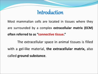
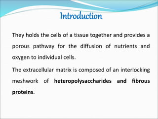
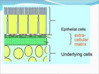
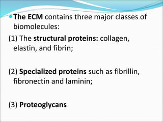
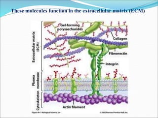
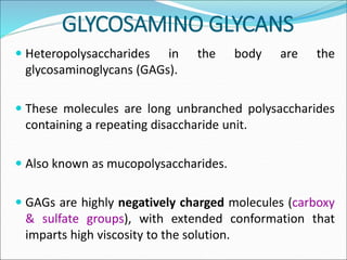
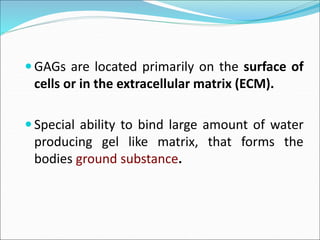
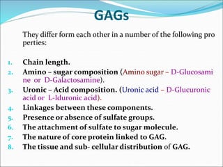
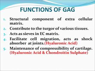
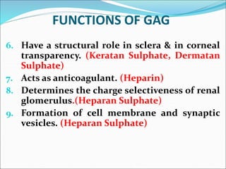
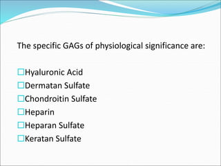
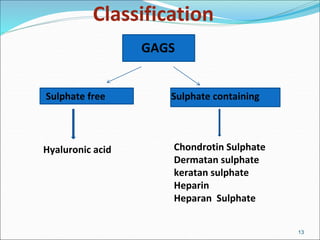
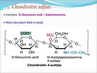
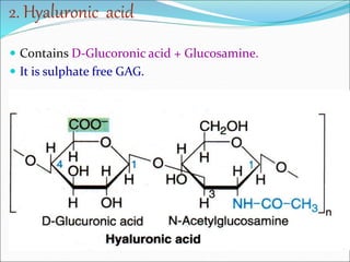
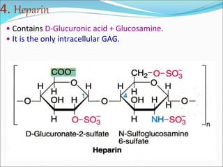
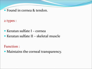
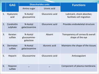
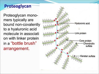
Dr. Astha Goyal
Dept. of Biochemistry
MGMC&H
Introduction
Most mammalian cells are located in tissues where they
are surrounded by a complex extracellular matrix (ECM)
often referred to as “connective tissue.”
The extracellular space in animal tissues is filled
with a gel-like material, the extracellular matrix, also
called ground substance.
Introduction
They holds the cells of a tissue together and provides a
porous pathway for the diffusion of nutrients and
oxygen to individual cells.
The extracellular matrix is composed of an interlocking
meshwork of heteropolysaccharides and fibrous
proteins.
Epithelial cells
extra-
cellular
matrix
Underlying cells cells
The ECM contains three major classes of
biomolecules:
(1) The structural proteins: collagen,
elastin, and fibrin;
(2) Specialized proteins such as fibrillin,
fibronectin and laminin;
(3) Proteoglycans
These molecules function in the extracellular matrix (ECM)
GLYCOSAMINO GLYCANS
 Heteropolysaccharides in the body are the
glycosaminoglycans (GAGs).
 These molecules are long unbranched polysaccharides
containing a repeating disaccharide unit.
 Also known as mucopolysaccharides.
 GAGs are highly negatively charged molecules (carboxy
& sulfate groups), with extended conformation that
imparts high viscosity to the solution.
 GAGs are located primarily on the surface of
cells or in the extracellular matrix (ECM).
 Special ability to bind large amount of water
producing gel like matrix, that forms the
bodies ground substance.
GAGs
They differ form each other in a number of the following pro
perties:
1. Chain length.
2. Amino – sugar composition (Amino sugar – D-Glucosami
ne or D-Galactosamine).
3. Uronic – Acid composition. (Uronic acid – D-Glucuronic
acid or L-Iduronic acid).
4. Linkages between these components.
5. Presence or absence of sulfate groups.
6. The attachment of sulfate to sugar molecule.
7. The nature of core protein linked to GAG.
8. The tissue and sub- cellular distribution of GAG.
FUNCTIONS OF GAG
1. Structural component of extra cellular
matrix.
2. Contribute to the turgor of various tissues.
3. Acts as sieves in EC matrix.
4. Facilitate cell migration, acts as shock
absorber at joints.(Hyaluronic Acid)
5. Maintenance of compressibility of cartilage.
(Hyaluronic Acid & Chondroitin Sulphate)
FUNCTIONS OF GAG
6. Have a structural role in sclera & in corneal
transparency. (Keratan Sulphate, Dermatan
Sulphate)
7. Acts as anticoagulant. (Heparin)
8. Determines the charge selectiveness of renal
glomerulus.(Heparan Sulphate)
9. Formation of cell membrane and synaptic
vesicles. (Heparan Sulphate)
The specific GAGs of physiological significance are:
Hyaluronic Acid
Dermatan Sulfate
Chondroitin Sulfate
Heparin
Heparan Sulfate
Keratan Sulfate
Classification
13
GAGS
Sulphate free Sulphate containing
Hyaluronic acid Chondrotin Sulphate
Dermatan sulphate
keratan sulphate
Heparin
Heparan Sulphate
• Contains D-Glucoronic acid + Galactosamine.
• Most abundant GAG in body.
1. Chondroitin sulfate
• Widely distributed in bone, cartilage & tendons.
• Function:
• In cartilage, it binds collagen & hold fibers in a tight
strong network.
• Role in Compressibility of cartilage in weight
bearing along with Hyaluronic acid.
• 2 types of chondroitin sulfate :
Sulphated at C4 or C6 group
2. Hyaluronic acid
 Contains D-Glucoronic acid + Glucosamine.
 It is sulphate free GAG.
 Ground substance of synovial fluid of joints, vitreous
humor of eyes and connective tissues, tendon.
 Hyaluronidase is an enzyme that breaks β-1 – 4 linkages.
 Present in high concentration in testes, seminal fluid, & in
certain snake and insect venoms.
Functions of Hyaluronic acid
 Serves as a lubricant and shock absorbant in joints.
 Acts as seives in extracelluar matrix.
 Permits cell migration during embryogenesis, morpho-
genesis & wound repair.
 Hyaluronidase enzyme of semen degrades the gel around
ovum & allows effective penetration of sperm into ovum.
3. DERMATAN SULFATe
 Contains L-iduronic acid + Galactosamine
 Present in skin, cardiac valves & tendon.
Function :
 Present in sclera of eye where it has important function
in maintaining overall shape of eye.
4. Heparin
• Contains D-Glucuronic acid + Glucosamine.
• It is the only intracellular GAG.
 It is an anticoagulant (prevents blood clotting ).
 Found in granules of mast cells that line the
arteries of lung, liver, kidney, spleen.
 Strongly acidic due to presence of more sulphate
group.
 Heparin helps in the release of the enzyme
lipoprotein lipase.
 Helps to clear the lipidemia after fatty meal –
also called clearing factor.
5. Heparan sulfate
 Same as heparin except some amino groups carry
acetyl group and % of sulphate groups are
smaller.
 It is extracellular GAG .
 Present in basement membrane & as a ubiquitous
component of cell surface.
6. KERATAN sulfate
• Contains D-Galactose + Glucosamine
•Only GAG with no uronic acid.
 Found in cornea & tendon.
2 types :
 Keratan sulfate І – cornea
 Keratan sulfate ІІ – skeletal muscle
Function :
 Maintains the corneal transparency.
GAG Disaccharides units Functions
Amino sugar Uronic acid
1. Hyaluronic
acid
N-Acetyl
glucosamine
Glucuronic acid Lubricant, shock absorber,
facilitate cell migration
2. Condroitin
sulphate
N-Acetyl
galactosamine
Glucuronic acid Provides endoskeletal structure
3. Keratan
sulfate
N-Acetyl
glucosamine-
galactose
Absent Transparency of cornea & overall
shape of the eye
4. Dermatan
sulfate
N-Acetyl
galactosamine
Iduronic acid Maintains the shape of the tissues
5. Heparin Glucosamine Glucuronic acid Anticoagulant
6. Heparan
sulfate
,, ,, Component of plasma membrane.
Proteoglycan
Proteoglycan mono-
mers typically are
bound non-covalently
to a hyaluronic acid
molecule in associati
on with linker protein
in a “bottle brush”
arrangement.
Thank you

Glycosaminoglycans

  • 1.
    Dr. Astha Goyal Dept.of Biochemistry MGMC&H
  • 2.
    Introduction Most mammalian cellsare located in tissues where they are surrounded by a complex extracellular matrix (ECM) often referred to as “connective tissue.” The extracellular space in animal tissues is filled with a gel-like material, the extracellular matrix, also called ground substance.
  • 3.
    Introduction They holds thecells of a tissue together and provides a porous pathway for the diffusion of nutrients and oxygen to individual cells. The extracellular matrix is composed of an interlocking meshwork of heteropolysaccharides and fibrous proteins.
  • 4.
  • 5.
    The ECM containsthree major classes of biomolecules: (1) The structural proteins: collagen, elastin, and fibrin; (2) Specialized proteins such as fibrillin, fibronectin and laminin; (3) Proteoglycans
  • 6.
    These molecules functionin the extracellular matrix (ECM)
  • 7.
    GLYCOSAMINO GLYCANS  Heteropolysaccharidesin the body are the glycosaminoglycans (GAGs).  These molecules are long unbranched polysaccharides containing a repeating disaccharide unit.  Also known as mucopolysaccharides.  GAGs are highly negatively charged molecules (carboxy & sulfate groups), with extended conformation that imparts high viscosity to the solution.
  • 8.
     GAGs arelocated primarily on the surface of cells or in the extracellular matrix (ECM).  Special ability to bind large amount of water producing gel like matrix, that forms the bodies ground substance.
  • 9.
    GAGs They differ formeach other in a number of the following pro perties: 1. Chain length. 2. Amino – sugar composition (Amino sugar – D-Glucosami ne or D-Galactosamine). 3. Uronic – Acid composition. (Uronic acid – D-Glucuronic acid or L-Iduronic acid). 4. Linkages between these components. 5. Presence or absence of sulfate groups. 6. The attachment of sulfate to sugar molecule. 7. The nature of core protein linked to GAG. 8. The tissue and sub- cellular distribution of GAG.
  • 10.
    FUNCTIONS OF GAG 1.Structural component of extra cellular matrix. 2. Contribute to the turgor of various tissues. 3. Acts as sieves in EC matrix. 4. Facilitate cell migration, acts as shock absorber at joints.(Hyaluronic Acid) 5. Maintenance of compressibility of cartilage. (Hyaluronic Acid & Chondroitin Sulphate)
  • 11.
    FUNCTIONS OF GAG 6.Have a structural role in sclera & in corneal transparency. (Keratan Sulphate, Dermatan Sulphate) 7. Acts as anticoagulant. (Heparin) 8. Determines the charge selectiveness of renal glomerulus.(Heparan Sulphate) 9. Formation of cell membrane and synaptic vesicles. (Heparan Sulphate)
  • 12.
    The specific GAGsof physiological significance are: Hyaluronic Acid Dermatan Sulfate Chondroitin Sulfate Heparin Heparan Sulfate Keratan Sulfate
  • 13.
    Classification 13 GAGS Sulphate free Sulphatecontaining Hyaluronic acid Chondrotin Sulphate Dermatan sulphate keratan sulphate Heparin Heparan Sulphate
  • 14.
    • Contains D-Glucoronicacid + Galactosamine. • Most abundant GAG in body. 1. Chondroitin sulfate
  • 15.
    • Widely distributedin bone, cartilage & tendons. • Function: • In cartilage, it binds collagen & hold fibers in a tight strong network. • Role in Compressibility of cartilage in weight bearing along with Hyaluronic acid. • 2 types of chondroitin sulfate : Sulphated at C4 or C6 group
  • 16.
    2. Hyaluronic acid Contains D-Glucoronic acid + Glucosamine.  It is sulphate free GAG.  It is sulphate free GAG.
  • 17.
     Ground substanceof synovial fluid of joints, vitreous humor of eyes and connective tissues, tendon.  Hyaluronidase is an enzyme that breaks β-1 – 4 linkages.  Present in high concentration in testes, seminal fluid, & in certain snake and insect venoms.
  • 18.
    Functions of Hyaluronicacid  Serves as a lubricant and shock absorbant in joints.  Acts as seives in extracelluar matrix.  Permits cell migration during embryogenesis, morpho- genesis & wound repair.  Hyaluronidase enzyme of semen degrades the gel around ovum & allows effective penetration of sperm into ovum.
  • 19.
    3. DERMATAN SULFATe Contains L-iduronic acid + Galactosamine
  • 20.
     Present inskin, cardiac valves & tendon. Function :  Present in sclera of eye where it has important function in maintaining overall shape of eye.
  • 21.
    4. Heparin • ContainsD-Glucuronic acid + Glucosamine. • It is the only intracellular GAG.
  • 22.
     It isan anticoagulant (prevents blood clotting ).  Found in granules of mast cells that line the arteries of lung, liver, kidney, spleen.  Strongly acidic due to presence of more sulphate group.  Heparin helps in the release of the enzyme lipoprotein lipase.  Helps to clear the lipidemia after fatty meal – also called clearing factor.
  • 23.
    5. Heparan sulfate Same as heparin except some amino groups carry acetyl group and % of sulphate groups are smaller.  It is extracellular GAG .  Present in basement membrane & as a ubiquitous component of cell surface.
  • 24.
    6. KERATAN sulfate •Contains D-Galactose + Glucosamine •Only GAG with no uronic acid.
  • 25.
     Found incornea & tendon. 2 types :  Keratan sulfate І – cornea  Keratan sulfate ІІ – skeletal muscle Function :  Maintains the corneal transparency.
  • 26.
    GAG Disaccharides unitsFunctions Amino sugar Uronic acid 1. Hyaluronic acid N-Acetyl glucosamine Glucuronic acid Lubricant, shock absorber, facilitate cell migration 2. Condroitin sulphate N-Acetyl galactosamine Glucuronic acid Provides endoskeletal structure 3. Keratan sulfate N-Acetyl glucosamine- galactose Absent Transparency of cornea & overall shape of the eye 4. Dermatan sulfate N-Acetyl galactosamine Iduronic acid Maintains the shape of the tissues 5. Heparin Glucosamine Glucuronic acid Anticoagulant 6. Heparan sulfate ,, ,, Component of plasma membrane.
  • 27.
    Proteoglycan Proteoglycan mono- mers typicallyare bound non-covalently to a hyaluronic acid molecule in associati on with linker protein in a “bottle brush” arrangement.
  • 28.