This document discusses the Wnt signaling system and its role in osteoporosis and other diseases. It provides an overview of the key components of the Wnt signaling pathway, including Frizzled receptors, LRP5/6 coreceptors, beta-catenin, and sclerostin. Mutations affecting these components can lead to high or low bone mass. The document also discusses how modulating the Wnt pathway may help develop new drugs for osteoporosis and reviews studies on Wnt's role in heart disease, cancer, and other conditions.
This document discusses Wnt signaling and provides an overview of research tools that can be used to study the pathway. It describes the discovery of Wnt proteins and their roles in canonical and noncanonical signaling. The document also reviews the functions of Wnt signaling in development, tissue homeostasis, and various pathological conditions. It proposes using PCR arrays, siRNA, methylation arrays, and reporter assays to develop a Wnt gene signature and measure pathway activity through changes in gene expression and protein levels.
This document provides an overview of Wnt signaling and research tools for studying the pathway. It begins with an introduction to Wnt proteins and receptors, and describes the canonical and noncanonical Wnt signaling pathways. The document then discusses the function of Wnt signaling in development, tissue homeostasis, and various pathological conditions. Various research tools for investigating Wnt signaling are presented, including whole genome expression profiling, siRNA inhibition of β-catenin, Wnt target gene PCR arrays, and EpiTech methylation arrays. The document proposes a workflow for developing a Wnt gene expression signature and demonstrates using custom PCR arrays and methylation analysis to understand differential Wnt responses between cell lines. It concludes by describing a combined Wnt signaling PCR
Seminar-Spring 2010-Role of Wnt signaling in Alzheimer's disease pathogenesisNisha Rizvi
1. Wnt ligands are involved in pre- and postsynaptic protein clustering and assembly at synapses and form functional new synapses.
2. Activation of Wnt signaling protects against Aβ toxicity in Alzheimer's disease by increasing neuronal survival, decreasing apoptosis, and stabilizing β-catenin levels. Inhibition of Wnt signaling can cause neuronal damage.
3. Wnt signaling interacts and forms a crosstalk with other pathways important in Alzheimer's like M1 muscarinic acetylcholine receptor activation and PPARγ activation, which may provide neuroprotective effects against Aβ toxicity.
The Wnt signaling pathway involves Wnt ligands binding to Frizzled receptors and regulating beta-catenin levels and activity. This pathway controls numerous cellular processes during development and has been implicated in diseases like cancer when dysregulated. Specifically, mutations in APC, a negative regulator of the pathway, lead to beta-catenin accumulation and are present in most colorectal cancers. Future research includes investigating epigenetic changes in this pathway and developing targeted therapies based on pathway components like APC.
This document discusses the role of the Wnt signaling pathway in cancer. It begins by describing how the Wnt pathway is normally regulated and how dysregulation can lead to cancer. It then summarizes key points about how the Wnt pathway is altered in cancer at the membrane, cytoplasmic, and nuclear levels. Mutations in genes that encode proteins in the Wnt pathway, such as APC, Axin, and β-catenin, have been implicated in various cancers like colorectal cancer. Understanding how the Wnt pathway is involved in cancer is important for developing new cancer therapeutics.
The Wnt signaling pathway controls cell-cell communication by transmitting signals from cell surface receptors to DNA expression in the nucleus. It regulates beta-catenin, which enters the nucleus to activate gene expression. Mutations that damage the pathway prevent proper control of beta-catenin, leading to over-expression of genes involved in diseases like cancer. Drugs targeting components of the Wnt pathway like beta-catenin show promise for treating cancers caused by alterations in this important signaling network.
The document describes the Wnt signaling pathway under normal and Wnt ligand present conditions, noting that in the presence of the Wnt ligand, β-catenin is stabilized and translocates to the nucleus to activate Wnt responsive genes. It also discusses how mutations in APC can lead to uncontrolled cell proliferation and cancer by dysregulating the Wnt pathway and allowing β-catenin to accumulate in the nucleus. Finally, it briefly outlines the important roles of the Wnt pathway in development.
Wnt signaling pathways play an important role in many endocrine functions and diseases. Wnt signaling regulates pancreatic beta cell proliferation, adipose tissue development, steroidogenesis in the adrenal cortex, bone mineral metabolism, and sex development. Therapies targeting Wnt signaling show promise for diseases like osteoporosis, with anti-sclerostin therapies approved to treat post-menopausal osteoporosis. Future therapies may target TCF7L2 to prevent diabetes or treat adrenal tumors by modulating Wnt signaling.
This document discusses the role of β-catenin in cancer therapy. It describes β-catenin as a dual function protein involved in cell adhesion and gene transcription. Mutations and overexpression of β-catenin are associated with many cancers. β-catenin is regulated by the destruction complex containing APC, and mutations in these genes lead to β-catenin accumulation and cancer development. The document reviews potential cancer therapeutic approaches targeting β-catenin, including inhibitors of its binding sites and upstream regulators in the WNT pathway.
NAD+ and NADH play roles in many important biological processes such as energy metabolism, mitochondrial function, calcium homeostasis, and gene expression. They have emerged as one of the most influential couples in life. Poly (ADP-ribose) polymerase 1 (PARP1) activation leads to NAD+ depletion, which mediates cell death. Intranasal administration of NAD+ can decrease ischemic brain injury by reducing PARP1-induced cell death and is a potential treatment for diseases involving PARP1.
The Wnt signaling pathway is an important developmental pathway that is activated upon binding of Wnt ligands to Frizzled and LRP receptors. This prevents degradation of beta-catenin, allowing it to enter the nucleus and activate target genes in conjunction with TCF transcription factors. Key components of the pathway include Wnt ligands, Frizzled and LRP receptors, cytoplasmic proteins like Dsh and Axin, and nuclear proteins like beta-catenin and TCF. Mutations in various components of this pathway can lead to developmental defects or diseases like cancer.
This document provides an overview of a study that aims to validate somatic mutations in cervical cancer reported by next-generation sequencing (NGS) using Sanger sequencing. The study involved selecting mutations from NGS data, designing primers, performing PCR and purification on tumor and blood samples, conducting Sanger sequencing, and analyzing the results. 16 out of 27 mutations selected were validated as somatic or germline by Sanger sequencing, for a validation rate of around 60%. Several of the validated mutations occurred in important cancer-related genes. The validated mutations were also found to be present in cervical cancer cell lines.
Postdoctoral work on Vangl2, a protein involved in cell polarity. Cell polarity is the capability of a cell to be able to orient itself in a particular direction.
The RET proto-oncogene encodes a receptor tyrosine kinase for members of the glial cell line-derived neurotrophic factor family of extracellular signalling molecules. RET loss of function mutations are associated with the development of Hirschsprung's disease, while gain of function mutations are associated with the development of various types of human cancer, including medullary thyroid carcinoma, multiple endocrine neoplasias type 2A and 2B, pheochromocytoma and parathyroid hyperplasia.
RET is an abbreviation for "rearranged during transfection", as the DNA sequence of this gene was originally found to be rearranged within a 3T3 fibroblast cell line following its transfection with DNA taken from human lymphoma cells. The human gene RET is localized to chromosome 10 (10q11.2) and contains 21 exons.
The natural alternative splicing of the RET gene results in the production of 3 different isoforms of the protein RET. RET51, RET43 and RET9 contain 51, 43 and 9 amino acids in their C-terminal tail respectively. The biological roles of isoforms RET51 and RET9 are the most well studied in-vivo as these are the most common isoforms in which RET occurs.
Common to each isoform is a domain structure. Each protein is divided into three domains: an N-terminal extracellular domain with four cadherin-like repeats and a cysteine-rich region, a hydrophobic transmembrane domain and a cytoplasmic tyrosine kinase domain, which is split by an insertion of 27 amino acids. Within the cytoplasmic tyrosine kinase domain, there are 16 tyrosines (Tyrs) in RET9 and 18 in RET51. Tyr1090 and Tyr1096 are present only in the RET51 isoform.
The extracellular domain of RET contains nine N-glycosylation sites. The fully glycosylated RET protein is reported to have a molecular weight of 170 kDa although it is not clear to which isoform this molecular weight relates.
Cellular Signaling Pathways have direct implications on our understanding of tumor cell behavior. A general overview is presented here followed by a brief discussion of some of the major pathways currently implicated in cancer progression : Ras/RAF/MAP kinase pathway and PI3K/AKT/mTOR pathway s
The document provides an overview of molecular biology concepts including:
1) DNA contains the genetic code arranged in chromosomes and is tightly packed in the nucleus.
2) Genes contain exons and introns and are used to make mRNA which is then used to make proteins through translation.
3) Proteins have many important functions in the cell and interact with DNA, RNA and other proteins through complex formation.
4) Experimental techniques like gene knockout are used to study gene function and protein interactions.
Presentation On Wnt 4 and rhe role of HDAC4 & SIRT1 in bone biologyGayathri Vijayakumar
Wnt proteins are a family of conserved signaling proteins that regulate various processes including cell proliferation, fate determination, and embryonic development. They act through both canonical and non-canonical pathways. Wnt4 specifically promotes female development and suppresses male development. It also contributes to kidney and adrenal cortex development. Studies show Wnt4 enhances stem cell osteogenic differentiation by activating non-canonical pathways like P38MAPK. The NASA-Orthofix project examines the effects of pulsed electromagnetic fields on osteoblast proliferation, differentiation and mineralization using rat primary osteoblasts. Genes analyzed include osteocalcin, BAP, collagen I and GAPDH. Results show PEMF stimulation increases cell count, differentiation markers and mineralization
1) NF-kB is constitutively activated in pancreatic cancer cells but not normal pancreatic cells, suggesting it plays a role in pancreatic tumorigenesis.
2) Expressing a mutant IkBa (IkBaM) that inhibits NF-kB suppressed the tumorigenicity of pancreatic cancer cells in a mouse model.
3) Inhibiting NF-kB reduced expression of pro-survival genes like Bcl-xL and Bcl-2, and reduced VEGF and IL-8 expression which are involved in angiogenesis. This suggests NF-kB inhibition can suppress tumorigenesis by reducing pro-survival and angiogenic factors.
This document summarizes the use of the nematode C. elegans as a model organism for studying human diseases. It discusses how CRISPR/Cas9 can be used to introduce disease-related mutations into C. elegans to model retinitis pigmentaria, cancer, and responses to chemotherapy. Drug screens and RNAi screens in these mutant worms have identified genetic modifiers and potential drug targets for treating human diseases. The small size, rapid life cycle, and genetic tractability of C. elegans make it a valuable pre-clinical model for validating targets and precision medicines before testing in mammalian systems.
The Wnt cascade has emerged as a critical regulator of stem cells. In many tissues, activation of Wnt signaling has also been found to be associated with cancer. Understanding the regulation by Wnt signaling may serve as a paradigm for understanding the dual nature of self-renewal signals.
Preimplantation Genetic Diagnosis using Next Generation Sequencing for Social...Maryam Rafati
The document discusses techniques for preimplantation genetic diagnosis (PGD), including PCR-based techniques, fluorescence in situ hybridization (FISH), and next generation sequencing (NGS). It summarizes misdiagnosis rates for different techniques and applications. NGS is presented as a rapid and low-cost method for comprehensive aneuploidy screening and simultaneous investigation of single gene disorders. Clinical experience using NGS-PGD is discussed, showing transfer of a single euploid embryo can increase pregnancy rates for patients with recurrent implantation failure. The European Society of Human Reproduction and Embryology (ESHRE) guidelines for PGD are also summarized.
This experiment aimed to identify genes that were overexpressed in BU.MPT cells resistant to apoptosis compared to normal BU.MPT cells. RNA from the two cell populations underwent subtractive hybridization to identify differences. Eight colonies were sequenced, with one gene of interest identified as cysteine and histidine rich protein 1 (Chrp). Chrp is known to bind specifically to galectin-3, keeping it localized in the cytoplasm where it can activate anti-apoptotic pathways and properties. This binding of Chrp to galectin-3 provides a potential explanation for the resistance to apoptosis in the selected BU.MPT cells.
This document discusses signal transduction and how it relates to cancer. It describes how growth factors and receptors contribute to normal signal transduction and how this process is deregulated in cancer. It explains that growth factors regulate growth, proliferation and survival, which are all altered in cancer. Several growth factors and receptors that can contribute to oncogenesis are identified. It also summarizes several key intracellular signaling pathways, like MAPK pathways, that are activated by growth factors and can result in the cancer phenotype if altered.
Presentation from the ECDC expert consultation on Whole Genome Sequencing organised by the European Centre of Disease Prevention and Control - Stockholm, 19 November 2015
This is the Powerpoint presentation from my recent presentation at the TTP LabTech US Acumen Users Group Meeting (UGM) held at the British Consulate-General in Cambridge, MA on May 18, 2010
This document summarizes research on Tuberous Sclerosis Complex (TSC), including key facts about the disease, progress that has been made in understanding the genetics and molecular mechanisms, development of treatments like Everolimus, and ongoing areas of research focus like clinical trials of new drugs and studying disease mechanisms using cellular and animal models.
Spatial and temporal gene expression in growth plate and articular cartilage is regulated through complex signaling pathways.
1) In the growth plate, chondrocytes in the resting, proliferative, and hypertrophic zones express different genes due to intrinsic and extrinsic spatial regulation involving BMP and PTHrP signaling gradients.
2) Temporal regulation causes growth plate proliferation and elongation to slow with age, associated with decreased IGF signaling. Prior growth inhibition can delay this senescence.
3) Articular and growth plate cartilage differ in progenitor cell populations and gene expression patterns that correlate inversely between zones.
Metastatic bone disease: An old dogma and a new insightMohamed Abdulla
Metastatic bone disease is a challenging condition that places a heavy burden on patients. New insights into the cellular and molecular mechanisms have led to improved treatments. Cancer cells interact with the bone microenvironment through factors like RANKL, RANK, and osteoprotegerin, inducing a "vicious cycle" of bone destruction. Emerging therapies target these interactions by inhibiting RANKL with drugs like denosumab. Radiopharmaceuticals like radium-223 also show promise by targeting areas of new bone growth in metastases. While radiation remains important for pain relief, combination therapies offer the potential for improved outcomes in metastatic bone disease.
1) Protein phosphatase 5 (PP5) regulates the differentiation of mesenchymal stem cells into either adipocytes or osteoblasts through phosphorylation of PPARγ and RUNX2. 2) Mice deficient in PP5 have increased bone formation and decreased fat accumulation in bone marrow. 3) Experiments analyzing mice with tissue-specific knockout of PP5 in adipose tissue or long bones found that loss of PP5 in adipose tissue increased trabecular bone, while loss in long bones increased cortical bone thickness, implicating PP5 in regulating the balance between bone and fat formation in different tissues.
1) Protein phosphatase 5 (PP5) regulates the differentiation of mesenchymal stem cells into either adipocytes or osteoblasts through phosphorylation of PPARγ and RUNX2.
2) Mice deficient in PP5 showed increased bone formation and decreased marrow adipocytes, suggesting PP5 promotes adipogenesis over osteogenesis.
3) Experiments on mice with tissue-specific PP5 knockout found loss of PP5 in bone led to increased cortical bone thickness while loss in adipose tissue led to increased trabecular bone formation, demonstrating PP5 regulates the balance between bone and fat formation in different tissues.
This document summarizes information presented at the 2008 ASBMR conference on new and emerging osteoporosis treatments. It discusses several drug classes and mechanisms of action, including SERMs, bisphosphonates, denosumab, cathepsin K inhibitors, calcilytics, sclerostin antibodies, PTH analogs, and strontium. It also summarizes clinical trial results for lasofoxifene, zoledronic acid after hip fracture, and the OPTAMISE study comparing teriparatide with prior bisphosphonate use. Odanacatib, a cathepsin K inhibitor, showed increases in bone mineral density in a phase II trial with once weekly dosing and no increased risk
The document discusses several novel treatments for osteoporosis, including denosumab, anti-sclerostin monoclonal antibodies, and lasofoxifene. Denosumab is an antibody to RANK ligand that inhibits bone resorption and reduces fracture risk. Blocking sclerostin may allow unlimited bone formation and a potential cure for osteoporosis. Lasofoxifene reduces fracture risk and also reduces risks of breast cancer and heart disease, with no increased risk of uterine cancer.
This study identified a mosaic activating mutation in the AKT1 gene associated with Proteus syndrome. Researchers sequenced DNA from tissue samples of 29 patients with Proteus syndrome and identified a recurrent somatic mutation, c.49G>A, in the AKT1 gene in 26 patients. This mutation leads to increased phosphorylation and activation of the AKT1 protein. Western blot analysis showed increased phosphorylation of AKT1 in tissues affected by Proteus syndrome. The findings link activation of the AKT1 signaling pathway through somatic mutations to overgrowth observed in Proteus syndrome patients.
STAT 3 and Other Target Proteins: New Concepts in Psoriasis Pathogenesis & Therapy
This document discusses STAT3 signaling and its role in psoriasis pathogenesis. It summarizes that:
1) STAT3 is activated by cytokines and growth factors and forms dimers that enter the nucleus and activate gene transcription.
2) STAT3 signaling is involved in processes like proliferation and differentiation of keratinocytes. Its dysregulation contributes to psoriasis pathogenesis.
3) Psoriasis is a chronic inflammatory skin disease involving excessive keratinocyte proliferation, abnormal differentiation, and immune system involvement. Understanding STAT3 signaling may provide novel therapeutic targets for psoriasis treatment.
Recent advances in osteoporosis new copyDr Sourya M
Osteoporosis is characterized by low bone mass and deterioration of bone structure, making bones fragile and prone to fractures. Key drugs used to treat osteoporosis include calcium, vitamin D, bisphosphonates, SERMs, calcitonin, PTH and teriparatide, and denosumab. Newer drugs under development include romosozumab, a sclerostin inhibitor that strongly increases bone mineral density, abaloparatide, and integrin antagonists. Non-drug approaches also show promise such as biomaterials and gut serotonin inhibitors.
Gregg L. Semenza discusses hypoxia-inducible factors (HIFs) and their role in physiology and medicine. HIFs control oxygen homeostasis by regulating erythropoietin production and red blood cell production. They also play a role in cardiovascular disease by mediating vascularization and in cancer by promoting tumor growth and metastasis. HIFs activate genes involved in angiogenesis, cancer stem cell maintenance, immune evasion, and premetastatic niche formation. Inhibiting HIF activity has been shown to reduce tumor growth and improve outcomes for cancer and cardiovascular disease.
1. The document discusses bone metabolism and prostate cancer, describing how factors like RANK ligand and osteoprotegerin regulate the balance between bone formation and resorption.
2. It summarizes a clinical trial comparing denosumab, a RANK ligand inhibitor, to zoledronic acid for treating bone metastases in prostate cancer patients. Denosumab delayed time to first skeletal-related event compared to zoledronic acid and reduced risk of multiple events.
3. Rates of adverse events were generally similar between the treatments, though denosumab resulted in fewer acute phase reactions and more cases of osteonecrosis of the jaw compared to zoledronic acid.
1) The study found that the long non-coding RNA TUG1 and bone morphogenetic protein BMP-7 were overexpressed and underexpressed, respectively, in clinical periprosthetic tissues and cobalt-chromium particle stimulated osteoblasts.
2) Knockdown of TUG1 significantly inhibited apoptosis of particle-stimulated osteoblasts by targeting BMP-7.
3) In vivo experiments in a mouse osteolysis model showed that knockdown of TUG1 increased bone mineral density, suggesting TUG1 inhibition may provide a new approach for treating periprosthetic osteolysis after hip arthroplasty.
This study aimed to validate an osteocyte-specific growth hormone receptor (GHR) knockout mouse model generated using the Cre/loxP system with Cre recombinase driven by the dentin matrix protein 1 (DMP1) promoter. The researchers found that while the DMP1-derived Cre mediated GHR knockout specifically in osteocytes as intended, GHR gene recombination was also detected in muscle tissue. They concluded that the DMP1-GHRKO mouse model is valid for studying the role of GHR in osteocytes, but the bone phenotype needs to be characterized with the knowledge that the gene recombination also occurred in muscle.
This document provides an overview of osteoimmunology and the key cells and pathways involved in bone remodeling. It begins by introducing osteoblasts, osteoclasts, and osteocytes as the main bone cells. It then discusses osteoclastogenesis and the central RANKL/RANK/OPG signaling pathway that regulates bone resorption. The document also explores the roles of cytokines and immune cells in modulating this pathway and bone remodeling. Overall, it covers the molecular and cellular interactions between the immune, skeletal, and endocrine systems in relation to osteoimmunology.
This document discusses sphingosine 1-phosphate (S1P) receptor signaling in multiple sclerosis (MS). It describes how S1P receptors signal in both the immune system and central nervous system (CNS), with effects on astrocytes in the CNS. Fingolimod, an FDA-approved drug for MS, acts as an S1P receptor modulator and has immune and CNS effects. The document provides contact information for Dr. Jerold Chun who studies lysophospholipid receptors including S1P receptors and their roles in diseases like MS.
This document summarizes key points from a lecture on chemotherapy for advanced colorectal cancer. It discusses various chemotherapy regimens including monotherapy, doublets, and triplets. It also reviews data on adding biological therapies like bevacizumab and cetuximab to treatment. Finally, it presents findings on the predictive marker TOPO1 and plans for the FOCUS-3 trial to further evaluate biomarkers.
This document summarizes molecular pathology of pre-mRNA splicing and discusses how mutations can affect splicing, leading to disease. It describes various methods used to identify pathological splicing mutations experimentally, including RT-PCR to detect mutations. It also discusses how RNA secondary structure and bioinformatics tools can influence splicing and be used to predict structure. Finally, it outlines emerging therapies targeting pre-mRNA splicing, such as antisense oligonucleotides, to treat diseases.
This document discusses different types of receptors and provides examples. It covers four main types of receptors:
1. G protein-coupled receptors such as those for epinephrine and serotonin.
2. Ion channel receptors such as the acetylcholine receptor.
3. Tyrosine kinase-linked receptors such as cytokine receptors.
4. Receptors with intrinsic enzymatic activity, also called receptor tyrosine kinases, which have their own catalytic domain like growth factor receptors. Examples of these include guanylate cyclase, serine-threonine kinases, receptor tyrosine phosphatases, and RTKs.
1) A study of 449 thrombotic events in patients with myeloproliferative neoplasms and thrombocythemia found that platelet count was higher at the time of thrombotic events compared to average counts during follow up.
2) Risk factors for thrombotic events included JAK2 mutation, inherited or acquired thrombophilia, hypertension, smoking, diabetes, and previous thrombosis. Higher white blood cell and hematocrit levels also increased thrombotic risk.
3) Treatment with hydroxyurea and aspirin dramatically reduced venous thrombotic events but arterial and microvascular events were reduced to a lesser extent.
4) The results suggest that higher platelet counts, rather than white blood
Similar to Seminar 08-10-2008 - wnt signaling system (20)
IWO Meeting 1 November 2023 - Stopping with Denosumab and Romosozumab, basic mechanisms and clinical aspects door Prof. dr. S. Ferrari, Geneva, Switzerland. (Engelstalige lezing)
1) DXA scanning is a reliable and low-radiation method to measure bone mineral density (BMD) at the lumbar spine, hip, and wrist to diagnose osteoporosis.
2) DXA can also detect vertebral fractures (VFA) and measure whole body composition, abdominal fat, and aortic calcification.
3) Interpretation of DXA results requires attention to potential variability between devices, accurate placement of regions of interest, and use of appropriate reference data since BMD can be under or overestimated in certain patients.
This document summarizes osteonecrosis of the jaw (ONJ) associated with antiresorptive agents. It defines ONJ and stages its severity. It discusses the pathogenesis of ONJ and risk factors like underlying disease, treatment duration, and dental procedures. Cancer patients on intravenous bisphosphonates have the highest ONJ risk of 1-8% due to higher drug doses and worse oral/general health. Management involves conservative measures like mouthwashes for early stages and surgery with antibiotics for later stages. Discontinuing antiresorptives may help healing but risks fractures. Teriparatide may help healing in some cases but its use in cancer is uncertain. More research is needed on preventing and treating established ON
This systematic review analyzed 895 cases of tumor-induced osteomalacia (TIO) from case reports. TIO is caused by tumors that produce excess fibroblast growth factor 23 (FGF23), which causes hypophosphatemia and osteomalacia. The review found that TIO mostly affects adults aged 40-60 years old, with long diagnostic delays of several years on average. The tumors were located variably but most commonly in the lower limbs or head and neck region. Higher FGF23 levels correlated with larger tumor size. Patients experienced significant bone fragility and fracture rates as high as 60% due to long-term hypophosphatemia. Early tumor detection and removal are important to improve outcomes for
This document discusses real-world evidence on denosumab for osteoporosis treatment and fracture prevention. It summarizes several studies, including one that found denosumab reduced fracture risk by 38% compared to placebo in over 25,000 postmenopausal women. Another study showed good long-term persistence with denosumab therapy in over 800 patients. Additional studies observed that zoledronic acid can prevent bone loss following denosumab discontinuation, and bisphosphonate treatment after denosumab provides protection against new vertebral fractures.
IWO Meeting 16 November 2022 - ASBMR young talent: Silvia Storoni (Amsterdam): Prevalence and Hospital Admissions in Patients With Osteogenesis Imperfecta in The Netherlands: A Nationwide Registry Study
The document appears to be a presentation on highlights from the ASBMR 2021 conference in San Diego. It discusses several topics that were covered at the conference, including fracture risk assessment, the effects of various osteoporosis treatments on bone mineral density, safety issues like osteonecrosis of the jaw and atypical femoral fractures, the role of vitamin D, and applications of artificial intelligence. The entire document is copyrighted by Prof. Dr. Joop van den Bergh.
This document discusses guidelines for medication to prevent fractures in patients using glucocorticoids. It notes that glucocorticoids significantly increase the risk of vertebral and non-vertebral fractures. While effective anti-osteoporosis drugs are available, many glucocorticoid-treated patients remain untreated. The document reviews new guidelines that simplify treatment criteria to improve implementation and outlines recommendations for when to start bone-sparing medications based on patient factors and glucocorticoid dose and duration. The goal is to optimize fracture prevention in glucocorticoid-treated patients.
This document discusses what actions should be taken when a vertebral fracture is discovered incidentally. It notes that vertebral fractures are very common fractures, especially in older individuals, and are often asymptomatic. Having a vertebral fracture significantly increases one's risk for future fractures both in the short and long term. If a vertebral fracture is found incidentally, such as on a CT scan, further investigation is warranted including assessing bone mineral density and checking for underlying bone diseases. Treatment options should also be considered, especially if the individual has low bone density in addition to the vertebral fracture, as this combination confers the highest risk. New automated detection algorithms aim to help identify vertebral fractures on scans to ensure appropriate follow up for individuals.
This document summarizes a cost-effectiveness model of Fracture Liaison Services (FLS) care in the Netherlands. The model found that FLS care would be highly cost-effective, with a cost of €9,076 per quality-adjusted life year gained. Total 5-year costs with FLS would be only 1.7% higher than current costs but would prevent fractures and improve health outcomes. The model can help decision-makers prioritize secondary fracture prevention and allow local payers and FLS to predict costs and benefits of implementation.
More from Stichting Interdisciplinaire Werkgroep Osteoporose (20)
Integrating Ayurveda into Parkinson’s Management: A Holistic ApproachAyurveda ForAll
Explore the benefits of combining Ayurveda with conventional Parkinson's treatments. Learn how a holistic approach can manage symptoms, enhance well-being, and balance body energies. Discover the steps to safely integrate Ayurvedic practices into your Parkinson’s care plan, including expert guidance on diet, herbal remedies, and lifestyle modifications.
Rasamanikya is a excellent preparation in the field of Rasashastra, it is used in various Kushtha Roga, Shwasa, Vicharchika, Bhagandara, Vatarakta, and Phiranga Roga. In this article Preparation& Comparative analytical profile for both Formulationon i.e Rasamanikya prepared by Kushmanda swarasa & Churnodhaka Shodita Haratala. The study aims to provide insights into the comparative efficacy and analytical aspects of these formulations for enhanced therapeutic outcomes.
One health condition that is becoming more common day by day is diabetes.
According to research conducted by the National Family Health Survey of India, diabetic cases show a projection which might increase to 10.4% by 2030.
Osteoporosis - Definition , Evaluation and Management .pdfJim Jacob Roy
Osteoporosis is an increasing cause of morbidity among the elderly.
In this document , a brief outline of osteoporosis is given , including the risk factors of osteoporosis fractures , the indications for testing bone mineral density and the management of osteoporosis
Clinic ^%[+27633867063*Abortion Pills For Sale In Tembisa Central19various
Clinic ^%[+27633867063*Abortion Pills For Sale In Tembisa Central Clinic ^%[+27633867063*Abortion Pills For Sale In Tembisa CentralClinic ^%[+27633867063*Abortion Pills For Sale In Tembisa CentralClinic ^%[+27633867063*Abortion Pills For Sale In Tembisa CentralClinic ^%[+27633867063*Abortion Pills For Sale In Tembisa Central
Muktapishti is a traditional Ayurvedic preparation made from Shoditha Mukta (Purified Pearl), is believed to help regulate thyroid function and reduce symptoms of hyperthyroidism due to its cooling and balancing properties. Clinical evidence on its efficacy remains limited, necessitating further research to validate its therapeutic benefits.
Does Over-Masturbation Contribute to Chronic Prostatitis.pptxwalterHu5
In some case, your chronic prostatitis may be related to over-masturbation. Generally, natural medicine Diuretic and Anti-inflammatory Pill can help mee get a cure.
Muscles of Mastication by Dr. Rabia Inam Gandapore.pptx
Seminar 08-10-2008 - wnt signaling system
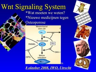
1. J. Coen NetelenbosJ. Coen Netelenbos
8 oktober 2008, IWO, Utrecht8 oktober 2008, IWO, Utrecht
Wnt Signaling SystemWnt Signaling System
*Wat moeten we weten?*Wat moeten we weten?
*Nieuwe medicijnen tegen*Nieuwe medicijnen tegen
OsteoporoseOsteoporose
2. Wnt signalingWnt signaling
WWg (wingless)g (wingless)
IntInt(egration)(egration)
The Wnt Homepage:
http://www.stanford.edu/~rnusse/wntwindow.html o.l.v.
Roel Nusse de ontdekker 1982 en naamgever Wnt in 1992
The Wnt Homepage:
http://www.stanford.edu/~rnusse/wntwindow.html o.l.v.
Roel Nusse de ontdekker 1982 en naamgever Wnt in 1992
3. plasterk
Nature 425, 633-637Nature 425, 633-637
(9 October 2003)(9 October 2003)
The Wnt/ -catenin pathwayThe Wnt/ -catenin pathway
regulates cardiac valveregulates cardiac valve
formationformation
Adam F. L. Hurlstone…….Adam F. L. Hurlstone…….
RonaldRonald H. A. PlasterkH. A. Plasterk11
andand
Hans CleversHans Clevers11
Wnt pathway componentsWnt pathway components ``
Wnt &Wnt &
4. Wnt & heart diseaseWnt & heart disease
• Wound healing response after myocardial infarction in ratsWound healing response after myocardial infarction in rats
and mice (and mice (Cardiovascular Research 2002 55(1):16-24)Cardiovascular Research 2002 55(1):16-24)
• Cardiac Hypertrophy and the Wnt PathwayCardiac Hypertrophy and the Wnt Pathway
(Cingolani OH et al. (Hypertension.(Cingolani OH et al. (Hypertension.
2007;49:427.)2007;49:427.)
.
5. Boyden LM et al. N Engl J Med 2002;346:1513-21Boyden LM et al. N Engl J Med 2002;346:1513-21
High bone density due to a gain-of-function mutationHigh bone density due to a gain-of-function mutation
inin LDL-Receptor Related Protein 5 (LRP5)LDL-Receptor Related Protein 5 (LRP5)
Wnt pathway speelt rolWnt pathway speelt rol
bij stimulatie humanebij stimulatie humane
osteoblasten met LRP5osteoblasten met LRP5
als co-receptor!als co-receptor!
6. Gong Y et al. Cell 2001;107:513-23Gong Y et al. Cell 2001;107:513-23
OsteoPorosis-PseudoGlioma syndrome (OPPG) due toOsteoPorosis-PseudoGlioma syndrome (OPPG) due to
a loss-of-function mutation in LDL-Receptor Relateda loss-of-function mutation in LDL-Receptor Related
Protein 5 (LRP5)Protein 5 (LRP5)
7. Wnt Signaling by Low-Density LipoproteinWnt Signaling by Low-Density Lipoprotein
Receptor-Related Protein 5 (LRP5),Receptor-Related Protein 5 (LRP5),
a coreceptor in Wnt signaling systema coreceptor in Wnt signaling system
LRP5(-/-) KO muis:LRP5(-/-) KO muis:
osteoporose enosteoporose en
pseudoglioom (OPPG)pseudoglioom (OPPG)
LRP5 gain of functionLRP5 gain of function
muation: hoge bot massamuation: hoge bot massa
8. Central Role of Canonical Wnt SignalingCentral Role of Canonical Wnt Signaling
Khosla S et al. JCI 2008;118:421-8
9. List target genes of Wnt/List target genes of Wnt/ßß-catenin signaling-catenin signaling
sept 2008sept 2008
10. Regulation Canonical Wnt SignalingRegulation Canonical Wnt Signaling
Piters E et al. Arch Biochem Biophysics 2008;473:112-6
11. Regulation Canonical Wnt Signaling (A)Regulation Canonical Wnt Signaling (A)
Frizzeld baconFrizzeld bacon
Low densityLow density
lipoproteinlipoprotein
Receptor-Receptor-
Related ProteinRelated Protein
=coreceptor=coreceptor
CatenaCatena
T-Cell-specificT-Cell-specific
transcriptiontranscription
FactorFactor
•ProliferatieProliferatie
•DifferentiatieDifferentiatie
•ApoptosisApoptosis
Dvl
DishevelledDishevelled
protein (Dvl)protein (Dvl)
Osteoblasten
12. Regulation Canonical Wnt Signaling (A)Regulation Canonical Wnt Signaling (A)
Dvl
GlycogeenGlycogeen
Synthetase KinaseSynthetase Kinase
LithiumLithium
&&
Small moleculesSmall molecules
met zelfde effectmet zelfde effect
op bot bij OVX-op bot bij OVX-
rat als PTH!rat als PTH!
•ProliferatieProliferatie
•DifferentiatieDifferentiatie
•ApoptosisApoptosis
Osteoblasten
13. Regulation Canonical Wnt Signaling (B)Regulation Canonical Wnt Signaling (B)
Piters E et al. Arch Biochem Biophysics 2008;473:112-6
DickkopfDickkopf (Dkk)(Dkk)
Dkk-1 InhibitorDkk-1 Inhibitor
(mAb) met(mAb) met
zelfde effectzelfde effect
op bot bij OVX-op bot bij OVX-
muis als PTH!muis als PTH!
15. ** SFRP KO (-/-) muisSFRP KO (-/-) muis::
O-blast ontwikkeling en functieO-blast ontwikkeling en functie
Apoptosis O-blast en O-cyteApoptosis O-blast en O-cyte
** rat met GIOP:rat met GIOP:
SFRP-1 expressieSFRP-1 expressie
Secreted Frizzled Related Protein-1 (sFRP-1) isSecreted Frizzled Related Protein-1 (sFRP-1) is
inhibitor of Wnt andinhibitor of Wnt and
Key Regulator Survival O-blast & O-cyteKey Regulator Survival O-blast & O-cyte
16. Sclerostin and Wnt SignalingSclerostin and Wnt Signaling
Buchem diseaseBuchem disease &&
Sclerostosis metSclerostosis met
mutaties SOST gene,mutaties SOST gene,
waardoor tekort aanwaardoor tekort aan
sclerostinsclerostin
toename botvormingtoename botvorming
First Case (1955)First Case (1955)
Antilichamen tegenAntilichamen tegen
Sclerostin (Scl-Ab)Sclerostin (Scl-Ab)
Phase 1 studyPhase 1 study::
BMD , BFR , BRRBMD , BFR , BRR =
17. Weekly s.c. antibody to sclerostin elevates BMDWeekly s.c. antibody to sclerostin elevates BMD
reversibly in OVX rats (1211 ASBMR 2008)reversibly in OVX rats (1211 ASBMR 2008)
Ab-Scerostin
18.
19. LRP5 in MechanotransductionLRP5 in Mechanotransduction
Sawakami K et al. J Biol Chem 2006;281:23698-711Sawakami K et al. J Biol Chem 2006;281:23698-711
21. PTHR1 Signaling in OsteocytesPTHR1 Signaling in Osteocytes
O’Brien CA et al. Plos ONE 2008;3:e
Bij LRP5-/- muisBij LRP5-/- muis
geen effect vangeen effect van
intermitt. PTH op HBMintermitt. PTH op HBM
Bij LRP5-/- transgeneBij LRP5-/- transgene
PTHR1 muis: geenPTHR1 muis: geen
verhoogde BM maarverhoogde BM maar
wel verhoogdewel verhoogde
remodeling dus andereremodeling dus andere
routeroute
Bij PTHR1- (gain ofBij PTHR1- (gain of
function) muis:function) muis:
verhoogde botmassaverhoogde botmassa
22. Wnt signaling: eating bone or adding it!Wnt signaling: eating bone or adding it!
Goldring SR et al. Nat Med 2007:13:133Goldring SR et al. Nat Med 2007:13:133
23. Wnt/Wnt/ß-catenin signaling systeem remt ontwikkelingß-catenin signaling systeem remt ontwikkeling
van mesenchymale stamcel tot kraakbeen en vetcelvan mesenchymale stamcel tot kraakbeen en vetcel
Baron R et al. Endocrinology 2008:148:2635-43Baron R et al. Endocrinology 2008:148:2635-43
24. PPARPPARγγ remt osteoblastgenesis &remt osteoblastgenesis &
bevordert osteoclastgenesisbevordert osteoclastgenesis
Takada I et al. IBMSBoneKEy 2008;5:258-61Takada I et al. IBMSBoneKEy 2008;5:258-61
ThiazolidinesThiazolidines
Role for SPPARMS:
good for bone and fat?
25. RANKL in osteoclastogenesis in InflammationRANKL in osteoclastogenesis in Inflammation
Lorenzo J et al. Endocr Rev 2008;29:403-440Lorenzo J et al. Endocr Rev 2008;29:403-440
Wnt?
26. DKK-1 in Osteoarthritis (OA),Rheumatoid ArthritisDKK-1 in Osteoarthritis (OA),Rheumatoid Arthritis
(RA) and Ankylosing Spondylitis (AS)(RA) and Ankylosing Spondylitis (AS)
Diarra D et al. Nat.Med 2007;13:156
Dickkopf-1 is a master regulator of joint remodeling
Effect anti-TNFEffect anti-TNF
on DKK-1on DKK-1
Effect disease activityEffect disease activity
on DKK-1on DKK-1
in RA and ASin RA and AS
Synovial tissueSynovial tissue
27. RANKL & Wnt in Rheumatoid ArthritisRANKL & Wnt in Rheumatoid Arthritis
Diarra D et al. Nat.Med 2007;13:156
Dickkopf-1 is a master regulator of joint remodeling
normaal rheum.arthritis
toepassen vantoepassen van
anti-DKK-1:
28. Role DDK1 Osteolysis in MyelomaRole DDK1 Osteolysis in Myeloma
Pinzone JJ et al. Blood 2008 sept on linePinzone JJ et al. Blood 2008 sept on line
KAHLER:KAHLER:
* Bortezomib* Bortezomib
remt DDK-1remt DDK-1
* Antistoffen* Antistoffen
tegen DDK1tegen DDK1
therapeutischtherapeutisch
op bot- enop bot- en
tumorlesietumorlesie
•DenosumabDenosumab
(Ab-RANKL)(Ab-RANKL)
alleen effectiefalleen effectief
op botlesiesop botlesies
29. Putative model involving Wnt/Putative model involving Wnt/ßcatenin in cancer cells;ßcatenin in cancer cells;
Kremen as possible gatekeeperKremen as possible gatekeeper
Nakamura T et al. J Cell Mol Med 2008;12:391-408Nakamura T et al. J Cell Mol Med 2008;12:391-408
32. Putative model involving Wnt/Putative model involving Wnt/ßcatenin in cancer cellsßcatenin in cancer cells
Nakamura T et al. J Cell Mol Med 2008;12:391-408Nakamura T et al. J Cell Mol Med 2008;12:391-408
33. Wnt in Prostate CancerWnt in Prostate Cancer
Hall CL et al. JCB 2006;97:661-72Hall CL et al. JCB 2006;97:661-72
• osteolytic phaseosteolytic phase: mediated by RANKL, PTHRP, and: mediated by RANKL, PTHRP, and
DKK-1DKK-1
• transitional phasetransitional phase: expression Wnts and decrease: expression Wnts and decrease
DKK-1DKK-1
• osteoblastic phaseosteoblastic phase: VEGF, endothelin-1: VEGF, endothelin-1
34. Dickkopf-1 [ DDK1]Dickkopf-1 [ DDK1]
• Remmer van Wnt/catenin signaling systeemRemmer van Wnt/catenin signaling systeem
(bindt aan LRPF5)(bindt aan LRPF5)
• Ontwikkeling skeletOntwikkeling skelet
• Homeostasis skeletHomeostasis skelet
• Hematopoeitische stamcel ?Hematopoeitische stamcel ?
• Osteolytische metastaseringOsteolytische metastasering
• Betrokken bij - botverlies door oestrogeentekortBetrokken bij - botverlies door oestrogeentekort
- GIOP- GIOP
- erosieve artritis- erosieve artritis
- teratogene effecten thalidomide- teratogene effecten thalidomide
35. Role DDK1 in boneRole DDK1 in bone
Pinzone JJ et al. Blood 2008 sept on linePinzone JJ et al. Blood 2008 sept on line
36. 19 Human Wnt Genes19 Human Wnt Genes
Link toLink to GeneGene cardscards SOURCESOURCE linklink HUGO linkHUGO link DiseaseDisease
WNT1
WNT1 WNT1
WNT2
WNT2 WNT2
WNT2B/13
WNT2B WNT2B
WNT3 WNT3 Tetra-Amelia (Niemann 2004)
WNT3A WNT3A
WNT4 WNT4
Mullerian-duct regression and virilization
(Biason-Lauber 2004)
SERKAL syndrome (Mandel, 2008)
WNT5A WNT5A WNT5A
WNT5B WNT5B
Associated with Susceptibility to type 2
diabetes (Kanazawa 2004)
WNT6 WNT6
WNT7A WNT7A WNT7A Fuhrmann syndrome
WNT7B WNT7B
WNT8A
WNT8A
WNT8B WNT8B WNT8B
WNT9A
(previously WNT14)
WNT9A WNT9A
WNT9B
Previously WNT15)
WNT9B
WNT10A WNT10A
Odonto-onycho-dermal dysplasia Adaimy
, 2007
WNT10B WNT10B WNT10B
 Mutations in Obesity patients
Christodoulides 2006
Split-Hand/Foot Malformation (Ugur 2008
)
WNT11 WNT11 WNT11
WNT16 WNT16
37. Human Diseases Caused by Abnormality Wnt SignalingHuman Diseases Caused by Abnormality Wnt Signaling
Nakamura T et al. J Cell Mol Med 2008;12:391-408Nakamura T et al. J Cell Mol Med 2008;12:391-408
39. DDK1 & Wnt Signaling in MSC differentiationDDK1 & Wnt Signaling in MSC differentiation
Pinzone JJ et al. Blood 2008 sept on linePinzone JJ et al. Blood 2008 sept on line
40. Biology of the Osteoclast in Bone MetabolismBiology of the Osteoclast in Bone Metabolism
Deftos LJ. NEJM 2005;353:872-5Deftos LJ. NEJM 2005;353:872-5
DenosDenosuumab
OdanaOdanacatib
Freedom TrialFreedom Trial::
3jaar bij 7868 PMP3jaar bij 7868 PMP
osteopor. vrouwenosteopor. vrouwen
•wervelFx 68%wervelFx 68%
•heupFx 40%heupFx 40%
•nonVertFX 20%nonVertFX 20%
41. Wnt & heart diseaseWnt & heart disease
• Wound healing response after myocardial infarction in rats and miceWound healing response after myocardial infarction in rats and mice
((Cardiovascular Research 2002 55(1):16-24)Cardiovascular Research 2002 55(1):16-24)
• Cardiac Hypertrophy and the Wnt/Frizzled PathwayCardiac Hypertrophy and the Wnt/Frizzled Pathway
(Cingolani OH et al. (Hypertension. 2007;49:427.)(Cingolani OH et al. (Hypertension. 2007;49:427.)
• LRP6 mutation in a family with early coronary disease and metabolicLRP6 mutation in a family with early coronary disease and metabolic
risk factors.risk factors. (Ani A et al.(Ani A et al. Science. 2007 MarScience. 2007 Mar
2;315(5816):1278-82 )2;315(5816):1278-82 )
.
Editor's Notes
opening
C elegans, rondworm nematode
Figure 1. Clinical and Radiographic Features of Affected Members of the Kindred. Photographs of an affected member at the ages of 12 years (Panel A) and 45 years (Panel B) show the development of the wide, deep mandible that was characteristic of all affected members of the kindred. A large, lobulated torus palatinus in an affected member (Panel C, arrow) was also characteristic of all affected kindred members. Characteristic radiographic findings included an abnormally thick mandibular ramus (arrow, Panel D); a markedly thickened cortex and narrowed medullary cavity (arrow) in the femur (Panel E), which was otherwise normal; and dense but otherwise normal- appearing vertebrae (Panel F).
Figure 1. Clinical Features of the Osteoporosis- Pseudoglioma Syndrome (A) Lower extremity photograph of a 40-yearold female OPPG patient demonstrating angular deformity of the tibia and fibula, the consequence of multiple fractures occurring during childhood. (B) Lateral lumbar spine radiograph of a 10- year-old male OPPG patient demonstrating abnormal flattening and concavity of the lumbar vertebrae. Affected individuals have normal growth during the first few years of life, but can fall below the normal range for height because of long bone fractures and vertebral collapse. (C) Photograph of the right eye of a 3-monthold male with OPPG demonstrating leukocoria (a white mass behind the pupil resembling retinoblastoma but actually a retrolental membrane). This individual had persistence of the tunica vasculosa partially covering the anterior lens surface, and evaluation of the dense retrolental membrane revealed a vascular central stalk that extended from the optic nerve into the vitreous cavity. (D and E) Iliac crest bone biopsies (both at 40 magnification) from an unaffected 2.5-year-old child (control) and from a 2.5-year-old child with OPPG, respectively. The control biopsy is substantially thicker than the OPPG biopsy. Note that the bone volume in the control (D) allows for only one cortical surface and a substantial trabecular bone network to be contained within the microscopic field. In contrast, both the inner and outer cortices and very little trabecular bone are contained within the field of the OPPG patient (E).
Figure 1. Prevention of Wnt Signaling by the Binding of Dickkopf (Dkk) Proteins to Low-Density Lipoprotein Receptor-Related Protein 5 (LRP5). In the absence of Dkk proteins, Wnt binding to its receptor and to its coreceptor, LRP5, activates intracellular signaling (Panel A). In the presence of Dkk proteins, the formation of an active signaling complex does not occur (Panel B).
Figure 2 The central role of canonical Wnt signaling in regulating osteoblast lineage specification, expansion, and terminal differentiation. Osteoblasts are derived from multipotent mesodermal or neural crest progenitors. Activation of the canonical (ca) Wnt signaling pathway, manifest through β-catenin stabilization, prevents the formation of cartilage (chondrogenesis). Wnt10b prevents adipogenesis (40). The canonical Wnt signaling pathway promotes survival of all cells of the osteoblast lineage and induces the proliferation of preosteoblasts. +, canonical Wnt signaling promotes the process; – , canonical Wnt signaling inhibits the process; AP, alkaline phosphatase; Cola1, collagen a1; DMP1, dentin matrix protein 1; OCN, osteocalcin; Osx, osterix; PTHR, receptor for PTH
Fig. 2. The canonical wnt/b-catenin signaling pathway and its extracellular regulation. (A) Extracellular binding of wnt to the Fz–LRP5/6 receptor complex causes intracellular accumulation of b-catenin that can induce the expression of target genes after translocation to the nucleus. (B) In the presence of Dkk and Krm a tertiary protein complex can be made with LRP5/6 for internalization thus inhibiting wnt signaling as b-catenin will no longer be stabilized but will be phosphorylated and subsequently degraded. (C) The extracellular sclerostin prevents by binding to LRP5/6–Fz further signaling.
Canonical wnt signaling can be regulated extracellularly, at the cell membrane, in the cytosol as well as in the nucleus. As shown in Fig. 2A, canonical wnt signaling is induced by binding of a wnt molecule to its receptor complex. This complex is composed of a member of the Fz receptor family, seven-transmembrane serpentine receptors, as well as a coreceptor either the LDL receptor related protein-5 (LRP5) or LRP6 [12–15]. Frizzled is a family of G protein-coupled receptor proteins [1] that serve as receptors in the Wnt signaling pathway and other signaling pathways. When activated, Frizzled leads to activation of Dishevelled in the cytosol. Frizzled proteins and the genes that encode them have been identified in an array of animals, from sponges to humans. Frizzled proteins also play key roles in governing cell polarity, embryonic development, formation of neural synapses , cell proliferation, and many other processes in developing and adult organisms. [2] Mutations in the human frizzled-4 receptor have been linked to familial exudative vitreoretinopathy, a rare disease affecting the retina at the back of the eye, and the vitreous, the clear fluid inside the eye.
Canonical wnt signaling can be regulated extracellularly, at the cell membrane, in the cytosol as well as in the nucleus. As shown in Fig. 2A, canonical wnt signaling is induced by binding of a wnt molecule to its receptor complex. This complex is composed of a member of the Fz receptor family, seven-transmembrane serpentine receptors, as well as a coreceptor either the LDL receptor related protein-5 (LRP5) or LRP6 [12–15]. Frizzled is a family of G protein-coupled receptor proteins[1] that serve as receptors in the Wnt signaling pathway and other signaling pathways. When activated, Frizzled leads to activation of Dishevelled in the cytosol. Frizzled proteins and the genes that encode them have been identified in an array of animals, from sponges to humans. Frizzled proteins also play key roles in governing cell polarity, embryonic development, formation of neural synapses, cell proliferation, and many other processes in developing and adult organisms.[2] Mutations in the human frizzled-4 receptor have been linked to familial exudative vitreoretinopathy, a rare disease affecting the retina at the back of the eye, and the vitreous, the clear fluid inside the eye.
One important mechanism of regulation involves Dickkopf (DKK) proteins that are secreted regulators which can bind to the coreceptor LRP5/6 preventing the formation of the LRP–Fz–wnt complex and thus inhibiting the induction of wnt signaling. Furthermore, in the presence of kremen1 or kremen2 (krm1/2), two-transmembrane proteins, a tertiary complex between krm, DKK and LRP5/6 can be formed which can be internalized reducing the availability of this coreceptor for its wnt-ligands (Fig. 2B) [16,17]. Alternative extracellular mechanisms to inhibit wnt signaling involve members of the secreted frizzled-related proteins (sFRPs) which have domains that can bind to wnts and thus prevent the binding of the latter to their receptor complex [18]. In a similar way wnt inhibitory factor-1 (Wif-1) can bind to wnts resulting in suppression of wnt signaling [18,19].
Recently, the two members of a gene family (SOST, encoding sclerostin, and WISE) are added to this list of extracellular wnt antagonists. As shown in Fig. 2C and discussed below, they are capable of binding to LRP5/6 and in this way modulating wnt signaling [20–23].
FIGURE 3. Lrp5/ mice have impaired osteogenic responses to mechanical loading. A , diagram of the noninvasive mouse ulna loading model, which applies cyclic compression to the forearm to produce mediolateral bending (due to natural curvature of the ulna) to mechanically stimulate ulnar bone tissue in adult mice in vivo . Midshaft ulnar tissue sections from control and loaded forearms among male Lrp5/ and Lrp5/ mice given fluorochrome injections after loading show a robust bone formation response on the medial ( inset ) and lateral surfaces of the loaded Lrp5/ ulna, yet almost no response can be observed in the loaded Lrp5/ ulna.
Figure 4 Potential cellular targets for the anabolic effects of PTH. PTH targets multiple cell types to mediate its bone anabolic effects. Specifically, PTH decreases apoptosis and proliferation of osteoblasts (OBs), as well as increasing their differentiation. It can activate bone-lining cells into functioning osteoblasts and decrease sclerostin production by osteocytes, which would be expected to increase Wnt signaling to osteoblasts. Finally, PTH is postulated to stimulate factors produced by osteoclasts (OCs) that stimulate osteoblasts.
Figure 7. PTHR1 Signaling in Osteocytes Leads to Increased Bone Mass and Remodeling via Distinct Mechanisms. In the proposed model, PTHR1 signaling in osteocytes activates at least two distinct pathways: one leading to increased bone mass and the other leading to increased bone remodeling. Suppression of Sost/sclerostin and activation of LRP5 signaling increase osteoblast numbers and are required for the increase in bone mass. The elevation of bone resorption is independent of the Sclerostin/LRP5 pathway. The question mark indicates uncertainty of the cellular source of RANKL and M-CSF (osteocytes versus stromal/osteoblastic cells). doi:10.1371/journal.pone.0002942.g007 PTHR1 Signaling in Osteocytes PLoS
Figure 1 Bifunctional role of the Wnt signaling pathway in regulation of osteoblast (bone-forming cell) and osteoclast (bone-resorbing cell) differentiation. Wnt signaling diverts the mesenchymal stem cells down the pathway of osteoblast differentiation. DKK-1 binds to the Wnt receptor complex on the surface of the osteoblast lineage cell and blocks Wnt signaling, arresting osteoblast proliferation and differentiation. The precursors of the mature osteoblast enhance bone resorption by boosting RANKLinduced osteoclastogenesis. Blockade of DKK-1 permits progression of osteoblast differentiation. Activation of the Wnt signaling pathway in the mature osteoblast upregulates OPG, which blocks RANKL-induced osteoclastogenesis, resulting in inhibition of bone resorption.
FIG. 2. Role of Wnt/-catenin signaling in determining the cell fate from mesenchymal progenitor cells. Wnt signaling plays a dual role in regulating chrondrocytic differentiation. The Wnt canonical pathway represses chondrocyte differentiation from progenitor cells, whereas it is required for chondrocyte hypertrophy. Wnt pathway activation also inhibits adipocyte differentiation and promotes osteoblast cell lineages by controlling proliferation, maturation, and terminal differentiation. Differentiated osteoblasts or osteocytes produce Wnt inhibitors such asDkk1 and sclerostin as a negative feedback control of osteoblast differentiation and/or function. Committed osteoblasts produce OPG to increase the OPG/RANKL ratio, thus decreasing osteoclast differentiation and activation
Fig. 1. PPAR-γ function in osteoclastogenesis and osteoblastogenesis. PPAR-γ inhibits osteoblastogenesis and promotes osteoclastogenesis. Cytokines such as IL-1, TNF-α and Wnt signals (Wnt-5a, Wnt-10b) suppress the transactivation function of PPAR-γ.
FIG. 3. Regulation of osteoclastogenesis in inflammation. In inflammatory states such as inflammatory arthritis, local production of proinflammatory cytokines (IL-1, IL-6, and TNF) as well as RANKL by inflamed tissues such as the synovium leads to stimulation of osteoclastogenesis and bone destruction. In addition, IL-17-producing TH17 T lymphocytes stimulate local production of RANKL by inflamed tissues and produce RANKL themselves, which enhances resorptive destruction of bone at sites adjacent to the inflammation.
( a ) In a physiological state, cortical bone formation and resorption next to joints are in balance. ( b ) Inflammatory arthritis such as rheumatoid arthritis leads to an imbalance between bone formation and resorption. Bone formation is hampered by TNF-mediated expression of DKK-1, which suppresses Wnt signals, whereas bone resorption is enhanced by expression of RANKL. ( c ) Blockade of DKK-1 met anti DKK-1 relieves Wnt signaling from DKK-1–mediated suppression and induces bone formation mirrored by the growth of osteophytes. Moreover, Wnt proteins induce OPG expression, which blocks RANKL-mediated bone resorption.
Figure 4. Metastatic cancer cells that secrete elevated levels of DKK1 disrupt the balance of osteoblastogenesis and osteoclastogenesis in favor of an osteolytic microenvironment that is conducive to tumor growth. MM has a unique and absolute requirement for the bone marrow microenvironment for its growth and survival and MM plasma cells may cultivate this ‘soil’ by synthesizing and secreting DKK1. The primary effect of DKK1 appears to be the disruption the differention of mesenchymal stem cells (MSC) in the osteoblasts (OB). RANK signaling regulates osteoclast development and Wnt signaling in MSC/OB differentially regulates RANK ligand (RANKL) and OPG, a RANKL decoy, which together modulate osteoclast development. DKK1 suppression of Wnt in MSC/OB leads to increased production of RANKL and IL-6 and decreased production of OPG. The shift in RANKL/OPG ratios leads to increased osteoclastogenesis and IL-6 is a potent survival factor MM cells. The subsequent loss of osteoblasts and increase in osteclasts causes lytic bone destruction, hyercalcemia, and loss of normal bone marrow function. A vicious cycle of bone destruction and tumor growth ensues. The absence of DKK1 in a subset of MM suggests that other soluble factors produced by MM cells may contribute to this process. These include MIP1a, sFRP-2, IL-3, RANKL and possibly sFRP-3. (see text for more details).
Figure 3. DKK1 is both anabolic and anti-catabolic in bone. TNFα enhances DKK1 secretion which inhibits MSC-derived osteoblastogenesis and lowers osteoprotegrin (OPG) levels, resulting in reduced bone accretion. In addition, DKK1 enhances receptor activator of NF-kappa B ligand (RANKL) levels, and the increased RANKL:OPG ratio activates osteoclast activity, leading to bone resorption.
Fig. 1. Overview of the different wnt signaling pathways. Binding of wnts to their receptor or receptor complex can result in two non-canonical pathways (Ca2+-dependent or the planar cell polarity (PCP) pathway) or the canonical pathway. The canonical or wnt/b-catenin pathway involves the induction of a cascade that results in the translocation of b-catenin to the nucleus where association with the lymphoid-enhancer binding factor (Lef)/T-cell specific transcription factors (Tcf’s) results in the expression of target genes (Fig. 1) [4,5], [http://www.stanford.edu/rnusse/wntwindow.html]. For intracellular accumulation and consecutive translocation to the nucleus, b-catenin needs to be hypophosphorylated. Glycogen synthase kinase 3 (GSK3) is, after complexation with Axin and adenomatous polyposis coli (APC), capable of phosphorylating b-catenin thus labeling it for proteasomal degradation (Fig. 1). However, binding of the wnt-ligand to one of the Frizzled (Fz) receptors results, through the protein Disheveled (Dvl), in inactivation of GSK3 and therefore prevents phosphorylation and consecutive degradation of b-catenin (For review see [4]). An alternative wnt pathway is calcium dependent and plays important roles during dorso-ventral patterning of the embryo, regulating cell migration, as well as heart development, and might play a role during tumor suppression. This wnt/calcium pathway relies on an intracellular release of Ca2+ to activate calcium sensitive enzymes like Ca2+-calmodulin dependent kinase II (CamKII), protein kinase C (PKC) or calcineurin (CaCN). TAK-1 (TGF-b activated kinase-1)/NLK (nemo-like kinase) was identified as a downstream target of CamKII for ventral development. Among the target proteins regulated by CaCN is the transcription factor Elk-1 and NF-AT (nuclear factor of activated T-cells). After activation, NF-AT enters the nucleus to regulate target gene expression [6]. Finally the planar cell polarity (PCP) pathway is also Dvl-dependent and results in coactivation of Rho and Rac, two small GTPases that are able to regulate cytoskeletal architecture. Wnt/Fz activation of Rac is independent of Rho and mediates activation of cjun NH2-terminal kinase (JNK) [7,8]. The JNK pathway is known to be required for embryonic morphogenesis and contributes to the regulation of cell proliferation and apoptosis. JNK also contributes to the functioning of some differentiated cells [9].
Figure 2. A prevailing model of the role of DKK1 and Wnt signaling in mesenchymal stem cell differentiation. Wnt3a, 5a, 7b and 10b, and Indian hedgehog (Ihh) cooperatively promote osteoblast differentiation, whereas DKK1 inhibits osteoblastogenesis and shifts the use only. From www.bloodjournal.org at VRIJE UNIVERSITEIT Medical Library 34942 on September 24, 2008. For personal 21 developmental program toward adipogenesis. Wnt-5A also inhibits adipogenesis by suppressing of PPAR-γactivation. β-catenin/TCF1 promotes early osteoblast lineage commitment through induction of the master bone development transcription factor Runt-related Transcription Factor 2/Runx2. Runx2 then promotes transcriptional activation of a second master bone development transcription factor Osterix (Osx), which utilizes DKK1 to repress Runx2 expression to further promote osteoblast progenitor maturation. Finally, phenotypic lineage-specific markers for osteoblast maturation are illustrated.