
Recommended
Recommended
More Related Content
Similar to IWO Meeting 1 November 2023 - Pitfalls DXA en VFA door Prof. dr. R. Slart (Groningen)
Similar to IWO Meeting 1 November 2023 - Pitfalls DXA en VFA door Prof. dr. R. Slart (Groningen) (20)
More from Stichting Interdisciplinaire Werkgroep Osteoporose
More from Stichting Interdisciplinaire Werkgroep Osteoporose (20)
Recently uploaded
Recently uploaded (20)
IWO Meeting 1 November 2023 - Pitfalls DXA en VFA door Prof. dr. R. Slart (Groningen)
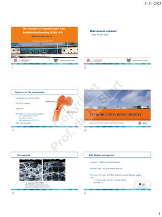
- 1. 5 -11 -2023 De waarde en beperkingen van botdichtheidmeting DXA/VFA Riemer Slart MD, PhD Nucleair geneeskundige, UMCG Disclosures speaker · None for this topic 1 2 Structure of the presentation · Introduction bone density (short) Cortical bone · The DXA – scanner · Applicants Trabecular bone · DXA/VFA: scoring, reporting, pitfalls ─ ‘the images behind the report’ ─ Examples (++pitfalls) ─ Other applications of DXA/VFA · Take home messages INTRODUCTION BONE DENSITY Department of Nuclear Medicine and Molecular Imaging 3 4 Osteoporosis Normal · Low bone mass (DXA: BMD) Osteoporosis Risk factors osteoporosis · Fracture ≥ 50 yrs & recent fracture · Persons long - term treatment with GC · Persons ≥ 60 years with RF, without a recent fracture and no GC ─ Low body weight , alcohol, smoking, falling , .. ─ Score RF ≥ 4 · Microarchitecture deterioration of bone · Increased bone fragility and risk on bone # 5 6 1 C o p y r i g h t P r o f . d r .
- 2. 5 -11 -2023 THE DXA - SCANNER Department of Nuclear Medicine and Molecular Imaging Principles of D(E)XA: UMCG Hologic Discovery · Dual-energy X -ray Absorptiometry (DXA) ─ 40 KeV, 78 KeV ─ 2D image ─ Bone mineral density = BMC/W (g/cm 2) · Bone mineral content = g/cm · W = width of the scanned line (cm) · Sites of measurement: ─ Lumbar spine ─ Hip ─ Wrist · Duration of examination: ─ Around 10 minutes · Radation dose: ─ < 0.01 mSv 7 8 Quality control · Quality control is essential ─ Daily phantom measurements · What to look for? ─ Day tot day variance ─ Trend analysis · Variants less ~1% Current QA phantom Lumbar spine DXA Hip DXA See also the online International Society for Clinical Densitometry (ISCD) 2019 document and the updated version of 2023 Courtesy of T. van den Wyngaert 9 10 APPLICANTS Department of Nuclear Medicine and Molecular Imaging Applicants DXA’s · Total DXA’s / yr UMCG: ± 5000 ( ± 2000 patients ) · Fracture out-patient-clinic ( traumatology ) · Internal medicine ( endocrinology / rheumatology ) · Gynaecology · Pediatrics · Others ─ GP ─ Organ transplantation (anti -rejection medication) ─ Oncology 11 12 2 C o p y r i g h t P r o f . d r .
- 3. 5 -11 -2023 ASSESSMENT AND INTERPRETATION CAPABILITIES OF DXA Department of Nuclear Medicine and Molecular Imaging · Assessment of the quality of the images ─ Quality ─ ‘Reconstruction’ · Correct positioning of the patient · Correct positioning of reference lines · Correct normal reference values used · Reporting: ─ Specific findings: · Degeneration · Fractures · Calcifications of the aorta · Rare findings (e.g. metal, X- ray contrast) · Report Z- score and T - score · VFA/IVA (Vertebral fracture assessment) · Trabecular bone score (TBS) · Conclusion ➢ WHO- classification: use lowest for diagnosis (hip/spine) → does not apply to patients < 18 years of age ➢ Recommendation 13 14 Components of DXA report (ISCD) 15 16 T – score and Z – score · T - score: compared with a young adult (29 yrs ) ─ Fracture risk vs. young ─ 2 SD < mean: increased # risk ─ Used for WHO scoring T – score and Z – score WHO Classification Normal T- score ≥ - 1.0 · Z - score: compared with same age, gender & race ─ Fracture risk vs. comparable subject Low bone mass ( osteopenia ) Osteoporosis (severe) < - 1.0 to > - 2.5 ≤ - 2.5 (+ recent fracture ) ─ NHANES III database BMD interpretation using T-score vs. Z-score* (The National Health and Nutrition Examination Survey) T-score In post/perimenopausal women and men age 50 years and older Z -score In healthy premenopausal women, men under age of 50 years, and children** Cannot be applied to healthy premenopausal women, men under age 50, and children Z -score -2.0 or less is defined as “below the expected range for age” Z -score above -2.0 is “within the expected range for age” WHO definition of osteoporosis. * ISCD (International Society for Clinical Densitometry) Official Positions ** **Beside BMD also other criteria should be included. 17 18 3 C o p y r i g h t P r o f . d r .
- 4. 5 -11 -2023 19 20 Wrist DXA · Non -dominant forearm · Hyperparathyroidism · No option for lumbar /hip DXA · Extreme obesitas CAPABILITIES OF DXA - BMD 21 22 Wrist DXA Motion GE Lunar Hologic 23 24 4 C o p y r i g h t P r o f . d r .
- 5. 5 -11 -2023 ROI Lumbar spine DXA 25 26 27 28 29 30 5 C o p y r i g h t P r o f . d r .
- 6. 5 -11 -2023 Artefact VP drains: hydrocephaly 31 32 Corrrect lumbar spine vertebrae: f.u . 33 34 Z-score and database: ethnicity W/B 35 36 6 C o p y r i g h t P r o f . d r .
- 7. 5 -11 -2023 Proximal femur DXA Hip position · Use total hip or neck for diagnosis 37 38 Lesser trochanter 39 40 Benign bone tumour IgG4 and prednison therapy 41 42 7 C o p y r i g h t P r o f . d r .
- 8. 5 -11 -2023 Femur head (2x) necrosis Children · Not WHO criteria · Only Z - score · No osteoporosis / osteopenia in the conclusion · Specific database X-pelvic MRI (T2) 43 44 DXA: take into account: · Variability between different vendors of scanner systems · No repeating DXA < 1 year ( variation + 1%) ─ UMCG test-retest trial (EANM 2023 presentation) · NHANES III (USA) · Artifacts ! · Bone volume assessment challenging: ─ Overestimation BDM in larger subjects ─ Underestimation BDM in smaller subjects ( children) 45 46 CAPABILITIES OF DXA - VFA Fractures caused by osteoporosis 1. Low bone density 2. Disrupted microstructure 3. Fractures! · Incidence vertebral fractures ─ 0.7% in all women > 50 yr ─ 0.2% in all men > 50 yr · 38.000 - 60.000 QUALYs are lost 47 48 8 C o p y r i g h t P r o f . d r .
- 9. 5 -11 -2023 Vertebral fractures · N = 2500 · 2 out of 3 patients with a vertebral fracture have no complaints · A fracture is a strong risk factor of more fractures Jager et al, Osteoporos Int.2011 Apr;22(4):1059 -68. VFA and reconstruction VFA = vertebral fracture assessment · Discovery A performed VFA lateral position ( patient supine) ─ No parallax and obliquity errors ─ Reproducible, easy and comfortable patient positioning ─ Simple and user friendly operating · Computer · Simple Point and click Genant criteria · Quantitative · Manual · Correction < 25% hoogte afname 25- 40% >40% hoogte afname. 49 50 Validation VFA DXA in UMCG · 250 patients, retrospective study · VFA & conventional X - ray thoracic and lumbar spine · Quantitative comparison (6 - point method) · Genant classification: grade & deformity · Result: Kappa = 0.87 Hospers et al, Radiology , 2009 51 52 Components of VFA report (ISCD) Example VFA · Patient with Cushing’s disease · BMD T -scores ─ Lumbar -0.6 ─ Hip -0.1 · VFA : ─ mild fracture L1 53 54 9 C o p y r i g h t P r o f . d r .
- 10. 5 -11 -2023 Example VFA: challenging Example VFA: artifact 55 56 Example VFA: artifact CAPABILITIES OF DXA - WBC 57 58 Whole body composition: bone, lean mass, fat % Whole body composition 59 60 10 C o p y r i g h t P r o f . d r .
- 11. 5 -11 -2023 Abdominal fat - InnerCore · Visceral Adipose Tissue (VAT) calculated from WB ─ No extra acquisition and analysis time ─ No additional dose ─ Easier access than CT ─ Equivalent to CT measurement · VAT is a strong parameter in Cardiometabolic risk evaluation ─ normal < 100 cm² ─ 100 < increased < 160 cm² ─ high > 160 cm² CAPABILITIES OF DXA ABDOMINALAORTA CALCIFICATION (AAC) 61 62 AAC on DXA ·L1 · L1 ·L2 · L2 ·L3 · L3 ·L4 · L4 DXA X-LWK 63 64 Advanced DXA Using TBS (>40 yrs) Kaplan-Meier curve for cumulative event -free survival in each group and population at risk at each time point. Golestani et al., Ann Med 2010 The Trabecular Bone Sore (TBS) report is generated simultaneously with the standard DXA spine printout. The report calculates an overall TBS, displays a texture image of the spine, and provides age-matched reference values. 65 66 11 C o p y r i g h t P r o f . d r .
- 12. 5 -11 -2023 Complete femur imaging DXA – NEW FEATURES 67 68 Solution – 3D -Shaper®: Advanced 3D Analysis A software solution for the 3D visualization and analysis of the cortical trabecular bone from 2D DXA scans using 3D-DXA technology. Creating 3D model and 3D -Shaper® Clinical Practice Integration Auto detection of all new DXA scans PACS Confidential © 3D- Shaper Medical 2023 Confidential © 3D- Shaper Medical 2023 69 70 International Working Group on DXA Bone Mineral Density Practice and Reporting Recommendations A partnership between the European Association of Nuclear Medicine (EANM), Canadian Association of Nuclear Medicine (CANM), International Osteoporosis Foundation (IOF), European Society for Clinical and Economic Aspects of Osteoporosis, Osteoarthritis and Musculoskeletal Diseases (ESCEO) International Society for Clinical Densitometry (ISCD), American Society for Bone and Mineral Research (ASBMR) and European Calcified Tissue Society (ECTS) 71 72 12 C o p y r i g h t P r o f . d r .
- 13. 5 -11 -2023 TAKE HOME MESSAGES Department of Nuclear Medicine and Molecular Imaging Take home messages · DXA imaging is reliable, patient friendly, easy method with a low radiation burden to quantify bone density · Beware of : variability (vendors , precision ), accurate ROIs needed , under/ overestimation BDM, small vertebrae # or variants , artifacts , databases (NHANES III),…. · Clear indications: Osteoporose en fractuurpreventie 2022 FMS · Updated Practice Guideline of Dual - energy X- ray Absorptiometry (DXA) appears soon · DXA imaging is more than bone density measurement alone. ─ VFA is of additional value and needs attention (Already included as a standard exam in the NL for Osteoporosis assessment) ─ TBS ─ Assessment of AAC with the aid of VFA has a prognostic value in terms of cardiovascular risk assessment ─ WBC · DXA developments: in software & quantification: AFF, 3D femur head/neck · Train (repeat it !) your (new) technologists 73 74 Vragen? GC en DXA r.h.j.a.slart@umcg.nl Department of Nuclear Medicine and Molecular Imaging 75 76 77 13 C o p y r i g h t P r o f . d r .