Resultados preliminares do implante de um novo anel associado ao PRK para pre...Ferrara Ophthalmics
Dr. Sandro Coscarelli, Dr. Pablo Rodrigues, Dr. Guilherme Rocha e Dr. Leonardo Torquetti compilaram e compartilham seus resultados com o uso de Segmentos de Anel de Ferrara HM associado ao PRK para a correção da miopia de pacientes com corneas finas e contra indicados para as técnicas de Excimer Laser apenas.
A multicentric nonrandomized study was conducted in which a new 320-ICRS was placed in 138 eyes of 130 patients with keratoconus. Uncorrected distance visual acuity (UDVA), corrected distance visual acuity (CDVA), keratometry, corneal volume, asphericity, lines of vision gain/loss, and vectorial analysis were assessed preoperatively and at the final follow-up visit after the procedure.
PURPOSE: To report the clinical outcomes of implantation of a new Ferrara intrastromal corneal ring segment (ICRS) with a 210-degree arc length in eyes with keratoconus.
Dr. Guilherme Rocha, Dr. Paulo Ferrara, Dr. Leonardo Torquetti, Dra. Luciene Barbosa analisam os resultados dos implantes de Anel de Ferrara de Arco longo no pós operatório de 6 meses
Doktor Vedat Kaya, Canan Aslı Utine, Sezen Harmancı Karakuş, Işılay Kavadarlı ve Ömer Faruk Yılmaz tarafından hazırlanmış olan bu makaleyi ilginize sunarız.
Comparative Study of Visual Outcome between Femtosecond Lasik with Excimer La...iosrjce
IOSR Journal of Dental and Medical Sciences is one of the speciality Journal in Dental Science and Medical Science published by International Organization of Scientific Research (IOSR). The Journal publishes papers of the highest scientific merit and widest possible scope work in all areas related to medical and dental science. The Journal welcome review articles, leading medical and clinical research articles, technical notes, case reports and others.
Excepcional artigo produzido pela Dra. Jordana Sandes do CEROF, GO mostra os efeitos dos arcos de 140º em Ceratocones com valores de astigmatismo elevados.
Avaliação das alterações na curvatura anterior e posterior da Córnea, sua paquimetria, resultados visuais e refrativos após o Implante de Anel de Ferrara com o auxilio do Galilei.
Micro Endodontics Training in Delhi| Advanced Endodontics | DelhiDr. Rajat Sachdeva
Course Schedule
Day 1 – Lecture Highlights
Orientation of Microscope in Endodontics.
Introduction to Microscope.
Indications of use of microscope in endodontics.
Parts of Microscope.
Magnification Step Vs Zoom Lenses – focal Length.
Parfocaling and IPD adjustments.
Beam splitter and its use for Documentation.
– Access; Basics of the operating microscope endodontics
– Rubber dam application & tricks.
– Live patient demonstration with aseptic techniques
– Treatment planning, instruments and techniques
– Working length determination; Principles of Nickel Titanium Instrumentation
– Hands-on for sequence of instrumentation & Live patient demonstration.
Day 2
– Principles, instruments and techniques for root canal obturation
-Treatment of post endodontic disease.
– Restoration of the Endodontically Treated Tooth— Lecture and Hands-on.
– Retreatment versus Endodontic Surgery.
– Hands-on and live patient demonstration for endodontic surgery.
– Five clinical cases will be given to each participant under Microendodontics .
How long is the course?
The course will be conducted over two full days .
How many students are there in a batch?
There will only be two students in a batch. This will ensure that each student gets personal attention.
What are the materials I need to get for the course?
All materials for the course will be provided by the center.
How is this course different from the microscope courses?
1) There are only two participants in a course. This will ensure that each participant gets complete attention.
2) The entire course is done in a clinical setting with a mannequin, natural teeth & 5 patients will be provided to each participant .
3) Each participant will have a separate microscope and dental chair.
4) Every participant will continue to get online mentoring after the completion of the course
When is the course held?
The course is held every month in delhi. Participants can register in advance for whichever month they prefer. Since the number of participants per batch is limited to only two, it is advisable to book your slot well in advance.
For more information, you can book an appointment at
Dr Sachdeva's Dental Aesthetic And Implant Institute,
I 101, Ashok Vihar Phase 1, Delhi- 110052
Contact us at
• Phone : +919818894041,01142464041
• Our Websites:
• www.sachdevadentalcare.com
• www.dentalclinicindelhi.com
• www.dentalimplantindia.co.in
• www.dentalcoursesdelhi.com
• www.facialaestheticsdelhi.com
• Google+ link: https://goo.gl/vqAmvr
• Facebook link: https://goo.gl/tui98A
• Youtube link: https://goo.gl/mk7jfm
• Linkedin link: https://goo.gl/PrPgpB
• Slideshare link : http://goo.gl/0HY6ep
• Twitter Page : https://goo.gl/tohkcI
• Instagram page : https://goo.gl/OOGVig
Terminologies
Introduction
Implant treatment options at the extraction site
Timing for immediate implants
Indications of immediate implants
Contraindications of immediate implants
Advantages of immediate implants
Disadvantages of immediate implants
Rule of 5 triangles
Deciding factors for immediate implant treatment modality in extraction socket
Armamentarium required for atraumatic extraction
Jumping distance or critical space
Immediate implantation in the extraction socket of anterior maxilla
Immediate implantation in the extraction socket of anterior mandible
Immediate implantation in the extraction socket of multi-rooted posterior teeth
Clinical guidelines for esthetic outcomes when using immediate implant protocol.
Hard tissue changes after immediate implant placement
Soft tissue changes after immediate implant placement
Criteria and guidelines for immediate implant placement site
Risk and complication in immediate implant placement
Loading options for the immediately inserted implant
Survival and success rate of immediate implants
Recent advances: socket shield
Review of Literature
Conclusion
References
Evaluation of Hard and Soft Tissue Dimensions Around Zirconium Oxide Implant–...Dr Ripunjay Tripathi
journal club Evaluation of Hard and Soft Tissue Dimensions Around Zirconium Oxide Implant–Supported Crowns: A 1-Year Retrospective Study
Kniha at el , J Periodontol 2016;87:511-518. �
Slides do curso avançado de atualização em implante de Anel de Ferrara, elaborado por Ferrara Ophtalmics. Para material completo, acesse www.aneldeferrara.com.br
Purpose: To evaluate the corneal volume (CV) before and after Ferrara intrastromal corneal ring segments (ICRS) implantation and its influence in clinical outcomes in keratoconus patients.
Background: Nowadays, ICRS are a step in the treatment of keratoconus. The purpose of this study was to evaluate the refractive effect and the tomographic and biomechanical parameters in keratoconus patients implanted with Ferrara ICRS, and their stability after 18 months.
PURPOSE: To evaluate the long-term safety and effica- cy of Ferrara intrastromal corneal ring segments (ICRS) (Ferrara Ring; AJL, Boecillo, Spain) in patients with kera- toconus.
Purpose: To evaluate the influence of age and severity of keratoconus in the clinical outcomes of implantation of Ferrara intrastromal corneal ring segments (ICRS).
PURPOSE: To evaluate the clinical outcomes of implantation of Ferrara intrastromal corneal ring segments (ICRS) in patients with corneal ectasia after refractive surgery.
One way to optimize Corneal Cross linking (CXL) !! DiyarAlzubaidy
Ophthalmology Lectures: Corneal crosslinking is the only way approved to stop progression of Keratoconus,,let's review the old & new methods of crosslinking
Double Rings - New approachs to fine tune clinical outcomes.pdfFerrara Ophthalmics
Inimaginável há poucos anos, a associação de segmentos concêntricos adjuvantes no tratamento da miopia e ceratocone tem trazido resultados praticamente refrativo no tratamento de pacientes com alta miopia e astigmatismo. A associação de segmentos de 5mm e 6mm se faz possível pela alta qualidade e precisão dos Tomógrafos e Laser de Femtosegundo que tornam a cirurgia extremamente precisa e os resultados reprodutíveis e passíveis de reintervenção e adequação do planejamento inicial.
Confira a apresentação competa com as indicações, citação do artigo científico, os resultados do estudo clinico inicial com 42 olhos, os resultados, a técnica cirúrgica e o nomograma do Ferrara HM para pacientes com miopia moderada a alta e contraindicação a cirurgia de PRK por limitação de espessura corneana.
Na edição 1991 do terceiro trimestre de 2021 a Revisa Oftalmologia em Foco destacou em sua capa o Surgimento de um Novo Anel intra corneano com foco nos pacientes com miopia moderada a alta com contra indicação ao PRK por limitação da espessura da córnea
Estudo de Casos Clínicos do Implante de Anel de Ferrara para o tratamento do Ceratocone disponibilizado pela Dra. Jordana Sandes para Drylab da Ferrara Ophthalmics no Curso refrativa RIO 2022
Estudos do comportamento da luz e de sua refração no prisma demonstram como o filtro amarelo atua minimizando a formação de halos e glare no corpo dos segmentos de Anel de Ferrara™.
O intuito do filtro é minimizar qualquer desconforto que a presença do ICRS - Intra Corneal Ring Segments - possam causar ao paciente.
Esta tecnologia é exclusiva da Ferrara Ophthalmics e utilizada em 87 países.
Desde sua patente em 1987, o Anel de Ferrara e a técnica de implante vêm se aprimorando. Hoje, com indicações seguras e reprodutíveis a técnica é realizada em 87 países e mais de 600.000 olhos já forma implantados.
Acompanhe este resumo do Implante de Anel de Ferrara para correção do Ceratocone. Característica, Mecanismo de Ação, técnica Cirúrgica, os diferentes arcos e efeitos são alguns dos tópicos abordados
A Aula apresenta os conceitos básicos sobre implantes intra corneanos, a evolução do nomograma da Ferrara Ophthalmics, a classificação morfologia do aspecto coreano e alguns casos clínicos.
Autor: Dr. Guilherme Rocha
Indicações de nomograma de utilização de segmentos de 320º de arco a partir de analise estatística de 26 casos operados e correlação com diferentes arcos existentes.
HOT NEW PRODUCT! BIG SALES FAST SHIPPING NOW FROM CHINA!! EU KU DB BK substit...GL Anaacs
Contact us if you are interested:
Email / Skype : kefaya1771@gmail.com
Threema: PXHY5PDH
New BATCH Ku !!! MUCH IN DEMAND FAST SALE EVERY BATCH HAPPY GOOD EFFECT BIG BATCH !
Contact me on Threema or skype to start big business!!
Hot-sale products:
NEW HOT EUTYLONE WHITE CRYSTAL!!
5cl-adba precursor (semi finished )
5cl-adba raw materials
ADBB precursor (semi finished )
ADBB raw materials
APVP powder
5fadb/4f-adb
Jwh018 / Jwh210
Eutylone crystal
Protonitazene (hydrochloride) CAS: 119276-01-6
Flubrotizolam CAS: 57801-95-3
Metonitazene CAS: 14680-51-4
Payment terms: Western Union,MoneyGram,Bitcoin or USDT.
Deliver Time: Usually 7-15days
Shipping method: FedEx, TNT, DHL,UPS etc.Our deliveries are 100% safe, fast, reliable and discreet.
Samples will be sent for your evaluation!If you are interested in, please contact me, let's talk details.
We specializes in exporting high quality Research chemical, medical intermediate, Pharmaceutical chemicals and so on. Products are exported to USA, Canada, France, Korea, Japan,Russia, Southeast Asia and other countries.
Flu Vaccine Alert in Bangalore Karnatakaaddon Scans
As flu season approaches, health officials in Bangalore, Karnataka, are urging residents to get their flu vaccinations. The seasonal flu, while common, can lead to severe health complications, particularly for vulnerable populations such as young children, the elderly, and those with underlying health conditions.
Dr. Vidisha Kumari, a leading epidemiologist in Bangalore, emphasizes the importance of getting vaccinated. "The flu vaccine is our best defense against the influenza virus. It not only protects individuals but also helps prevent the spread of the virus in our communities," he says.
This year, the flu season is expected to coincide with a potential increase in other respiratory illnesses. The Karnataka Health Department has launched an awareness campaign highlighting the significance of flu vaccinations. They have set up multiple vaccination centers across Bangalore, making it convenient for residents to receive their shots.
To encourage widespread vaccination, the government is also collaborating with local schools, workplaces, and community centers to facilitate vaccination drives. Special attention is being given to ensuring that the vaccine is accessible to all, including marginalized communities who may have limited access to healthcare.
Residents are reminded that the flu vaccine is safe and effective. Common side effects are mild and may include soreness at the injection site, mild fever, or muscle aches. These side effects are generally short-lived and far less severe than the flu itself.
Healthcare providers are also stressing the importance of continuing COVID-19 precautions. Wearing masks, practicing good hand hygiene, and maintaining social distancing are still crucial, especially in crowded places.
Protect yourself and your loved ones by getting vaccinated. Together, we can help keep Bangalore healthy and safe this flu season. For more information on vaccination centers and schedules, residents can visit the Karnataka Health Department’s official website or follow their social media pages.
Stay informed, stay safe, and get your flu shot today!
Pulmonary Thromboembolism - etilogy, types, medical- Surgical and nursing man...VarunMahajani
Disruption of blood supply to lung alveoli due to blockage of one or more pulmonary blood vessels is called as Pulmonary thromboembolism. In this presentation we will discuss its causes, types and its management in depth.
Prix Galien International 2024 Forum ProgramLevi Shapiro
June 20, 2024, Prix Galien International and Jerusalem Ethics Forum in ROME. Detailed agenda including panels:
- ADVANCES IN CARDIOLOGY: A NEW PARADIGM IS COMING
- WOMEN’S HEALTH: FERTILITY PRESERVATION
- WHAT’S NEW IN THE TREATMENT OF INFECTIOUS,
ONCOLOGICAL AND INFLAMMATORY SKIN DISEASES?
- ARTIFICIAL INTELLIGENCE AND ETHICS
- GENE THERAPY
- BEYOND BORDERS: GLOBAL INITIATIVES FOR DEMOCRATIZING LIFE SCIENCE TECHNOLOGIES AND PROMOTING ACCESS TO HEALTHCARE
- ETHICAL CHALLENGES IN LIFE SCIENCES
- Prix Galien International Awards Ceremony
Lung Cancer: Artificial Intelligence, Synergetics, Complex System Analysis, S...Oleg Kshivets
RESULTS: Overall life span (LS) was 2252.1±1742.5 days and cumulative 5-year survival (5YS) reached 73.2%, 10 years – 64.8%, 20 years – 42.5%. 513 LCP lived more than 5 years (LS=3124.6±1525.6 days), 148 LCP – more than 10 years (LS=5054.4±1504.1 days).199 LCP died because of LC (LS=562.7±374.5 days). 5YS of LCP after bi/lobectomies was significantly superior in comparison with LCP after pneumonectomies (78.1% vs.63.7%, P=0.00001 by log-rank test). AT significantly improved 5YS (66.3% vs. 34.8%) (P=0.00000 by log-rank test) only for LCP with N1-2. Cox modeling displayed that 5YS of LCP significantly depended on: phase transition (PT) early-invasive LC in terms of synergetics, PT N0—N12, cell ratio factors (ratio between cancer cells- CC and blood cells subpopulations), G1-3, histology, glucose, AT, blood cell circuit, prothrombin index, heparin tolerance, recalcification time (P=0.000-0.038). Neural networks, genetic algorithm selection and bootstrap simulation revealed relationships between 5YS and PT early-invasive LC (rank=1), PT N0—N12 (rank=2), thrombocytes/CC (3), erythrocytes/CC (4), eosinophils/CC (5), healthy cells/CC (6), lymphocytes/CC (7), segmented neutrophils/CC (8), stick neutrophils/CC (9), monocytes/CC (10); leucocytes/CC (11). Correct prediction of 5YS was 100% by neural networks computing (area under ROC curve=1.0; error=0.0).
CONCLUSIONS: 5YS of LCP after radical procedures significantly depended on: 1) PT early-invasive cancer; 2) PT N0--N12; 3) cell ratio factors; 4) blood cell circuit; 5) biochemical factors; 6) hemostasis system; 7) AT; 8) LC characteristics; 9) LC cell dynamics; 10) surgery type: lobectomy/pneumonectomy; 11) anthropometric data. Optimal diagnosis and treatment strategies for LC are: 1) screening and early detection of LC; 2) availability of experienced thoracic surgeons because of complexity of radical procedures; 3) aggressive en block surgery and adequate lymph node dissection for completeness; 4) precise prediction; 5) adjuvant chemoimmunoradiotherapy for LCP with unfavorable prognosis.
These lecture slides, by Dr Sidra Arshad, offer a quick overview of physiological basis of a normal electrocardiogram.
Learning objectives:
1. Define an electrocardiogram (ECG) and electrocardiography
2. Describe how dipoles generated by the heart produce the waveforms of the ECG
3. Describe the components of a normal electrocardiogram of a typical bipolar leads (limb II)
4. Differentiate between intervals and segments
5. Enlist some common indications for obtaining an ECG
Study Resources:
1. Chapter 11, Guyton and Hall Textbook of Medical Physiology, 14th edition
2. Chapter 9, Human Physiology - From Cells to Systems, Lauralee Sherwood, 9th edition
3. Chapter 29, Ganong’s Review of Medical Physiology, 26th edition
4. Electrocardiogram, StatPearls - https://www.ncbi.nlm.nih.gov/books/NBK549803/
5. ECG in Medical Practice by ABM Abdullah, 4th edition
6. ECG Basics, http://www.nataliescasebook.com/tag/e-c-g-basics
MANAGEMENT OF ATRIOVENTRICULAR CONDUCTION BLOCK.pdfJim Jacob Roy
Cardiac conduction defects can occur due to various causes.
Atrioventricular conduction blocks ( AV blocks ) are classified into 3 types.
This document describes the acute management of AV block.
Tom Selleck Health: A Comprehensive Look at the Iconic Actor’s Wellness Journeygreendigital
Tom Selleck, an enduring figure in Hollywood. has captivated audiences for decades with his rugged charm, iconic moustache. and memorable roles in television and film. From his breakout role as Thomas Magnum in Magnum P.I. to his current portrayal of Frank Reagan in Blue Bloods. Selleck's career has spanned over 50 years. But beyond his professional achievements. fans have often been curious about Tom Selleck Health. especially as he has aged in the public eye.
Follow us on: Pinterest
Introduction
Many have been interested in Tom Selleck health. not only because of his enduring presence on screen but also because of the challenges. and lifestyle choices he has faced and made over the years. This article delves into the various aspects of Tom Selleck health. exploring his fitness regimen, diet, mental health. and the challenges he has encountered as he ages. We'll look at how he maintains his well-being. the health issues he has faced, and his approach to ageing .
Early Life and Career
Childhood and Athletic Beginnings
Tom Selleck was born on January 29, 1945, in Detroit, Michigan, and grew up in Sherman Oaks, California. From an early age, he was involved in sports, particularly basketball. which played a significant role in his physical development. His athletic pursuits continued into college. where he attended the University of Southern California (USC) on a basketball scholarship. This early involvement in sports laid a strong foundation for his physical health and disciplined lifestyle.
Transition to Acting
Selleck's transition from an athlete to an actor came with its physical demands. His first significant role in "Magnum P.I." required him to perform various stunts and maintain a fit appearance. This role, which he played from 1980 to 1988. necessitated a rigorous fitness routine to meet the show's demands. setting the stage for his long-term commitment to health and wellness.
Fitness Regimen
Workout Routine
Tom Selleck health and fitness regimen has evolved. adapting to his changing roles and age. During his "Magnum, P.I." days. Selleck's workouts were intense and focused on building and maintaining muscle mass. His routine included weightlifting, cardiovascular exercises. and specific training for the stunts he performed on the show.
Selleck adjusted his fitness routine as he aged to suit his body's needs. Today, his workouts focus on maintaining flexibility, strength, and cardiovascular health. He incorporates low-impact exercises such as swimming, walking, and light weightlifting. This balanced approach helps him stay fit without putting undue strain on his joints and muscles.
Importance of Flexibility and Mobility
In recent years, Selleck has emphasized the importance of flexibility and mobility in his fitness regimen. Understanding the natural decline in muscle mass and joint flexibility with age. he includes stretching and yoga in his routine. These practices help prevent injuries, improve posture, and maintain mobilit
These simplified slides by Dr. Sidra Arshad present an overview of the non-respiratory functions of the respiratory tract.
Learning objectives:
1. Enlist the non-respiratory functions of the respiratory tract
2. Briefly explain how these functions are carried out
3. Discuss the significance of dead space
4. Differentiate between minute ventilation and alveolar ventilation
5. Describe the cough and sneeze reflexes
Study Resources:
1. Chapter 39, Guyton and Hall Textbook of Medical Physiology, 14th edition
2. Chapter 34, Ganong’s Review of Medical Physiology, 26th edition
3. Chapter 17, Human Physiology by Lauralee Sherwood, 9th edition
4. Non-respiratory functions of the lungs https://academic.oup.com/bjaed/article/13/3/98/278874
Ozempic: Preoperative Management of Patients on GLP-1 Receptor Agonists Saeid Safari
Preoperative Management of Patients on GLP-1 Receptor Agonists like Ozempic and Semiglutide
ASA GUIDELINE
NYSORA Guideline
2 Case Reports of Gastric Ultrasound
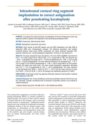
FR and the astigmatism after penetrating keratoplasty
1. Intrastromal corneal ring segment
implantation to correct astigmatism
after penetrating keratoplasty
Sandro Coscarelli, MD, Guilherme Ferrara, MD, Jose F. Alfonso, MD, PhD, Paulo Ferrara, MD, PhD,
Jesus Merayo-Lloves, MD, PhD, Luana P.N. Araujo, MD, Aydano P. Machado, PhD,
Jo~ao Marcelo Lyra, MD, PhD, Leonardo Torquetti, MD, PhD
PURPOSE: To evaluate the clinical outcomes of implantation of Ferrara intrastromal corneal ring
segments (ICRS) in patients with astigmatism after penetrating keratoplasty (PKP).
SETTING: Private clinic, Belo Horizonte, Brazil.
DESIGN: Retrospective consecutive case series.
METHODS: Chart records of post-PKP patients who had ICRS implantation from May 2005 to
September 2009 were retrospectively reviewed. The following parameters were studied:
corrected distance visual acuity (CDVA), keratometry (K) values, spherical equivalent (SE),
spherical refractive error, corneal topographic astigmatism, minimum K, and maximum K.
RESULTS: The study evaluated 59 eyes (54 patients). The mean CDVA (logMAR) improved from
0.45 G 0.17 (SD) (range 0.18 to 1.00) to 0.30 G 0.17 (range 0.00 to 1.00). The mean SE was
À6.34 G 3.40 diopters (D) (range 0.37 to À16.50 D) preoperatively and À2.66 G 2.52 D (range
0.87 to À10.50 D) postoperatively. The mean spherical refractive error decreased from À7.10 G
3.07 D (range 2.15 to 16.68 D) preoperatively to À3.46 G 2.05 D (range 0.88 to 10.79 D) postop-
eratively. No patient lost visual acuity. The mean corneal topographic astigmatism decreased
from 3.37 G 1.51 D preoperatively to 1.69 G 1.04 D postoperatively. The mean maximum
K value decreased from 48.09 G 2.56 D to 44.17 G 2.67 D and the mean minimum K value,
from 44.90 G 2.54 D to 42.46 G 2.63 D. All changes were statistically significant (P.0001).
CONCLUSION: Intrastromal corneal ring segments effectively reduced corneal cylinder in patients
with astigmatism after PKP.
Financial Disclosure: Drs. Ferrara and Merayo-Lloves have proprietary interest in the Ferrara ring.
Drs. Coscarelli, Torquetti, and Alfonso have no financial or proprietary interest in any material or
method mentioned.
J Cataract Refract Surg 2012; 38:1006–1013 Q 2012 ASCRS and ESCRS
The postoperative astigmatism associated with pene-
trating keratoplasty (PKP) is a common condition in
clinical practice. The reason for this could be related
to factors inherent to the receptor, such as previous
corneal trauma or keratoconus.1
Additional contrib-
uting factors may include the trephination technique,
inadequate fixation of the eye during surgery with
compression or deformation of the ocular globe,
the suture technique, and postoperative issues such
as the patient’s age and receptor corneal disease,
time on topical corticosteroids, and early suture
removal.2
Previous studies2–4
report several risk factors that
may increase the incidence of post-PKP astigmatism
and its management. Contact lenses are a better choice
than spectacles to correct astigmatism because they
provide better quality of vision.5,6
Moreover, the
corrected distance visual acuity (CDVA) with contact
lenses is usually better than with spectacles in this
type of astigmatism. When optical methods fail to
achieve satisfactory visual rehabilitation, surgical
treatment may be necessary. Whereas some authors
have published that incisions and wedge resection of
the cornea could be useful to correct the
Q 2012 ASCRS and ESCRS 0886-3350/$ - see front matter
Published by Elsevier Inc. doi:10.1016/j.jcrs.2011.12.037
1006
ARTICLE
2. astigmatism,7–9
other authors report that laser in situ
keratomileusis (LASIK)10,11
or the implantation of
toric phakic intraocular lenses (pIOLs) could achieve
better and more predictable results.12,13
In the present study, we evaluated the use of intra-
stromal corneal ring segments (ICRS) as an alternative
surgical option for the treatment of astigmatism in
patients who had PKP for keratoconus, bullous kerat-
opathy, radial keratotomy (RK), post-LASIK ectasia,
or stromal scarring. The outcome analysis comprised
the CDVA, spherical equivalent (SE), refractive error,
corneal topographic astigmatism, and minimum and
maximum keratometry (K) values. To our knowledge,
this study has the largest sample of patients with ICRS
implantation to correct post-PKP astigmatism in the
literature.
PATIENTS AND METHODS
This retrospective study included consecutive post-PKP
patients who had ICRS implantation (Ferrara ring, Ferrara
e Hijos) from May 2005 to September 2009 to correct residual
astigmatism. All patients were informed about the possible
intraoperative and postoperative complications and gave
written informed consent in accordance with institutional
guidelines and the Declaration of Helsinki.
Inclusion criteria were a clear and transparent corneal
graft, a minimum of 2.50 diopters (D) and a maximum of
8.00 D of astigmatism, and contact lens intolerance. All pa-
tients had at least 2 years of follow-up after PKP before
ICRS implantation. Patients who did not meet the inclusion
criteria were not evaluated in this study.
Surgical Technique
The same surgeon (S.C.) performed all surgeries using
a manual technique as previously described.14
The surgery
was performed using topical anesthesia after miosis was
achieved with pilocarpine 2.0%. The visual axis was marked
by pressing a Sinskey hook on the central corneal epithelium
while asking the patient to fixate on the corneal light reflex of
the microscope light. Using a marker tinted with gentian
violet, a 5.0 mm optical zone and incision site were aligned
to the desired axis in which the incision would be made.
This incision site was always at the steepest topographic
axis of the cornea given by the topographer.
A square diamond blade was set at 80% of corneal thick-
ness at the incision site, and this blade was used to make
the incision. A pocket was formed in each side of the incision
using a stromal spreader. Two (clockwise and counterclock-
wise) 270-degree semicircular dissecting spatulas were
consecutively inserted through the incision and gently
pushed with quick rotary back-and-forth tunneling move-
ments. After channel creation, the ring segments were
inserted using a modified McPherson forceps. The rings
were properly positioned with the aid of a Sinskey hook.
Postoperative Regimen and Assessment
The postoperative regimen consisted of tobramycin 0.3%
and dexamethasone 0.1% eyedrops 4 times a day for
1 week, after which the dose was tapered over 3 weeks. In
addition, patients received topical lubricants 4 times a day
for at least 3 months.
Postoperative examinations were performed at 1 and
7 days, after 1 and 6 months, and then every year. Measure-
ment of CDVA, slitlamp evaluation, refraction, corneal
topography, fundoscopy, and tonometry were performed
at each control visit. Visual acuity was determined on
a Snellen chart and then converted to logMAR notation.
The K values were obtained by corneal topography
(CT4000 Corneal Topographer, Eyetech, Inc.).
To evaluate the CDVA, refractive error, and corneal topo-
graphic astigmatism, the nonparametric Mann-Whitney test
was used because at least 1 datum from the sample did not
have a Gaussian distribution. The SE was corrected by Welch
transformation because of significant difference between
2 standard deviations (SDs).
Analysis of Astigmatism
The astigmatism results were analyzed arithmetically
(nonvector analysis) and with regard to the cylindrical axis
using vector analysis. Although empirical changes in cylin-
ders are commonly reported, they do not accurately reflect
the true nature of the change in cylinder. Cylinders have
a magnitude and an axis, which are related to the spherical
power.15
To take into account all 3 components, the data in
this study were transformed into Cartesian coordinates (ie,
x and y coordinates) to allow mathematic analyses. The re-
sult in the Cartesian coordinate form was then reconverted
into polar coordinates (sphere, cylinder, axis). To distinguish
the mean value of the cylinder calculated in this manner, the
term centroid has been proposed.16
Refractions before and 12 months after ICRS insertion
were assessed for astigmatism using the power vector
method.17
Any spherocylindrical refractive error was ex-
pressed by 3 dioptric powers: M, J0, J45, with M being the
aspheric lens equal to the SE of the given refractive error
and J0 and J45 being 2 Jackson cross-cylinders equivalent
to the conventional cylinders. These numbers are the coordi-
nates of a point in a 3-dimensional dioptric space, being the
power vector from the origin of this space to the point (M, J0,
J45). Thus, the length of the vector is a measure of the overall
blurring strength of the spherocylindrical refractive error.
Changes in refractive error induced by the surgery were
computed by the ordinary rules of vector extraction.
The target astigmatism is the intended astigmatic correc-
tion in each individual eye. The ideal target astigmatism
was zero (ie, intention to correct the full magnitude of the
cylinder).
Submitted: May 23, 2011.
Final revision submitted: November 18, 2011.
Accepted: December 5, 2011.
From Ennio Coscarelli Eye Clinic (Coscarelli), Paulo Ferrara Eye
Clinic (G. Ferrara, P. Ferrara, Torquetti), Belo Horizonte and
Brazilian Study Group of Artificial Intelligence and Cornea (Araujo,
Machado, Lyra, Torquetti), Maceio, Brazil; Fernandez-Vega Eye
Institute (Alfonso, Merayo-Lloves), Oviedo, Spain.
Corresponding author: Guilherme Ferrara, MD, Avenida Brasil,
1312 – Santa Efigenia – Belo Horizonte, MG 30140-001, Brazil.
E-mail: guilherme@ferrararing.com.br.
1007ICRS FOR ASTIGMATISM CORRECTION AFTER PKP
J CATARACT REFRACT SURG - VOL 38, JUNE 2012
3. Statistical Analysis
Data reported here are from the 12-month examination
after ICRS implantation. Statistical analysis included the
Student t test, Welch transformation, and Mann-Whitney
nonparametric test and was performed using Instat for Mac-
intosh software (version 3.1a, Graphpad Software). Vectorial
analysis was performed using SigmaPlot software (SPSS
Inc.). Internet-Based Refractive Analysis software (Zubisoft
GmbH) was used for clinical outcomes analysis.
RESULTS
This study included 59 eyes of 54 patients. The mean
age of the 28 women (51.85%) and 26 men (48.14%)
was 36.01 years G 11.02 (SD) (range 19 to 72 years).
The indications for PKP were keratoconus in 49 eyes,
post-LASIK ectasia in 5 eyes, progressive hyperopia
secondary to RK in 2 eyes, stromal scarring in 2 eyes,
and bullous keratopathy in 1 eye. Forty-nine patients
had a single eye treatment, whereas 5 patients had
both eyes treated.
All patients completed at least 1 year of follow-up.
The mean follow-up was 14 months.
Table 1 shows the preoperative and last follow-up
examination data. The preoperative corneal astigma-
tism ranged from 3.00 to 5.00 D. Figures 1 and 2
show the preoperative and postoperative CDVA. No
patient lost visual acuity. Of the patients, 28 (47.4%)
gained 2 or more lines of CDVA (Figure 3). The
improvements in CDVA, SE, and refractive error
were statistically significant (P!.0001).
Intended Correction
Regarding the predictability of the postoperative SE,
43 eyes (72.8%) presented with undercorrection and
9 (15.2%) with overcorrection. There was concordance
between the attempted refraction and achieved refrac-
tion in 7 eyes (13.0%) (Figure 4).
The decrease in the mean corneal topographic
astigmatism at 3.0 mm from preoperatively to post-
operatively was statistically significant (P!.0001).
Most eyes had more than 3.0 D of refractive astigma-
tism preoperatively (Figure 5). Approximately half
the eyes remained with more than 3.00 D of ref-
ractive astigmatism postoperatively; the rest had
less than 3.00 D (Figure 6). The decrease in K values
from preoperatively to postoperatively was
Table 1. Preoperative and last follow-up examination data.
Parameter Preoperative Postoperative
P
Value
Eyes (n) 59 d d
Patients (n) 54 d d
Sex (n)
Male 26 d d
Female 28 d d
Mean age (y) 36.01 G 11.02 d d
Mean follow-up (mo) 14 d d
CDVA (logMAR) .001
Mean G SD 0.45 G 0.17 0.30 G 0.17
Range 0.18, 1.00 0.00, 1.00
SE (D) .001
Mean G SD À6.34 G 3.40 À2.66 G 2.52
Range 0.37, À16.50 0.87, À10.50
Spherical refractive
error (D)
.001
Mean G SD À7.10 G 3.07 À3.46 G 2.05
Range 2.15 to 16.68 0.88 to 10.79
Mean TA at 3.0 mm (D) 3.37 G 1.51 1.69 G 1.04 .001
Mean maximum K (D) 48.09 G 2.56 44.17 G 2.67 .001
Mean minimum K (D) 44.90 G 2.54 42.46 G 2.63 .001
CDVA Z corrected distance visual acuity; K Z keratometry; SE Z spher-
ical equivalent; TA Z topographic astigmatism
Figure 1. The CDVA (logMAR) before and after the ICRS implanta-
tion (unpaired t test) (CDVA Z corrected distance visual acuity).
Figure 2. Preoperative and postoperative CDVA (logMAR) (CDVA
Z corrected distance visual acuity).
1008 ICRS FOR ASTIGMATISM CORRECTION AFTER PKP
J CATARACT REFRACT SURG - VOL 38, JUNE 2012
4. statistically significant (P!.0001) (Table 1). Vectorial
analysis showed that most eyes had a statistically
significant reduction in spherocylinder refractive
error (Figure 7).
Double-Angle Plot
Figure 8 shows the double-angle plots of the indi-
vidual cylinders, providing an overview of the cylin-
der magnitude (diopter) and axis (degree) of each
data point. The radius from the center of the plot to
each individual point represents the magnitude of
the cylinder. After ICRS implantation, the refractive
astigmatism centroid was 1.00 D closer to zero and
the SD of the astigmatism was reduced by a factor of
1.66 (3.83 D/2.31 D). The relocation of the centroid
closer to the origin and the contraction of the ellipse
on the doubled-angle plots show the amount of
improvement.
Figure 9 shows the doubled-angle plot of the preop-
erative and postoperative keratometric astigmatism.
Although there was a reduction in the mean kerato-
metric astigmatism, it was considerably less than the
reduction in refractive astigmatism.
There were no vision-threatening complications.
The ICRS were deeply implanted in all eyes
(Figure 10). In 3 eyes (5%), the surgical procedure
was interrupted due to dehiscence of the inner layers
of corneal transplantation, even 2 years after
transplantation.
Figure 3. Lines of CDVA lost and gained. Figure 4. Predictability of SE correction. The blue dots represent
undercorrection, the green dots represent full correction, and the
red dots represent overcorrection (Cor. Z correlation; Coeff. Z coef-
ficient; Lin. Z linear; SE Z spherical equivalent; Res. Var. Z re-
sponse variable).
Figure 5. Preoperative refractive astigmatism. Figure 6. Postoperative refractive astigmatism.
1009ICRS FOR ASTIGMATISM CORRECTION AFTER PKP
J CATARACT REFRACT SURG - VOL 38, JUNE 2012
5. DISCUSSION
Post-PKP residual astigmatism and refractive error are
frequently observed,11,18
and their management may
be a challenge for anterior segment surgeons. In this
retrospective study, we evaluated 59 eyes of 49 pa-
tients who had ICRS implantation to correct irregular
astigmatism after previous PKP. Despite proper
wound healing and good anatomic results, high
and/or irregular astigmatism can preclude satisfac-
tory vision in these patients.
Many factors inherent to the patient, host cornea,
surgical technique, and postoperative management
may influence the astigmatism.2
Peripheral disorders,
such as keratoconus,1
can persist on the corneal host
and cause irregular astigmatism. This cause possibly
explains the high prevalence of keratoconus patients
in our study. Although contact lenses and excimer la-
ser refractive surgery are viable options in this group
of eyes,19–21
contact lens–related problems, such as
dry eye, neovascularization, and rejection of donor
cornea, must be considered,22
as well as contraindica-
tions to PRK or LASIK because of high ametropia, low
baseline corneal thickness, and young age that make
these patients unsuitable for corneal laser refractive
surgery.23
The visual outcomes in our study were satisfactory.
The CDVA was unchanged in 16 eyes (27.2%)
Figure 7. Astigmatic vectors (J0 and J45) before and 12 months after
ICRS implantation. The more central location (0,0) of postoperative
data represents the reduction of preoperative astigmatism by the im-
plantation of the ICRS.
Figure 8. Double-angle plot of refractive astigmatism. Individual
cylinders demonstrate the cylinder magnitude (D) and axis (de-
grees). The radius from the center of the plot to each individual point
represents the magnitude of the cylinder.
Figure 9. Double-angle plot of keratometric astigmatism. Figure 10. Anterior segment optical coherence tomography shows
the ICRS deeply implanted in the cornea stroma and graft–host inter-
face (yellow arrow).
1010 ICRS FOR ASTIGMATISM CORRECTION AFTER PKP
J CATARACT REFRACT SURG - VOL 38, JUNE 2012
6. postoperatively, whereas 43 eyes (72.8%) improved at
least 1 line. The mean SE value decreased from À6.34
G 3.40 D to À2.66 G 2.52 D, and the K values also
decreased, improving the corneal irregularity.
Alfonso et al.24
and Morshirfar et al.12
evaluated the
results of pIOL implantation in young patients for the
correction of refractive errors after PKP and found
safe, predictable, and stable visual and refractive
outcomes. Alfonso et al.24
describe the efficacy, pre-
dictability, and safety of Implantable Collamer Lens
posterior chamber pIOL implantation for the correc-
tion of post-PKP refractive error in 15 eyes of 15 pa-
tients; there was a large reduction in the refractive
error and CDVA improvement. The postoperative
CDVA was 20/40 or better in 12 eyes (80%) and
20/25 in 6 eyes (40%). No eye lost more than 1 line
of acuity, 2 eyes gained 1 line, and 5 eyes gained
more than 2 lines; 8 eyes were unchanged. Morshirfar
et al.12
evaluated the Artisan iris-supported pIOL to
treat high myopia after PKP in 2 patients; there was
improvement in uncorrected distance visual acuity
and CDVA and no significant endothelial cell density
loss 6 months postoperatively.
Our findings are in agreement with results in other
studies.25–27
In a case report, Coskunseven et al.25
advocate the use of ICRS, a minimally invasive proce-
dure, to correct high astigmatism after PKP. According
to Coskunseven et al., eyes with thin corneal grafts and
recurrent keratoconus are unsuitable for laser refrac-
tive corrections because of the possibility of postoper-
ative complications. In addition, Arriola-Villalobos
et al.,5
in a series of 9 patients, found that ICRS implan-
tation improved the CDVA in all eyes and decreased
the topographic mean, minimum, and maximum
K values. They conclude that ICRS implantation might
be a good surgical choice to correct high astigmatism
after PKP and yields good visual, refractive, and
topographic outcomes.
Chang and Hardten27
recommend that ICRS im-
plantation after PKP not be performed until at least
1 year after transplantation and at least 3 months after
suture removal. We proceeded as Arriola-Villalobos
et al.5
suggest; that is, we waited at least 2 years after
corneal transplantation and a minimum of 6 months
after suture removal to avoid damaging the interface
by the traction generated by the dissectors used during
surgery in the manual technique.
The use of Ferrara ICRS with a 5.0 mm optical zone
has 2 advantages over the use of larger optical zone
ICRS. First, the central cornea flattening is theoretically
greater because the refractive outcome is inversely
proportional to the diameter of the segment.28
The
second benefit is that a small diameter ensures greater
distance between the rings and the graft–host junction.
This reduces the risk for interface dehiscence or
vascularization of the stromal channel by vessels ex-
tending from the limbus and host cornea. Thus, in pa-
tients with a corneal transplant with a diameter of
7.5 mm or smaller, the ICRS with larger optical zones
(6.0 or 7.0 mm) should not be used because the seg-
ments would be very close to the graft–host junction.
Potential disadvantages of ICRS with a small optical
zone are halos and glare. Some patients, especially
those with large pupil diameters in dim-light condi-
tions, occasionally report halos and glare.
All patients had PKP performed by the same
surgeon (S.C.), who always used a discrepancy of
0.50 mm in trephination between the donor graft
(8.0 mm) and the host (7.5 mm). Because the Ferrara
ICRS is placed at a 5.0 mm optical zone, it is always
located far from the graft–host junction, which makes
the procedure safer for small-optical-zone ICRS. How-
ever, in cases of small trephinations or decentered
grafts, care must be taken to avoid excessive torque,
which could open the previous keratoplasty wound,
thus requiring sutures and postponing ICRS im-
plantation. In our study, ICRS implantation had to
be postponed in 5% of cases (3/59 eyes) because
wound dehiscence occurred during dissection. All
patients had at least 2 years of follow-up after PKP,
which indicates that in some cases the graft–host inter-
face strength can be permanently reduced. This may
be related to the depth (too shallow or too deep) of
the passage of the 10-0 needle during the keratoplasty
and early removal of sutures. Tunnel creation using
the femtosecond laser in these cases could not only
facilitate the surgical procedure but also prevent this
type of complication.26,29
Implantation of ICRS after PKP may yield results
different than those when the technique is used for
keratoconus treatment. Keratoconic corneas are thin-
ner and more elastic than healthy corneas, whereas
corneas that had PKP are more rigid, with normal
thickness and elasticity. Theoretically, this could
explain why 73% of eyes presented with undercorrec-
tion and there was a low concordance between the
attempted refraction and the achieved refraction. The
nomogram of the Ferrara ICRS is designed for kerato-
conus treatment; thus, the ring thickness should be
adjusted when using ICRS for post-keratoplasty cases.
This means that thicker segments should be implanted
in post-PKP patients given the same K values as in
keratoconus patients. The main purpose of ICRS
implantation is to regularize the corneal surface and
improve visual acuity; the refraction reduction after
the surgery could be considered a secondary goal.
There are several potential advantages of ICRS
implantation over other surgical techniques in eyes
with high astigmatism after PKP. First, ICRS implanta-
tion avoids excimer laser treatment, eliminating
1011ICRS FOR ASTIGMATISM CORRECTION AFTER PKP
J CATARACT REFRACT SURG - VOL 38, JUNE 2012
7. central corneal wound healing, which could be unsat-
isfactory in post-PKP corneas. This leaves the optical
center of the cornea untouched, enhancing refractive
outcomes. Second, the technique is reversible in cases
of unsatisfactory refractive or clinical outcomes. Third,
adjustment can be performed using thinner or thicker
rings. In cases of unexpected corneal shape changes,
1 segment can be removed or exchanged. Fourth, it
avoids the complications of intraocular surgery.
The results in our study suggested that ICRS
implantation is a promising treatment for post-PKP
astigmatism, especially in eyes with thin and irregular
corneas. Long-term randomized comparative prospec-
tive studies are needed to better evaluate this tech-
nique as a treatment for irregular astigmatism in
post-PKP patients.
WHAT WAS KNOWN
Surgical treatments such as wedge resection of the
cornea, LASIK, and pIOL implantation are often necessary
to manage high astigmatism after PKP when nonsurgical
methods fail to achieve satisfactory visual acuity.
Intrastromal corneal ring segment implantation for the
treatment of post-PKP astigmatism has been described
in a case report and a 9-patient case series.
WHAT THIS PAPER ADDS
In a larger clinical series, ICRS implantation improved
CDVA in 73% of eyes and produced significant reduction
in topographic astigmatism. Dehiscence of the graft–
host junction with mechanical dissection occurred in 5%
of eyes.
REFERENCES
1. Lim L, Pesudovs K, Goggin M, Coster DJ. Late onset post-
keratoplasty astigmatism in patients with keratoconus. Br
J Ophthalmol 2004; 88:371–376. Available at: http://www.ncbi.
nlm.nih.gov/pmc/articles/PMC1772053/pdf/bjo08800371.pdf.
Accessed January 28, 2012
2. Barraquer RI, Alvarez de Toledo JP, Alvarez Fisher M, Martınez
Grau G. Prevencion y tratamiento del astigmatismo en querato-
plastia penetrante. Ann Oftalmol 2002; 10:69–80. Available at:
http://www.nexusediciones.com/pdf/ao2002_2/of-10-2-003.pdf.
Accessed January 28, 2012
3. Landau D, Siganos CS, Mechoulam H, Solomon A, Frucht-
Pery J. Astigmatism after mersilene and nylon suture use for
penetrating keratoplasty. Cornea 2006; 25:691–694
4. Alvarez de Toledo J, de la Paz MF, Barraquer RI, Barraquer J.
Long-term progression of astigmatism after penetrating
keratoplasty for keratoconus; evidence of late recurrence.
Cornea 2003; 22:317–323
5. Arriola-Villalobos P, Dıas-Valle D, G€uel JL, Iradier-Urrutia MT,
Jimenez-Alfaro I, Cui~na-Sardi~na R, Benıtez-del-Castillo JM.
Intrastromal corneal ring segment implantation for high astigma-
tism after penetrating keratoplasty. J Cataract Refract Surg
2009; 35:1878–1884
6. Price FW Jr, Whitson WE, Marks RG. Progression of visual
acuity after penetrating keratoplasty. Ophthalmology 1991;
98:1177–1185
7. Burillon C, Durand L, Hachmanian KF. La resection cuneiforme,
traitement correctif des astigmatismes corneens geants [Wedge
resection, corrective treatment of giant corneal astigmatism].
J Fr Ophtalmol 1989; 12:447–453
8. Saragoussi JJ, Abenhaim A, Pouliquen Y. Resultats des
incisions transverses dans la correction chirurgicale des forts
astigmatismes post-keratoplastie [Results of transverse
incisions in surgical correction of severe post-keratoplasty
astigmatism]. J Fr Ophtalmol 1990; 13:492–499
9. Coscarelli SA, Figueiredo P, Ramos Miranda MDF. Delam-
inac‚ ~ao lımbica: uma nova tecnica para correc‚ ~ao de astigmatis-
mo pos-transplante [Limbal delamination: a new technique for
postkeratoplasty astigmatism correction]. Arq Bras Oftalmol
2003; 66:189–192. Available at: http://www.scielo.br/pdf/abo/
v66n2/15472.pdf. Accessed January 28, 2012
10. Aguilar-Montes G, Perez-Balbuena L, Naranjo-Tackman R.
Manejo quirurgico con tecnica de lasik en la correccion del
astigmatismo miopico secundario a transplante corneal [Lasik
technique for myopic astigmatism correction after corneal
transplantation]. Rev Mex Oftalmol 2000; 74:221–236
11. Campos M, Hertzog L, Garbus J, Lee M, McDonnell PJ. Photo-
refractive keratectomy for severe postkeratoplasty astigmatism.
Am J Ophthalmol 1992; 114:429–436
12. Moshirfar M, Barsam CA, Parker JW. Implantation of an Artisan
phakic intraocular lens for the correction of high myopia after
penetrating keratoplasty. J Cataract Refract Surg 2004;
30:1578–1581
13. Moshirfar M, Gregoire FJ, Mirzaian G, Whitehead GF,
Kang PC. Use of Verisyse iris-supported phakic intraocular
lens for myopia in keratoconic patients. J Cataract Refract
Surg 2006; 32:1227–1232
14. Torquetti L, Berbel RF, Ferrara P. Long-term follow-up of intra-
stromal corneal ring segments in keratoconus. J Cataract
Refract Surg 2009; 35:1768–1773
15. Bennett AG, Rabbetts RB. Proposed new method of analysis. In:
Bennett AG, Rabbetts RB, eds, Clinical Visual Optics. London,
England, Butterworth, 1984; 437
16. Holladay JT, Dudeja DR, Koch DD. Evaluating and reporting
astigmatism for individual and aggregate data. J Cataract
Refract Surg 1998; 24:57–65
17. Thibos LN, Horner D. Power vector analysis of the optical out-
come of refractive surgery. J Cataract Refract Surg 2001;
27:80–85
18. Serdarevic ON, Renard GJ, Pouliquen Y. Randomized clinical
trial comparing astigmatism and visual rehabilitation after
penetrating keratoplasty with and without intraoperative suture
adjustment. Ophthalmology 1994; 101:990–999
19. Lipener C, Kwitko S, Uras R, Zamboni F, Lewinski R, Araujo A,
Pereira Lima R Jr. Adaptac‚ €ao de lentes de contato pos-trans-
plante de cornea [Fitting contact lenses after penetrating kerato-
plasty]. Arq Bras Oftalmol 1990; 53:41–44
20. Huang PYC,Huang PT, Astle WF, IngramAD, Hebert A,HuangJ,
Ruddell S. Laser-assisted subepithelial keratectomy and photore-
fractive keratectomy for post-penetrating keratoplasty myopia
and astigmatism in adults. J Cataract Refract Surg 2011;
37:335–340
21. Malecha MA, Holland EJ. Correction of myopia and astigmatism
after penetrating keratoplasty with laser in situ keratomileusis.
Cornea 2002; 21:564–569
1012 ICRS FOR ASTIGMATISM CORRECTION AFTER PKP
J CATARACT REFRACT SURG - VOL 38, JUNE 2012
8. 22. Genvert GI, Cohen EJ, Arentsen JJ, Laibson PR. Fitting
gas-permeable contact lenses after penetrating keratoplasty.
Am J Ophthalmol 1985; 99:511–514
23. Lee HS, Kim MS. Factors related to the correction of astigma-
tism by LASIK after penetrating keratoplasty. J Refract Surg
2010; 26:960–965
24. Alfonso JF, Lisa C, Abdelhamid A, Montes-Mico R, Poo-
Lopez A, Ferrer-Blasco T. Posterior chamber phakic intraocular
lenses after penetrating keratoplasty. J Cataract Refract Surg
2009; 35:1166–1173
25. Coskunseven E, Kymionis GD, Talu H, Aslan E, Diakonis VF,
Bouzoukis DI, Pallikaris I. Intrastromal corneal ring implantation
with the femtosecond laser in a post-keratoplasty patient with
recurrent keratoconus. J Cataract Refract Surg 2007;
33:1808–1810
26. Prazeres TMB, Cezar da Luz Souza A, Pereira NC, Ursulino F,
Grupenmacher L, Barbosa de Souza L. Intrastromal corneal ring
segment implantation by femtosecond laser for the correction of
residual astigmatism after penetrating keratoplasty. Cornea
2011; 30:1293–1297
27. Chang DH, Hardten DR. Refractive surgery after corneal
transplantation. Curr Opin Ophthalmol 2005; 16:251–255
28. Barraquer JI. Modification of refraction by means of intracorneal
inclusions. Int Ophthalmol Clin 1966; 6(1):53–78
29. Coskunseven E, Kymionis GD, Jankov MR II. Complications of
intra corneal ring segment implantation with femtosecond laser
channel creation in patients with keratoconus (explanations
and solutions). J Emmetropia 2010; 1:221–228. Available at:
http://www.journalofemmetropia.org/2171-4703/jemmetropia.
2010.1.221.228.pdf. Accessed January 28, 2012
1013ICRS FOR ASTIGMATISM CORRECTION AFTER PKP
J CATARACT REFRACT SURG - VOL 38, JUNE 2012