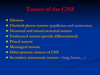
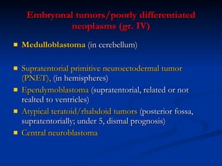
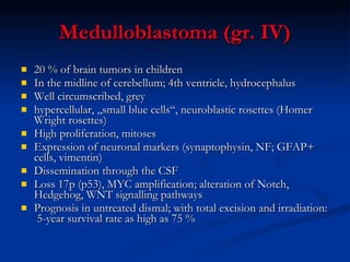
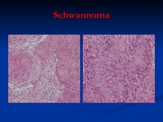
The Central Nervous System: Tumors The peripheral nervous system Markéta Hermanová
Tumor of the CNS Gliomas Chorioid plexus tumors  (papillomas and carcinomas) Neuronal and mixed neuronal tumors Embryonal tumors (poorly differentiated) Pineal tumors Meningeal tumors Other primary tumors of CNS Secondary (metastatic tumors –  lung, breast,…)
Gliomas Astrocytomas (AC) Diffuse astrocytomas (grade II) – LG AC (static, with slow progression) Variants: fibrillary, protoplasmic, gemistocytic, mixed Anaplastic astrocytomas (III) Glioblastoma multiforme (grade (IV) Pilocytic astrocytoma (I) Pleomorphic xanthoastrocytoma (II-III) Oligodendrogliomas Oligondendroglioma (II) Anaplastic oligodendroglioma (III) Mixed gliomas  Mixed oligoastrocytoma (II) and other mixed gliomas  Ependymal tumors Ependymoma (II) Anaplastic ependymoma (III) Myxopapillary ependymoma (I) Subependymoma (I)
High grade (HG) astrocytomas (AC) Anaplastic astrocytoma (III) Increased cellularity Increased degree of anaplasia, nuclear pleomorphism, increased proliferative activity Glioblastoma multiforme (IV) Necrosis and pseudopalisading Vascular endothelial proliferation Primary (in older) and secondary (in younger with history of LG AC)  Very poor prognosis  + Gliomatosis cerebri (III-IV)  – multiple regions, in some cases the entire brain infiltrated by neoplastic astrocytes
Glioblastoma multiforme
Pilocytic astrocytoma (I) Often cystic, also solid Usually circumscribed, arising from optic nerve to conus medullaris  Bipolar cells („hair cells“) + Rosenthal fibers and eosinophilic granular bodies Often biphasic (fibrillary areas + loose microcystic pattern) Usually first two decades Pleomorphic xantoastrocytoma (II-III) Temporal lobe of children and young adults Neoplastic occasionally bizarre astrocytes, also lipidized Necrosis and mitotic activity indicate higher grade
Oligodendroglioma (II) White matter of cerebral hemispheres (most frequently frontal lobes) Well circumscribed, gelatinous, gray masses, with cysts, hemorrhage, calcification Sheets of regular cells, clear halo of cytoplasm Delicate network of anastomosing capillaries Perineuronal satellitosis  LOH for 1p and 9q Better prognosis than AC +  anaplastic oligodendroglioma (III):  hypercellularity, nuclear anaplasia,  mitotic activity, necrosis
Oligodendroglioma
Ependymoma (II) Next to ependyma-lined ventricular system, 4th ventricle First 2 decades affected Solid or papillary masses; complete extirpation due to localization impossible  Small „blue“ cells, granular chromatin, dense fibrillary background, perivascular pseudorosettes and rosettes Anaplastic ependymoma (III) Subependymoma (I) benign, slowly growing, intraventricular Myxopapillary ependymoma (I) Filum terminale of spinal cord
Ependymoma
Neuronal and mixed neuronal tumors Gangliogliomas (I-II) Dysembryoblastic neuroepithelial tumor (DNET) In temporal lobe Associated with epilepsy Usually grade I; gangliogliomas may be gr. II/III  Dysplastic gangliocytoma of the cerebellum (I) Central neurocytoma (II) LG neuronal neoplasms Within vetricular system
Ganglioglioma
DNET
Embryonal tumors/poorly differentiated neoplasms (gr. IV) Medulloblastoma  (in cerebellum) Supratentorial primitive neuroectodermal tumor (PNET),  (in hemispheres)  Ependymoblastoma  (supratentorial, related or not realted to ventricles) Atypical teratoid/rhabdoid tumors  (posterior fossa, supratentorially; under 5, dismal prognosis)  Central neuroblastoma
Medulloblastoma (gr. IV) 20 % of brain tumors in children In the midline of cerebellum; 4th ventricle, hydrocephalus Well circumscribed, grey  hypercellular, „small blue cells“, neuroblastic rosettes (Homer Wright rosettes) High proliferation, mitoses Expression of neuronal markers (synaptophysin, NF; GFAP+ cells, vimentin) Dissemination through the CSF Loss 17p (p53), MYC amplification; alteration of Notch, Hedgehog, WNT signalling pathways Prognosis in untreated dismal; with total excision and irradiation:  5-year survival rate as high as 75 %
Medulloblastoma
Other tumors of CNS Primary CNS lymphomas (DLBCL) Germ cell tumors  Midline structures, pineal region, suprasellar region Teratomas; germinomas (similar to seminomas),…   Pineal parenchymal tumors Pinealoblastomas (high grade tumors) Pineocytomas (well differentiated) Gliomas in pineal region
Tumors of the meninges Meningioma  (meningothelial) - nonmeningothelial Meningeal hemangiopericytoma (so-called) Solitary fibrous tumors
Meningioma (gr. I- III ) Usually well defined rounded masses, adjacent to dura; encapsulated, extension into bone (reactive hyperostotic changes); less common „en plaque“ growth  Grade I meningiomas:  -  meningothelial  fibroblastic  transitional psammomatous  microcystic, secretory, angiomatous,…. Grade II meningiomas: atypical, clear cell, chordoid Grade III meningiomas: - anaplastic (malignant), rhabdoid, papillary
Meningioma
Craniopharyngeoma: Arise from squamous cell rests (derived from Rathke pouch) in sellar region Benign (gr. I), partly cystic epithelial tumor  Hemangioblastoma: Sporadic or ass. with VHL sy (in younger) Cerebellum (medulla, spinal cord,…., supratentorial, retinal in VHL) Well circumscribed, cystic, with mural nodule(s) Capillary-size and larger thin-walled vessels with intervening neoplastic „stromal cells“ (large polygonal, vaculated, lipid-rich, PAS+)
Familial tumor syndromes with involvement of tumor suppressor gene (AD) Cowden syndrome  PTEN  mutation Dysplastic gangliocytoma of the cerebellum Li Fraumeni syndrome Inactivation of p53 Medulloblastoma Turcot syndrome Mutations in  APC  or mismatch repair gene Medulloblastoma or glioblastoma Gorlin syndrome PTCH  mutations, upregulation of SHH medulloblastoma
Neurofibromatosis type I AD; neurofibromas (plexiform and solitary)+gliomas of optic nerve+pigmented nodules of iris-cutaneous hyperpigmented macules  (café au lait spots) Malignant transformation of neurofibromas NF1  gene (17q11.2); neurofibromin Neurofibromatosis type II AD; 8th nerve schwannomas and multiple meningiomas + gliomas, ependymomas of spinal cord + non-neoplastic lesions of Schwann cells, meningeal cells, hamartia NF2  gene (22q12); merlin Tuberous sclerosis complex AD; hamartomas and benign tumors of the brain and other tissues: cortical tubers (epileptogenic), subependymal nodules, subependymal giant cell astrocytomas,…, + renal angiomyolipomas, retinal glial hamartomas, pulmonary lympangioleiomyomatosis, cardiac rhabdomyoma + cysts – cutaneous lesions (angiofibromas, subungual fibromas, hypopigmented lesions) tuberin or hamartin genes mutated Von Hippel Lindau Disease AD; hemangioblastomas + cysts (pancreas, liver, kidney) + renal carcinomas, pheochromocytomas tumor suppressor gene – pVHL – 3p25-p26
Peripheral nerve sheath tumors Schwannoma benign, from neural crest-derived Schwann cell, component of NF2 well circumscribed, encapsulated, attached to nerve; 2 patterns: Antoni A and Antoni B often vestibular branch of 8th nerve; sensory nerves preferentially involved (trigeminus, dorsal roots,..); extradurally – large nerve trunks  Malignant peripheral nerve sheath tumor Highly malignant, medium and large nerves affected; in NF1   Neurofibroma:  Cutaneous:  localized, in dermis or subcucateously Plexiform:  infiltrating lesion growing within and expanding a peripheral nerve; NF1; potential for malignant transofrmation; significant neurologic deficits
Schwannoma
Diseases of peripheral nerves Inflammatory neuropathies Infectious polyneuropathies Hereditary neuropathies Acquired metabolic and toxic neuropathies Traumatic neuropathies
Inflammatory neuropathies Immune mediated neuropathies:   Guillain-Barré syndrome – GBS (acute inflammatory demyelinating polyradiculoneuropathy) Weakness in distal limbs, ascending paralysis, hospital intensive care before recovering normal function (up to 20 % long term disability); in some patients followed by a subacute or chronic course Inflammation and demyelination of spinal nerve roots and peripheral nerves (radiculoneuropathy) Infections or prior vaccination ass. with GBS  T-cell mediated immune response
Infectious polyneuropathies Leprosy (Hansen disease) Lepromatous leprosy:   Mycobacterium leprae invading Schwann cells Segmental demyelination, remyelination, loss of axons; endoneurial fibrosis and multilayered thickening of perineurial sheats Symmetric polyneuropthy; pain fibers (loss of sensation) Tuberculoid leprosy:  cell-mediated immune response to M. leprae – granulomatous inflammation in dermis, cutaneous nerves affected  Diphteria  (diphteria exotoxin; selective demyelination of axons) Varicella zoster virus  (varicella zoster virus; following chickenpox virus persists in neurons and sesory ganglia with potential ractivation)
Hereditary neuropathies Hereditary motor and sensory neuropathies (HSMN I-III,….) Hereditary sensory and autonomic neuropathies (HSANs) Familial amyloid polyneuropathies Peripheral neuropathy accompanying inherited metabolic disorders
HSMN HSMN - Charcot-Marie-Tooth  (peripheral myelin protein 22, myelin, connexin,… Demyelinating neuropathy; usually AD Repetitive de- and remyelinations (onion bulbs – Schwann cell hyperplasia) Slowly progressive, progressive muscular atrophy (legs) , uscle weakness, pes cavus HSMN II  (kinesin family member KIF1B) Axonal form – loss of myelinated axons HSMN III – Dejerine-Sottas neuropathy AR, genetically heterogeneous (the same genes as in HSMN I)  Enlarged peripheral nerves, trunk and limb muscles affected
Acquired metabolic and toxic neuropathies Peripheral neuropathy in adult onset diabetes mellitus  (polyol pathway and nonenzymatic glycation of proteins involved) Distal symmetric sensory or sensorimotor neuropathy Autonomic neuropathy Focal or multifocal asymmetric neuropathy Loss of small myelinated fibers, also unmyelinated fibers  Thickening of endoneurial arterioles Metabolic and nutritional neuropathies Uremic neuropathy Chronic liver disease, respiratory insuf., thyroid dysfunction Thiamine deficiency (neuropathic beriberi) Avitaminosis B 12 , B 6 , and E  Neuropathies associated with malignancy Brachial plexopathy (apex of a lung), obturator palsy (pelvic tumors), cranial verve palsies (intracranial tumors,….) Paraneoplastic effect (small cell ca of lungs, plasmocytoma)  Toxic neuropathies  Heavy metals, lead, arsenic
Tumors of autonomic nervous system Extraadrenal paragangliomas  (carotid body paragangliomas, vagal and other paragangliomas) non-chromaffin paragangliomas, usually related to parasympathetic nervous system Alveolar pattern, cell nests; chief cells and sustentakular cells Also malignant forms Extraadrenal paragangliomas of sympathoadreal neuroendocrine system  (anywhere from the pelvic floor to the neck) Pheochromocytomas (adrenal paraganglioma)  (production of katecholamins, hypertension, usually benign) Gangliocytic paraganglioma  (benign, in duodenum) Neuroblastoma and ganglioneuroblastoma In children under 4 ys (85 %) In adrenal gland or intra-abdominal sympathetic chain (70 %) and in thorax (at least 20 %) „ Small blue cell“ tumor, bulky, multinodular, hemorrhages and necrosis often, calcification, also pseudocystic, lobular or nesting pattern, fibrillary material between cells (neuritic cell processes) – neurofibrillary matrix, rosettes, chromatin: „salt-and- pepper“ appearance Ganglioneuroblastoma – some cytodifferentiation or maturation with recognizable ganglion cells  Ganglioneuroma In posterior mediastinum or retroperitoneum; some arising in adrenal gland Patient over 10 ys Well, circumscribed, with no necrosis or hemorrhages, on cut surface whorled or trabecular pattern Spindle cell matrix and mature ganglion cells
Pheochromocytoma
Neuroblastoma
Thank you for your attention …

Cns tumors and pns

  • 1.
    The Central NervousSystem: Tumors The peripheral nervous system Markéta Hermanová
  • 2.
    Tumor of theCNS Gliomas Chorioid plexus tumors (papillomas and carcinomas) Neuronal and mixed neuronal tumors Embryonal tumors (poorly differentiated) Pineal tumors Meningeal tumors Other primary tumors of CNS Secondary (metastatic tumors – lung, breast,…)
  • 3.
    Gliomas Astrocytomas (AC)Diffuse astrocytomas (grade II) – LG AC (static, with slow progression) Variants: fibrillary, protoplasmic, gemistocytic, mixed Anaplastic astrocytomas (III) Glioblastoma multiforme (grade (IV) Pilocytic astrocytoma (I) Pleomorphic xanthoastrocytoma (II-III) Oligodendrogliomas Oligondendroglioma (II) Anaplastic oligodendroglioma (III) Mixed gliomas Mixed oligoastrocytoma (II) and other mixed gliomas Ependymal tumors Ependymoma (II) Anaplastic ependymoma (III) Myxopapillary ependymoma (I) Subependymoma (I)
  • 4.
    High grade (HG)astrocytomas (AC) Anaplastic astrocytoma (III) Increased cellularity Increased degree of anaplasia, nuclear pleomorphism, increased proliferative activity Glioblastoma multiforme (IV) Necrosis and pseudopalisading Vascular endothelial proliferation Primary (in older) and secondary (in younger with history of LG AC) Very poor prognosis + Gliomatosis cerebri (III-IV) – multiple regions, in some cases the entire brain infiltrated by neoplastic astrocytes
  • 5.
  • 6.
    Pilocytic astrocytoma (I)Often cystic, also solid Usually circumscribed, arising from optic nerve to conus medullaris Bipolar cells („hair cells“) + Rosenthal fibers and eosinophilic granular bodies Often biphasic (fibrillary areas + loose microcystic pattern) Usually first two decades Pleomorphic xantoastrocytoma (II-III) Temporal lobe of children and young adults Neoplastic occasionally bizarre astrocytes, also lipidized Necrosis and mitotic activity indicate higher grade
  • 7.
    Oligodendroglioma (II) Whitematter of cerebral hemispheres (most frequently frontal lobes) Well circumscribed, gelatinous, gray masses, with cysts, hemorrhage, calcification Sheets of regular cells, clear halo of cytoplasm Delicate network of anastomosing capillaries Perineuronal satellitosis LOH for 1p and 9q Better prognosis than AC + anaplastic oligodendroglioma (III): hypercellularity, nuclear anaplasia, mitotic activity, necrosis
  • 8.
  • 9.
    Ependymoma (II) Nextto ependyma-lined ventricular system, 4th ventricle First 2 decades affected Solid or papillary masses; complete extirpation due to localization impossible Small „blue“ cells, granular chromatin, dense fibrillary background, perivascular pseudorosettes and rosettes Anaplastic ependymoma (III) Subependymoma (I) benign, slowly growing, intraventricular Myxopapillary ependymoma (I) Filum terminale of spinal cord
  • 10.
  • 11.
    Neuronal and mixedneuronal tumors Gangliogliomas (I-II) Dysembryoblastic neuroepithelial tumor (DNET) In temporal lobe Associated with epilepsy Usually grade I; gangliogliomas may be gr. II/III Dysplastic gangliocytoma of the cerebellum (I) Central neurocytoma (II) LG neuronal neoplasms Within vetricular system
  • 12.
  • 13.
  • 14.
    Embryonal tumors/poorly differentiatedneoplasms (gr. IV) Medulloblastoma (in cerebellum) Supratentorial primitive neuroectodermal tumor (PNET), (in hemispheres) Ependymoblastoma (supratentorial, related or not realted to ventricles) Atypical teratoid/rhabdoid tumors (posterior fossa, supratentorially; under 5, dismal prognosis) Central neuroblastoma
  • 15.
    Medulloblastoma (gr. IV)20 % of brain tumors in children In the midline of cerebellum; 4th ventricle, hydrocephalus Well circumscribed, grey hypercellular, „small blue cells“, neuroblastic rosettes (Homer Wright rosettes) High proliferation, mitoses Expression of neuronal markers (synaptophysin, NF; GFAP+ cells, vimentin) Dissemination through the CSF Loss 17p (p53), MYC amplification; alteration of Notch, Hedgehog, WNT signalling pathways Prognosis in untreated dismal; with total excision and irradiation: 5-year survival rate as high as 75 %
  • 16.
  • 17.
    Other tumors ofCNS Primary CNS lymphomas (DLBCL) Germ cell tumors Midline structures, pineal region, suprasellar region Teratomas; germinomas (similar to seminomas),… Pineal parenchymal tumors Pinealoblastomas (high grade tumors) Pineocytomas (well differentiated) Gliomas in pineal region
  • 18.
    Tumors of themeninges Meningioma (meningothelial) - nonmeningothelial Meningeal hemangiopericytoma (so-called) Solitary fibrous tumors
  • 19.
    Meningioma (gr. I-III ) Usually well defined rounded masses, adjacent to dura; encapsulated, extension into bone (reactive hyperostotic changes); less common „en plaque“ growth Grade I meningiomas: - meningothelial fibroblastic transitional psammomatous microcystic, secretory, angiomatous,…. Grade II meningiomas: atypical, clear cell, chordoid Grade III meningiomas: - anaplastic (malignant), rhabdoid, papillary
  • 20.
  • 21.
    Craniopharyngeoma: Arise fromsquamous cell rests (derived from Rathke pouch) in sellar region Benign (gr. I), partly cystic epithelial tumor Hemangioblastoma: Sporadic or ass. with VHL sy (in younger) Cerebellum (medulla, spinal cord,…., supratentorial, retinal in VHL) Well circumscribed, cystic, with mural nodule(s) Capillary-size and larger thin-walled vessels with intervening neoplastic „stromal cells“ (large polygonal, vaculated, lipid-rich, PAS+)
  • 22.
    Familial tumor syndromeswith involvement of tumor suppressor gene (AD) Cowden syndrome PTEN mutation Dysplastic gangliocytoma of the cerebellum Li Fraumeni syndrome Inactivation of p53 Medulloblastoma Turcot syndrome Mutations in APC or mismatch repair gene Medulloblastoma or glioblastoma Gorlin syndrome PTCH mutations, upregulation of SHH medulloblastoma
  • 23.
    Neurofibromatosis type IAD; neurofibromas (plexiform and solitary)+gliomas of optic nerve+pigmented nodules of iris-cutaneous hyperpigmented macules (café au lait spots) Malignant transformation of neurofibromas NF1 gene (17q11.2); neurofibromin Neurofibromatosis type II AD; 8th nerve schwannomas and multiple meningiomas + gliomas, ependymomas of spinal cord + non-neoplastic lesions of Schwann cells, meningeal cells, hamartia NF2 gene (22q12); merlin Tuberous sclerosis complex AD; hamartomas and benign tumors of the brain and other tissues: cortical tubers (epileptogenic), subependymal nodules, subependymal giant cell astrocytomas,…, + renal angiomyolipomas, retinal glial hamartomas, pulmonary lympangioleiomyomatosis, cardiac rhabdomyoma + cysts – cutaneous lesions (angiofibromas, subungual fibromas, hypopigmented lesions) tuberin or hamartin genes mutated Von Hippel Lindau Disease AD; hemangioblastomas + cysts (pancreas, liver, kidney) + renal carcinomas, pheochromocytomas tumor suppressor gene – pVHL – 3p25-p26
  • 24.
    Peripheral nerve sheathtumors Schwannoma benign, from neural crest-derived Schwann cell, component of NF2 well circumscribed, encapsulated, attached to nerve; 2 patterns: Antoni A and Antoni B often vestibular branch of 8th nerve; sensory nerves preferentially involved (trigeminus, dorsal roots,..); extradurally – large nerve trunks Malignant peripheral nerve sheath tumor Highly malignant, medium and large nerves affected; in NF1 Neurofibroma: Cutaneous: localized, in dermis or subcucateously Plexiform: infiltrating lesion growing within and expanding a peripheral nerve; NF1; potential for malignant transofrmation; significant neurologic deficits
  • 25.
  • 26.
    Diseases of peripheralnerves Inflammatory neuropathies Infectious polyneuropathies Hereditary neuropathies Acquired metabolic and toxic neuropathies Traumatic neuropathies
  • 27.
    Inflammatory neuropathies Immunemediated neuropathies: Guillain-Barré syndrome – GBS (acute inflammatory demyelinating polyradiculoneuropathy) Weakness in distal limbs, ascending paralysis, hospital intensive care before recovering normal function (up to 20 % long term disability); in some patients followed by a subacute or chronic course Inflammation and demyelination of spinal nerve roots and peripheral nerves (radiculoneuropathy) Infections or prior vaccination ass. with GBS T-cell mediated immune response
  • 28.
    Infectious polyneuropathies Leprosy(Hansen disease) Lepromatous leprosy: Mycobacterium leprae invading Schwann cells Segmental demyelination, remyelination, loss of axons; endoneurial fibrosis and multilayered thickening of perineurial sheats Symmetric polyneuropthy; pain fibers (loss of sensation) Tuberculoid leprosy: cell-mediated immune response to M. leprae – granulomatous inflammation in dermis, cutaneous nerves affected Diphteria (diphteria exotoxin; selective demyelination of axons) Varicella zoster virus (varicella zoster virus; following chickenpox virus persists in neurons and sesory ganglia with potential ractivation)
  • 29.
    Hereditary neuropathies Hereditarymotor and sensory neuropathies (HSMN I-III,….) Hereditary sensory and autonomic neuropathies (HSANs) Familial amyloid polyneuropathies Peripheral neuropathy accompanying inherited metabolic disorders
  • 30.
    HSMN HSMN -Charcot-Marie-Tooth (peripheral myelin protein 22, myelin, connexin,… Demyelinating neuropathy; usually AD Repetitive de- and remyelinations (onion bulbs – Schwann cell hyperplasia) Slowly progressive, progressive muscular atrophy (legs) , uscle weakness, pes cavus HSMN II (kinesin family member KIF1B) Axonal form – loss of myelinated axons HSMN III – Dejerine-Sottas neuropathy AR, genetically heterogeneous (the same genes as in HSMN I) Enlarged peripheral nerves, trunk and limb muscles affected
  • 31.
    Acquired metabolic andtoxic neuropathies Peripheral neuropathy in adult onset diabetes mellitus (polyol pathway and nonenzymatic glycation of proteins involved) Distal symmetric sensory or sensorimotor neuropathy Autonomic neuropathy Focal or multifocal asymmetric neuropathy Loss of small myelinated fibers, also unmyelinated fibers Thickening of endoneurial arterioles Metabolic and nutritional neuropathies Uremic neuropathy Chronic liver disease, respiratory insuf., thyroid dysfunction Thiamine deficiency (neuropathic beriberi) Avitaminosis B 12 , B 6 , and E Neuropathies associated with malignancy Brachial plexopathy (apex of a lung), obturator palsy (pelvic tumors), cranial verve palsies (intracranial tumors,….) Paraneoplastic effect (small cell ca of lungs, plasmocytoma) Toxic neuropathies Heavy metals, lead, arsenic
  • 32.
    Tumors of autonomicnervous system Extraadrenal paragangliomas (carotid body paragangliomas, vagal and other paragangliomas) non-chromaffin paragangliomas, usually related to parasympathetic nervous system Alveolar pattern, cell nests; chief cells and sustentakular cells Also malignant forms Extraadrenal paragangliomas of sympathoadreal neuroendocrine system (anywhere from the pelvic floor to the neck) Pheochromocytomas (adrenal paraganglioma) (production of katecholamins, hypertension, usually benign) Gangliocytic paraganglioma (benign, in duodenum) Neuroblastoma and ganglioneuroblastoma In children under 4 ys (85 %) In adrenal gland or intra-abdominal sympathetic chain (70 %) and in thorax (at least 20 %) „ Small blue cell“ tumor, bulky, multinodular, hemorrhages and necrosis often, calcification, also pseudocystic, lobular or nesting pattern, fibrillary material between cells (neuritic cell processes) – neurofibrillary matrix, rosettes, chromatin: „salt-and- pepper“ appearance Ganglioneuroblastoma – some cytodifferentiation or maturation with recognizable ganglion cells Ganglioneuroma In posterior mediastinum or retroperitoneum; some arising in adrenal gland Patient over 10 ys Well, circumscribed, with no necrosis or hemorrhages, on cut surface whorled or trabecular pattern Spindle cell matrix and mature ganglion cells
  • 33.
  • 34.
  • 35.
    Thank you foryour attention …