SlideShare a Scribd company logo
1 of 45
The
Schistosomes
The Blood fluke
Outline
– General feature of the blood fluke
– List of the blood flukes of medical importance
– For each species of blood flukes
• Geographical distribution
• Morphology and differential characteristics,
• Transmission and life Cycles,
• Laboratory diagnosis and
• Prevention and control
1- A genus of Trematodes, Schistosoma, commonly known as blood-fluke, are parasitic
flatworms responsible for a highly significant group of infections in humans termed
schistosomiasis.
.
2- The sexes are separate (they are dioecious)
3- They reside in the blood vessels of the definitive host in their life cycles. Hence the
common name “Blood Flukes.”
4- Male worm has a split body called the gynecophoral canal. The female is usually found
within this canal “safe in the arms of her lover.” She leaves only during the egg laying period.
Males are broader and females are filiform and larger than male
General feature of the blood fluke
Snail hosts
The different species of Schistosoma have different types of snails serving as their intermediate
hosts; these hosts are as follows:
1- Biomphalaria for S. Mansoni ‫البولية‬ ‫المجاري‬ ‫بلهارسيا‬ ‫قوقع‬
.))
2
- Oncomelania for S. japonicum.
3- Bulinus for S. haematobium. ‫المستقيم‬ ‫بلهارسيا‬ ‫قوقع‬
)
Symptoms of schistosomiasis are caused by the body’s reaction to the worms’ eggs.
Intestinal schistosomiasis
1- abdominal pain,
2- diarrhoea
3- and blood in the stool.
4- Liver enlargement is common in advanced cases, and is frequently associated with an
accumulation of fluid in the peritoneal cavity and hypertension of the abdominal blood vessels.
In such cases there may also be enlargement of the
The classic sign of urogenital schistosomiasis is:
1-haematuria (blood in urine).
2- Fibrosis of the bladder and ureter,
3- and kidney damage.
Learning objective
• At the end of this section the student will be
able to
– Explain the general feature of blood fluke
– List the blood flukes of medical importance
– Explain the geographical distribution,
Morphology, differential characteristics,
transmission, life Cycles and pathogenesis of each
species
– Apply the necessary laboratory procedure for
detection and identification of the liver fluke
Blood fluke
• S. mansoni
• S. hematobium
• S. japonicum
• S. intrecalatum
• S. mekongi
• Animal schistosome
which occasionally
infect humans
– Schistosoma mattheel
– Schistosoma bovis
– Schistosoma rodhaini
– Schistosoma
margrebowiei
The Schistosomes
• General feature:
– They reside in the blood
vessels of the definitive host
in their life cycles. Hence
the common name “Blood
Flukes.”
– The sexes are separate (they
are dioecious)
– They are long cylindrical(~
20mm) and adopted to life in
blood vessel
• Male worm has a split body
called the gynecophoral
canal. The female is usually
found within this canal “safe
in the arms of her lover.”
She leaves only during the
egg laying period.
• Males are broader and
females are filiform and
larger than male
– Humans are the only or most significant host for
most of the species
– Snail host is required as intermediate host to
complete their life cycle
– There are no second intermediate hosts
– Reproduction takes in the sporocyst stage in the
snail.
– No redia and metacercariae stages
– Cercaria is the infective stage to humans in water
and the infective route is by skin
– egg without operculum, but with spine
– The eggs are main pathogenic stage.
Epidemology:
• Wide spread species
– Schistosoma mansoni causes intestinal schistosomiasis and
is prevalent in 52 countries and territories of Africa,
Caribbean, the Eastern Mediterranean and South America
– Schistosoma haematobium causes urinary schistosomiasis
and affects 54 countries in Africa and the Eastern
Mediterranean
– Schistosoma japonicum cause intestinal schistosomiasis
and are Common in parts of Japan, China, Taiwan,
Philippines, Thailand, and other parts of Southeast Asia
• Less wide spread species
– Schistosoma mekongi cause intestinal schistosomiasis and
are prevalent in 7 African countries and the Pacific region
– Schistosoma intercalatum is found in ten African countries
Distribution of Schistosomiasis
Life cycle
Transmissions
Life Cycle of Schistosoma spp.
• The eggs escape from
the body by penetrating
the walls of the veins
and small intestine or
urinary bladder (where
adults reside), and they
are passed in the feces
or urine. Example of a
Schistosoma mansoni egg
Life Cycle of Schistosoma spp.
Miracidium Swimming towards Snail Intermediate Host
• The eggs, if and when they reach fresh water,
will quickly hatch.
• The miracidium swims ceaselessly until
finds a snail host (die in 3-6 hours).
Life cycle in Snail
• After miracidum enters, the parasite goes through two
asexual developmental stages: mother and daughter
sporocysts.
• Mother sporocysts contain the daughters, which are
then released and found in the snail’s digestive and
reproductive organs.
• The daughter sporocysts hold the cercaiae,
• Continues producing sporocystes for up to seven
weeks within snail
Intermediate Hosts of Schistosoma spp.
S. mansoni only infect snails of the genus Biomphalaria.
Intermediate Hosts of Schistosoma spp.
S. japonicum are found in Oncomelania snails
Intermediate Hosts of Schistosoma spp.
S. haematobium persist in species of Bulinus.
Life cycle of Schistosoma spp.: Cercariae
• Cercaiae start to
emerge four
weeks after
penetration by
miracidium
• There is NO
second
intermediate
host
Life cycle of Schistosoma spp.: Cercariae
• Cercariae swim up and down in the water column until
finding host; without host die after three days.
• Move around and then enter and can disappear below surface
in 10 to 30 seconds, and into circulation system within 24
hours.
5 Minutes after penetration of the human skin by cercaria,
the newly transformed schistosomule has penetrated the
outer layer of the epidermis, and is positioned just
beneath the skin.
20 minutes after penetration of
the skin. Here the
schistosomule is migrating
through the dermis. This it will
do until it locates a blood
vessel. The schistosomule will
then break through into the
blood vessel, to be transported
in the circulatory system to the
heart and then the lungs.
Life cycle of
Schistosoma spp.: In
Human
• Various ways of migrating
through circulatory system
(heart, liver). End up in
veins draining liver where
they develop for three
weeks
• Pair up in these veins and
migrate to walls of guts or
bladder, depending on
species to produce eggs.
Adults can live 20
to 30 years!!
Life cycle of Schistosoma spp.: In
Human
• Species differences in Site Preferences of
Adults (male and female in copula)
– S. mansoni –veins of Large Intestine
– S. haematobium – veins of bladder
– S. japonicum – veins of small intestine
• The infective inhabitation of
Schistosoma in mesenteric
vein
Life cycle of Schistosoma spp.:
In Human
• Unusual in that eggs are the damaging
stage
– Traverse the walls of veins, tissues and mucosa
– Egg promotes growth of granuloma and this is
moved out into lumen of intestine or bladder where
expelled.
Granuloma of Schistosoma
2/3 of eggs remain in abdomen granuloma
Intestinal schistosomiasis
• Penetration of skin:- Penetration of of skin by
cercariae causes transient dermatitis
(swimmers' itch)
• Migratory phase - 4-10 weeks after infection.
– Is characterized by fever and toxic or allergic
reactions resulting from migration of immature
organisms.
– Often results in bronchitis, hepatomegaly,
splenomegaly, and diarrhea.
• Chronic phase - persons living in endemic
regions are often asymptotic.
– May have mild, chronic bloody stools or urine.
– Often have formation of granulomas.
Hepatomegaly, Spleenomegaly, Ascites
(accumulation of fluid in abdominal cavity
– Terminal stage is characterized by portal vein
hypertension syndrome, common saying, abdomen
distention looks like a big drum
– Ectopic lesion: The damage to the central nervous
system ( brain, spinal ) may cause paralysis
(monoplegia, hemiplegia ).
• In less than 10% of
cases, granulomas can
cause blockage of blood
flow in liver causing
enlargement of the
spleen and fluid
retention in abdomen.
abdomen distention looks like
a big drum, Ascites,
emaciation, varicosity, and
splenomegaly
abdomen distention looks like a big drum,
emaciation looks like a fire wood.
A patient of
schistosomiasis
Laboratory diagnosis
• Intestinal schistosomiasis
– Finding the eggs in faeces by direct examination or more
commonly by using concentration ;
– Mucus and blood are often present in the faecal specimen
1. Formol-ether conc. Tech
2. Formol detergent gravity sed tech
3. Kotho-katz technique
– Examining a rectal biopsy for eggs when they cannot be
found in faeces.
– occasionally eggs may also be found in urine often
following faecal contamination
Egg:S.mansoni
• Size : 114-17µm 45-68µm
• Shape: Oval, with one well
rounded pole and one more
conical pole
• Colour: pale yellow-brown
• Spine: large, triangular lateral
spine near the rounded end
• Shell: smooth, very thin
• Content: fully embryonated
(developed miracidium) when
discharged with the faeces
Egg: S japonicum
• Size: 70-80m
• Shape: oval, almost
round
• Colour: transparent or
pale-yellow
• Spine: very small hook-
like spine laterally
• Contain a fully
developed miracidium
• Ab-detection
– Immunodiagnosis using ELISA, RIA, Latex
`agglutination are helpful particularly in prepatent
period, and in chronic and ectopic cases in which
eggs are difficult to be demonstrated in the faeces.
• Ag-detection.
• EIA- detect circulating schistosoma antigens
Urinary schistosomiasis
• Finding eggs or occasionally the hatched miracidia in urine.
• Quantitative report is required, no of eggs/10ml urine
– Urine contains blood and appears red or red-brown and cloudy.
• Hematuria
• Protienuria
• Eosinphil
– Eggs may not be present in the urine all the time; it is neccessary to
examine urine collected over several days.
• Less frequently detecting eggs in faeces, rectal biopsy or
bladder mucosal biopsy when an infection is light.
• Immunodiagnosis: a variety of serodiagnostic methods are
currently available. These include: RIA, ELISA IHA.
Egg:S.hematobium
• Size : 120 - 170 µm by 40-70µm
• Shape: oval, with one well rounded
pole
• Spine : Terminal spine at one pole
• Shell: Smooth, very thin except minute
spines on the sucker
• Shell is not acid fast in Ziehl-Neelsen
staining, but the egg shell of other
terminally spined Schistosoma species
is acid fast
• Colour: pale yellow-brown
• Contain fully developed miracidium
when laid
lab of medical and health sciences by Abd Al Salam Najm

More Related Content

Similar to lab of medical and health sciences by Abd Al Salam Najm

Schistosoma ppt dr somesh 2015 - Parasitology - Trematodes
Schistosoma ppt dr somesh 2015 - Parasitology - TrematodesSchistosoma ppt dr somesh 2015 - Parasitology - Trematodes
Schistosoma ppt dr somesh 2015 - Parasitology - TrematodesSOMESHWARAN R
 
Trematodes kb
Trematodes kbTrematodes kb
Trematodes kbdotadota
 
Schistosoma.ppt
Schistosoma.pptSchistosoma.ppt
Schistosoma.pptKashiAli7
 
HUMAN SCHISTOSOMIASIS STUDS.ppt
HUMAN                SCHISTOSOMIASIS STUDS.pptHUMAN                SCHISTOSOMIASIS STUDS.ppt
HUMAN SCHISTOSOMIASIS STUDS.pptAnthonyMatu1
 
Trematodes MW.ppt
Trematodes MW.pptTrematodes MW.ppt
Trematodes MW.pptObsa2
 
Chapter 11 Trematodes.pptx
Chapter 11 Trematodes.pptxChapter 11 Trematodes.pptx
Chapter 11 Trematodes.pptxDebasish Sahoo
 
SCHISTOSOMES - TREMATODES - by shanmukh tunuguntla
SCHISTOSOMES - TREMATODES - by shanmukh tunuguntlaSCHISTOSOMES - TREMATODES - by shanmukh tunuguntla
SCHISTOSOMES - TREMATODES - by shanmukh tunuguntlatunuguntla0811
 
TREMATODES_-_finals.pdf
TREMATODES_-_finals.pdfTREMATODES_-_finals.pdf
TREMATODES_-_finals.pdfMaeRabaria3
 
Schistosomiasis
SchistosomiasisSchistosomiasis
SchistosomiasisWani Insha
 
HCM 124 lect 3.pptx
HCM 124 lect 3.pptxHCM 124 lect 3.pptx
HCM 124 lect 3.pptxelphaswalela
 
Unveiling the Intricacies of Leishmania donovani: Structure, Life Cycle, Path...
Unveiling the Intricacies of Leishmania donovani: Structure, Life Cycle, Path...Unveiling the Intricacies of Leishmania donovani: Structure, Life Cycle, Path...
Unveiling the Intricacies of Leishmania donovani: Structure, Life Cycle, Path...Dr. Asif Anas
 
2.phylum sarcomastigophora classes zoomastigota 3
2.phylum sarcomastigophora classes zoomastigota 32.phylum sarcomastigophora classes zoomastigota 3
2.phylum sarcomastigophora classes zoomastigota 3SaudhaFathimath1
 
Class Trematoda. Tapeworms as parasites of human being
Class Trematoda. Tapeworms as parasites of human beingClass Trematoda. Tapeworms as parasites of human being
Class Trematoda. Tapeworms as parasites of human beingEneutron
 

Similar to lab of medical and health sciences by Abd Al Salam Najm (20)

Schistosoma ppt dr somesh 2015 - Parasitology - Trematodes
Schistosoma ppt dr somesh 2015 - Parasitology - TrematodesSchistosoma ppt dr somesh 2015 - Parasitology - Trematodes
Schistosoma ppt dr somesh 2015 - Parasitology - Trematodes
 
Trematodes kb
Trematodes kbTrematodes kb
Trematodes kb
 
Schistosoma.ppt
Schistosoma.pptSchistosoma.ppt
Schistosoma.ppt
 
HUMAN SCHISTOSOMIASIS STUDS.ppt
HUMAN                SCHISTOSOMIASIS STUDS.pptHUMAN                SCHISTOSOMIASIS STUDS.ppt
HUMAN SCHISTOSOMIASIS STUDS.ppt
 
Trematodes MW.ppt
Trematodes MW.pptTrematodes MW.ppt
Trematodes MW.ppt
 
Chapter 11 Trematodes.pptx
Chapter 11 Trematodes.pptxChapter 11 Trematodes.pptx
Chapter 11 Trematodes.pptx
 
SCHISTOSOMES - TREMATODES - by shanmukh tunuguntla
SCHISTOSOMES - TREMATODES - by shanmukh tunuguntlaSCHISTOSOMES - TREMATODES - by shanmukh tunuguntla
SCHISTOSOMES - TREMATODES - by shanmukh tunuguntla
 
Strongyloides stercoralis
Strongyloides stercoralisStrongyloides stercoralis
Strongyloides stercoralis
 
TREMATODES_-_finals.pdf
TREMATODES_-_finals.pdfTREMATODES_-_finals.pdf
TREMATODES_-_finals.pdf
 
Nematoda
NematodaNematoda
Nematoda
 
Schistosomiasis
SchistosomiasisSchistosomiasis
Schistosomiasis
 
HCM 124 lect 3.pptx
HCM 124 lect 3.pptxHCM 124 lect 3.pptx
HCM 124 lect 3.pptx
 
Malaria
MalariaMalaria
Malaria
 
Unveiling the Intricacies of Leishmania donovani: Structure, Life Cycle, Path...
Unveiling the Intricacies of Leishmania donovani: Structure, Life Cycle, Path...Unveiling the Intricacies of Leishmania donovani: Structure, Life Cycle, Path...
Unveiling the Intricacies of Leishmania donovani: Structure, Life Cycle, Path...
 
Schistosomiasis
SchistosomiasisSchistosomiasis
Schistosomiasis
 
2.phylum sarcomastigophora classes zoomastigota 3
2.phylum sarcomastigophora classes zoomastigota 32.phylum sarcomastigophora classes zoomastigota 3
2.phylum sarcomastigophora classes zoomastigota 3
 
29. parasites
29. parasites29. parasites
29. parasites
 
Trematode
TrematodeTrematode
Trematode
 
Schistosomiasis
SchistosomiasisSchistosomiasis
Schistosomiasis
 
Class Trematoda. Tapeworms as parasites of human being
Class Trematoda. Tapeworms as parasites of human beingClass Trematoda. Tapeworms as parasites of human being
Class Trematoda. Tapeworms as parasites of human being
 

Recently uploaded

Crayon Activity Handout For the Crayon A
Crayon Activity Handout For the Crayon ACrayon Activity Handout For the Crayon A
Crayon Activity Handout For the Crayon AUnboundStockton
 
CARE OF CHILD IN INCUBATOR..........pptx
CARE OF CHILD IN INCUBATOR..........pptxCARE OF CHILD IN INCUBATOR..........pptx
CARE OF CHILD IN INCUBATOR..........pptxGaneshChakor2
 
“Oh GOSH! Reflecting on Hackteria's Collaborative Practices in a Global Do-It...
“Oh GOSH! Reflecting on Hackteria's Collaborative Practices in a Global Do-It...“Oh GOSH! Reflecting on Hackteria's Collaborative Practices in a Global Do-It...
“Oh GOSH! Reflecting on Hackteria's Collaborative Practices in a Global Do-It...Marc Dusseiller Dusjagr
 
Solving Puzzles Benefits Everyone (English).pptx
Solving Puzzles Benefits Everyone (English).pptxSolving Puzzles Benefits Everyone (English).pptx
Solving Puzzles Benefits Everyone (English).pptxOH TEIK BIN
 
Types of Journalistic Writing Grade 8.pptx
Types of Journalistic Writing Grade 8.pptxTypes of Journalistic Writing Grade 8.pptx
Types of Journalistic Writing Grade 8.pptxEyham Joco
 
How to Make a Pirate ship Primary Education.pptx
How to Make a Pirate ship Primary Education.pptxHow to Make a Pirate ship Primary Education.pptx
How to Make a Pirate ship Primary Education.pptxmanuelaromero2013
 
Roles & Responsibilities in Pharmacovigilance
Roles & Responsibilities in PharmacovigilanceRoles & Responsibilities in Pharmacovigilance
Roles & Responsibilities in PharmacovigilanceSamikshaHamane
 
Interactive Powerpoint_How to Master effective communication
Interactive Powerpoint_How to Master effective communicationInteractive Powerpoint_How to Master effective communication
Interactive Powerpoint_How to Master effective communicationnomboosow
 
internship ppt on smartinternz platform as salesforce developer
internship ppt on smartinternz platform as salesforce developerinternship ppt on smartinternz platform as salesforce developer
internship ppt on smartinternz platform as salesforce developerunnathinaik
 
Meghan Sutherland In Media Res Media Component
Meghan Sutherland In Media Res Media ComponentMeghan Sutherland In Media Res Media Component
Meghan Sutherland In Media Res Media ComponentInMediaRes1
 
Proudly South Africa powerpoint Thorisha.pptx
Proudly South Africa powerpoint Thorisha.pptxProudly South Africa powerpoint Thorisha.pptx
Proudly South Africa powerpoint Thorisha.pptxthorishapillay1
 
CELL CYCLE Division Science 8 quarter IV.pptx
CELL CYCLE Division Science 8 quarter IV.pptxCELL CYCLE Division Science 8 quarter IV.pptx
CELL CYCLE Division Science 8 quarter IV.pptxJiesonDelaCerna
 
call girls in Kamla Market (DELHI) 🔝 >༒9953330565🔝 genuine Escort Service 🔝✔️✔️
call girls in Kamla Market (DELHI) 🔝 >༒9953330565🔝 genuine Escort Service 🔝✔️✔️call girls in Kamla Market (DELHI) 🔝 >༒9953330565🔝 genuine Escort Service 🔝✔️✔️
call girls in Kamla Market (DELHI) 🔝 >༒9953330565🔝 genuine Escort Service 🔝✔️✔️9953056974 Low Rate Call Girls In Saket, Delhi NCR
 
18-04-UA_REPORT_MEDIALITERAСY_INDEX-DM_23-1-final-eng.pdf
18-04-UA_REPORT_MEDIALITERAСY_INDEX-DM_23-1-final-eng.pdf18-04-UA_REPORT_MEDIALITERAСY_INDEX-DM_23-1-final-eng.pdf
18-04-UA_REPORT_MEDIALITERAСY_INDEX-DM_23-1-final-eng.pdfssuser54595a
 
DATA STRUCTURE AND ALGORITHM for beginners
DATA STRUCTURE AND ALGORITHM for beginnersDATA STRUCTURE AND ALGORITHM for beginners
DATA STRUCTURE AND ALGORITHM for beginnersSabitha Banu
 
Framing an Appropriate Research Question 6b9b26d93da94caf993c038d9efcdedb.pdf
Framing an Appropriate Research Question 6b9b26d93da94caf993c038d9efcdedb.pdfFraming an Appropriate Research Question 6b9b26d93da94caf993c038d9efcdedb.pdf
Framing an Appropriate Research Question 6b9b26d93da94caf993c038d9efcdedb.pdfUjwalaBharambe
 
Alper Gobel In Media Res Media Component
Alper Gobel In Media Res Media ComponentAlper Gobel In Media Res Media Component
Alper Gobel In Media Res Media ComponentInMediaRes1
 

Recently uploaded (20)

Crayon Activity Handout For the Crayon A
Crayon Activity Handout For the Crayon ACrayon Activity Handout For the Crayon A
Crayon Activity Handout For the Crayon A
 
CARE OF CHILD IN INCUBATOR..........pptx
CARE OF CHILD IN INCUBATOR..........pptxCARE OF CHILD IN INCUBATOR..........pptx
CARE OF CHILD IN INCUBATOR..........pptx
 
“Oh GOSH! Reflecting on Hackteria's Collaborative Practices in a Global Do-It...
“Oh GOSH! Reflecting on Hackteria's Collaborative Practices in a Global Do-It...“Oh GOSH! Reflecting on Hackteria's Collaborative Practices in a Global Do-It...
“Oh GOSH! Reflecting on Hackteria's Collaborative Practices in a Global Do-It...
 
Solving Puzzles Benefits Everyone (English).pptx
Solving Puzzles Benefits Everyone (English).pptxSolving Puzzles Benefits Everyone (English).pptx
Solving Puzzles Benefits Everyone (English).pptx
 
Types of Journalistic Writing Grade 8.pptx
Types of Journalistic Writing Grade 8.pptxTypes of Journalistic Writing Grade 8.pptx
Types of Journalistic Writing Grade 8.pptx
 
How to Make a Pirate ship Primary Education.pptx
How to Make a Pirate ship Primary Education.pptxHow to Make a Pirate ship Primary Education.pptx
How to Make a Pirate ship Primary Education.pptx
 
Roles & Responsibilities in Pharmacovigilance
Roles & Responsibilities in PharmacovigilanceRoles & Responsibilities in Pharmacovigilance
Roles & Responsibilities in Pharmacovigilance
 
Interactive Powerpoint_How to Master effective communication
Interactive Powerpoint_How to Master effective communicationInteractive Powerpoint_How to Master effective communication
Interactive Powerpoint_How to Master effective communication
 
internship ppt on smartinternz platform as salesforce developer
internship ppt on smartinternz platform as salesforce developerinternship ppt on smartinternz platform as salesforce developer
internship ppt on smartinternz platform as salesforce developer
 
Meghan Sutherland In Media Res Media Component
Meghan Sutherland In Media Res Media ComponentMeghan Sutherland In Media Res Media Component
Meghan Sutherland In Media Res Media Component
 
Model Call Girl in Bikash Puri Delhi reach out to us at 🔝9953056974🔝
Model Call Girl in Bikash Puri  Delhi reach out to us at 🔝9953056974🔝Model Call Girl in Bikash Puri  Delhi reach out to us at 🔝9953056974🔝
Model Call Girl in Bikash Puri Delhi reach out to us at 🔝9953056974🔝
 
9953330565 Low Rate Call Girls In Rohini Delhi NCR
9953330565 Low Rate Call Girls In Rohini  Delhi NCR9953330565 Low Rate Call Girls In Rohini  Delhi NCR
9953330565 Low Rate Call Girls In Rohini Delhi NCR
 
Proudly South Africa powerpoint Thorisha.pptx
Proudly South Africa powerpoint Thorisha.pptxProudly South Africa powerpoint Thorisha.pptx
Proudly South Africa powerpoint Thorisha.pptx
 
CELL CYCLE Division Science 8 quarter IV.pptx
CELL CYCLE Division Science 8 quarter IV.pptxCELL CYCLE Division Science 8 quarter IV.pptx
CELL CYCLE Division Science 8 quarter IV.pptx
 
call girls in Kamla Market (DELHI) 🔝 >༒9953330565🔝 genuine Escort Service 🔝✔️✔️
call girls in Kamla Market (DELHI) 🔝 >༒9953330565🔝 genuine Escort Service 🔝✔️✔️call girls in Kamla Market (DELHI) 🔝 >༒9953330565🔝 genuine Escort Service 🔝✔️✔️
call girls in Kamla Market (DELHI) 🔝 >༒9953330565🔝 genuine Escort Service 🔝✔️✔️
 
TataKelola dan KamSiber Kecerdasan Buatan v022.pdf
TataKelola dan KamSiber Kecerdasan Buatan v022.pdfTataKelola dan KamSiber Kecerdasan Buatan v022.pdf
TataKelola dan KamSiber Kecerdasan Buatan v022.pdf
 
18-04-UA_REPORT_MEDIALITERAСY_INDEX-DM_23-1-final-eng.pdf
18-04-UA_REPORT_MEDIALITERAСY_INDEX-DM_23-1-final-eng.pdf18-04-UA_REPORT_MEDIALITERAСY_INDEX-DM_23-1-final-eng.pdf
18-04-UA_REPORT_MEDIALITERAСY_INDEX-DM_23-1-final-eng.pdf
 
DATA STRUCTURE AND ALGORITHM for beginners
DATA STRUCTURE AND ALGORITHM for beginnersDATA STRUCTURE AND ALGORITHM for beginners
DATA STRUCTURE AND ALGORITHM for beginners
 
Framing an Appropriate Research Question 6b9b26d93da94caf993c038d9efcdedb.pdf
Framing an Appropriate Research Question 6b9b26d93da94caf993c038d9efcdedb.pdfFraming an Appropriate Research Question 6b9b26d93da94caf993c038d9efcdedb.pdf
Framing an Appropriate Research Question 6b9b26d93da94caf993c038d9efcdedb.pdf
 
Alper Gobel In Media Res Media Component
Alper Gobel In Media Res Media ComponentAlper Gobel In Media Res Media Component
Alper Gobel In Media Res Media Component
 

lab of medical and health sciences by Abd Al Salam Najm

  • 2. Outline – General feature of the blood fluke – List of the blood flukes of medical importance – For each species of blood flukes • Geographical distribution • Morphology and differential characteristics, • Transmission and life Cycles, • Laboratory diagnosis and • Prevention and control
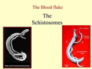
  • 3. 1- A genus of Trematodes, Schistosoma, commonly known as blood-fluke, are parasitic flatworms responsible for a highly significant group of infections in humans termed schistosomiasis. . 2- The sexes are separate (they are dioecious) 3- They reside in the blood vessels of the definitive host in their life cycles. Hence the common name “Blood Flukes.” 4- Male worm has a split body called the gynecophoral canal. The female is usually found within this canal “safe in the arms of her lover.” She leaves only during the egg laying period. Males are broader and females are filiform and larger than male General feature of the blood fluke
  • 4. Snail hosts The different species of Schistosoma have different types of snails serving as their intermediate hosts; these hosts are as follows: 1- Biomphalaria for S. Mansoni ‫البولية‬ ‫المجاري‬ ‫بلهارسيا‬ ‫قوقع‬ .)) 2 - Oncomelania for S. japonicum. 3- Bulinus for S. haematobium. ‫المستقيم‬ ‫بلهارسيا‬ ‫قوقع‬ ) Symptoms of schistosomiasis are caused by the body’s reaction to the worms’ eggs. Intestinal schistosomiasis 1- abdominal pain, 2- diarrhoea 3- and blood in the stool. 4- Liver enlargement is common in advanced cases, and is frequently associated with an accumulation of fluid in the peritoneal cavity and hypertension of the abdominal blood vessels. In such cases there may also be enlargement of the The classic sign of urogenital schistosomiasis is: 1-haematuria (blood in urine). 2- Fibrosis of the bladder and ureter, 3- and kidney damage.
  • 5. Learning objective • At the end of this section the student will be able to – Explain the general feature of blood fluke – List the blood flukes of medical importance – Explain the geographical distribution, Morphology, differential characteristics, transmission, life Cycles and pathogenesis of each species – Apply the necessary laboratory procedure for detection and identification of the liver fluke
  • 6. Blood fluke • S. mansoni • S. hematobium • S. japonicum • S. intrecalatum • S. mekongi • Animal schistosome which occasionally infect humans – Schistosoma mattheel – Schistosoma bovis – Schistosoma rodhaini – Schistosoma margrebowiei
  • 7. The Schistosomes • General feature: – They reside in the blood vessels of the definitive host in their life cycles. Hence the common name “Blood Flukes.” – The sexes are separate (they are dioecious) – They are long cylindrical(~ 20mm) and adopted to life in blood vessel
  • 8. • Male worm has a split body called the gynecophoral canal. The female is usually found within this canal “safe in the arms of her lover.” She leaves only during the egg laying period. • Males are broader and females are filiform and larger than male
  • 9. – Humans are the only or most significant host for most of the species – Snail host is required as intermediate host to complete their life cycle – There are no second intermediate hosts – Reproduction takes in the sporocyst stage in the snail. – No redia and metacercariae stages – Cercaria is the infective stage to humans in water and the infective route is by skin – egg without operculum, but with spine – The eggs are main pathogenic stage.
  • 10. Epidemology: • Wide spread species – Schistosoma mansoni causes intestinal schistosomiasis and is prevalent in 52 countries and territories of Africa, Caribbean, the Eastern Mediterranean and South America – Schistosoma haematobium causes urinary schistosomiasis and affects 54 countries in Africa and the Eastern Mediterranean – Schistosoma japonicum cause intestinal schistosomiasis and are Common in parts of Japan, China, Taiwan, Philippines, Thailand, and other parts of Southeast Asia • Less wide spread species – Schistosoma mekongi cause intestinal schistosomiasis and are prevalent in 7 African countries and the Pacific region – Schistosoma intercalatum is found in ten African countries
  • 14. Life Cycle of Schistosoma spp. • The eggs escape from the body by penetrating the walls of the veins and small intestine or urinary bladder (where adults reside), and they are passed in the feces or urine. Example of a Schistosoma mansoni egg
  • 15. Life Cycle of Schistosoma spp. Miracidium Swimming towards Snail Intermediate Host • The eggs, if and when they reach fresh water, will quickly hatch. • The miracidium swims ceaselessly until finds a snail host (die in 3-6 hours).
  • 16. Life cycle in Snail • After miracidum enters, the parasite goes through two asexual developmental stages: mother and daughter sporocysts. • Mother sporocysts contain the daughters, which are then released and found in the snail’s digestive and reproductive organs. • The daughter sporocysts hold the cercaiae, • Continues producing sporocystes for up to seven weeks within snail
  • 17. Intermediate Hosts of Schistosoma spp. S. mansoni only infect snails of the genus Biomphalaria.
  • 18. Intermediate Hosts of Schistosoma spp. S. japonicum are found in Oncomelania snails
  • 19. Intermediate Hosts of Schistosoma spp. S. haematobium persist in species of Bulinus.
  • 20. Life cycle of Schistosoma spp.: Cercariae • Cercaiae start to emerge four weeks after penetration by miracidium • There is NO second intermediate host
  • 21. Life cycle of Schistosoma spp.: Cercariae • Cercariae swim up and down in the water column until finding host; without host die after three days. • Move around and then enter and can disappear below surface in 10 to 30 seconds, and into circulation system within 24 hours.
  • 22. 5 Minutes after penetration of the human skin by cercaria, the newly transformed schistosomule has penetrated the outer layer of the epidermis, and is positioned just beneath the skin.
  • 23. 20 minutes after penetration of the skin. Here the schistosomule is migrating through the dermis. This it will do until it locates a blood vessel. The schistosomule will then break through into the blood vessel, to be transported in the circulatory system to the heart and then the lungs.
  • 24. Life cycle of Schistosoma spp.: In Human • Various ways of migrating through circulatory system (heart, liver). End up in veins draining liver where they develop for three weeks • Pair up in these veins and migrate to walls of guts or bladder, depending on species to produce eggs.
  • 25. Adults can live 20 to 30 years!!
  • 26. Life cycle of Schistosoma spp.: In Human • Species differences in Site Preferences of Adults (male and female in copula) – S. mansoni –veins of Large Intestine – S. haematobium – veins of bladder – S. japonicum – veins of small intestine
  • 27. • The infective inhabitation of Schistosoma in mesenteric vein
  • 28. Life cycle of Schistosoma spp.: In Human • Unusual in that eggs are the damaging stage – Traverse the walls of veins, tissues and mucosa – Egg promotes growth of granuloma and this is moved out into lumen of intestine or bladder where expelled.
  • 29. Granuloma of Schistosoma 2/3 of eggs remain in abdomen granuloma
  • 30.
  • 31. Intestinal schistosomiasis • Penetration of skin:- Penetration of of skin by cercariae causes transient dermatitis (swimmers' itch) • Migratory phase - 4-10 weeks after infection. – Is characterized by fever and toxic or allergic reactions resulting from migration of immature organisms. – Often results in bronchitis, hepatomegaly, splenomegaly, and diarrhea.
  • 32. • Chronic phase - persons living in endemic regions are often asymptotic. – May have mild, chronic bloody stools or urine. – Often have formation of granulomas. Hepatomegaly, Spleenomegaly, Ascites (accumulation of fluid in abdominal cavity – Terminal stage is characterized by portal vein hypertension syndrome, common saying, abdomen distention looks like a big drum – Ectopic lesion: The damage to the central nervous system ( brain, spinal ) may cause paralysis (monoplegia, hemiplegia ).
  • 33. • In less than 10% of cases, granulomas can cause blockage of blood flow in liver causing enlargement of the spleen and fluid retention in abdomen.
  • 34. abdomen distention looks like a big drum, Ascites, emaciation, varicosity, and splenomegaly
  • 35. abdomen distention looks like a big drum, emaciation looks like a fire wood.
  • 37.
  • 38.
  • 39. Laboratory diagnosis • Intestinal schistosomiasis – Finding the eggs in faeces by direct examination or more commonly by using concentration ; – Mucus and blood are often present in the faecal specimen 1. Formol-ether conc. Tech 2. Formol detergent gravity sed tech 3. Kotho-katz technique – Examining a rectal biopsy for eggs when they cannot be found in faeces. – occasionally eggs may also be found in urine often following faecal contamination
  • 40. Egg:S.mansoni • Size : 114-17µm 45-68µm • Shape: Oval, with one well rounded pole and one more conical pole • Colour: pale yellow-brown • Spine: large, triangular lateral spine near the rounded end • Shell: smooth, very thin • Content: fully embryonated (developed miracidium) when discharged with the faeces
  • 41. Egg: S japonicum • Size: 70-80m • Shape: oval, almost round • Colour: transparent or pale-yellow • Spine: very small hook- like spine laterally • Contain a fully developed miracidium
  • 42. • Ab-detection – Immunodiagnosis using ELISA, RIA, Latex `agglutination are helpful particularly in prepatent period, and in chronic and ectopic cases in which eggs are difficult to be demonstrated in the faeces. • Ag-detection. • EIA- detect circulating schistosoma antigens
  • 43. Urinary schistosomiasis • Finding eggs or occasionally the hatched miracidia in urine. • Quantitative report is required, no of eggs/10ml urine – Urine contains blood and appears red or red-brown and cloudy. • Hematuria • Protienuria • Eosinphil – Eggs may not be present in the urine all the time; it is neccessary to examine urine collected over several days. • Less frequently detecting eggs in faeces, rectal biopsy or bladder mucosal biopsy when an infection is light. • Immunodiagnosis: a variety of serodiagnostic methods are currently available. These include: RIA, ELISA IHA.
  • 44. Egg:S.hematobium • Size : 120 - 170 µm by 40-70µm • Shape: oval, with one well rounded pole • Spine : Terminal spine at one pole • Shell: Smooth, very thin except minute spines on the sucker • Shell is not acid fast in Ziehl-Neelsen staining, but the egg shell of other terminally spined Schistosoma species is acid fast • Colour: pale yellow-brown • Contain fully developed miracidium when laid