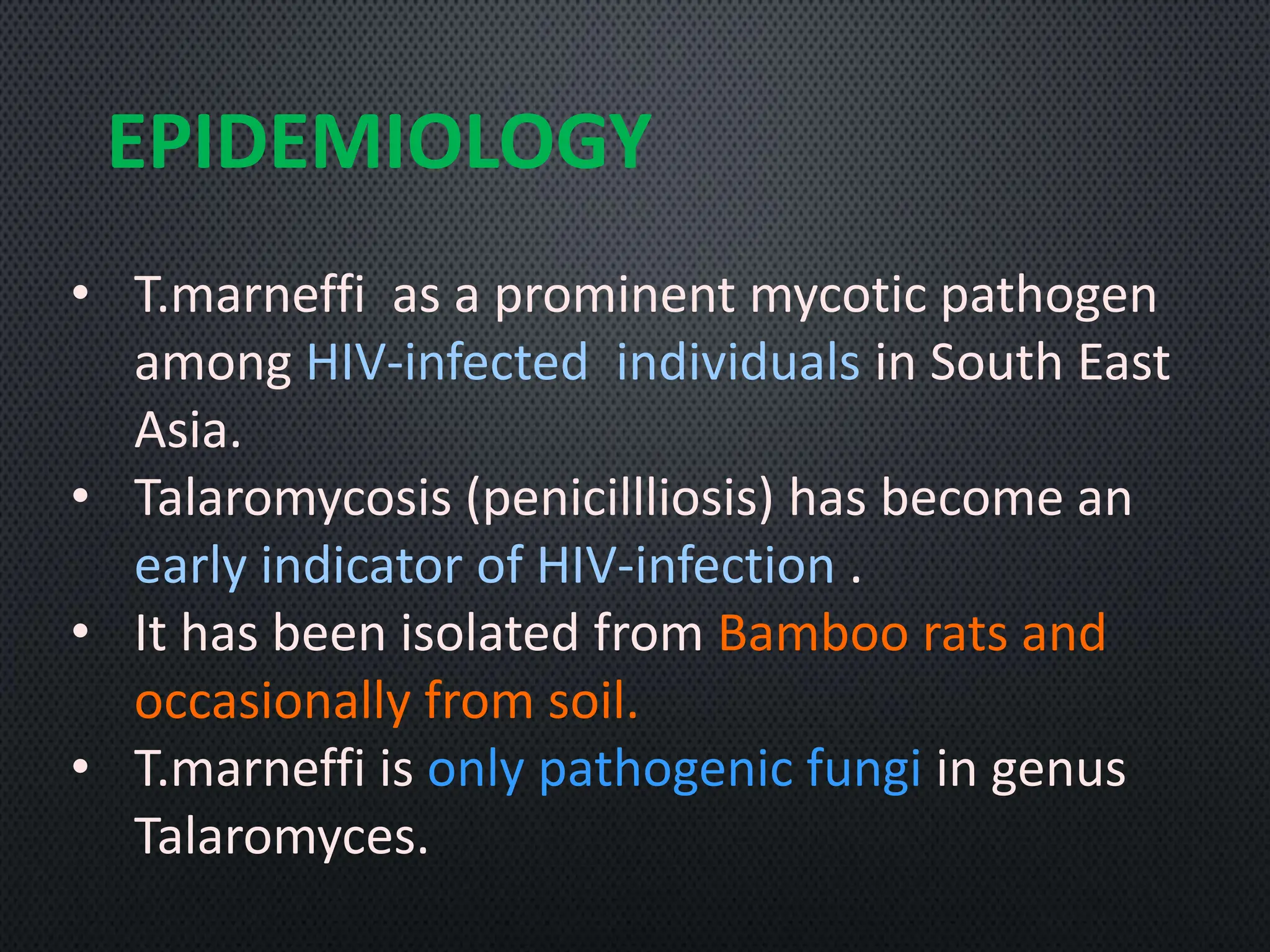
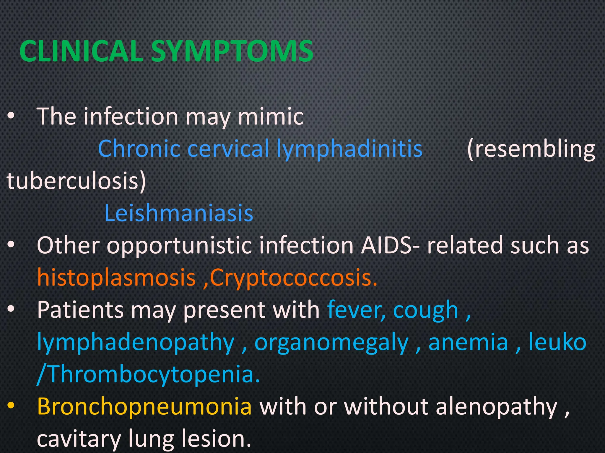
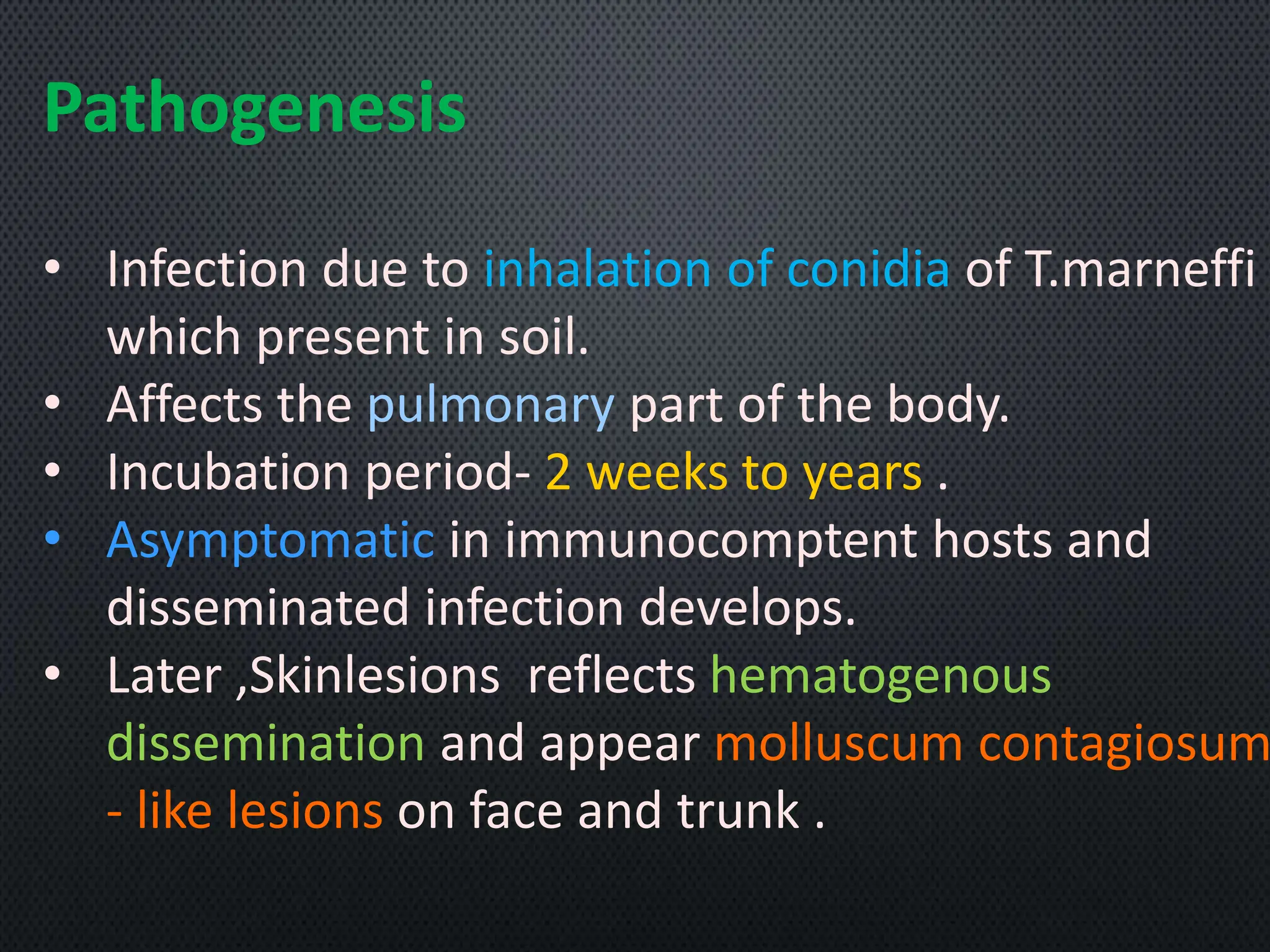
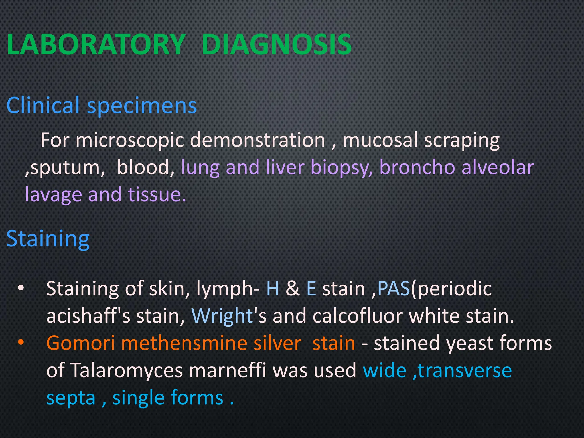
INTRODUCTIO
N
• Talaromycosis is a Dimorphic fungus
and Endemic fungi .
• Caused by Talaromyces marneffi
(formerly penicillium marneffi ).
• Infections involved in mononulear
phagocytic system and occurs primarily
in HIV - infected individuals.
• It's a Disseminated mycosis.
MORPHOLOGY
Saprobic phase[ Mold in 25°C]
• Mold with conidiophores
terminating in conspicuous
,penicillus - bearing ,
ellispsodial,Smooth conidia .
• Formation od Red pigment that
diffuses into agar .
• It exhibits sporulating structures
that are typical of the genus.
Parasitic phase [yeast in 37°c]
• Pleomorphic elongated yeast (1-8
micrometer ) with transverssepta.
Morphology in tissue
• Globose to elongated sausage -
shaped yeast (3-5 micrometer ) that
are intracellular ,divided by Fission .
EPIDEMIOLOGY
• T.marneffi as a prominent mycotic pathogen
among HIV-infected individuals in South East
Asia.
• Talaromycosis (penicillliosis) has become an
early indicator of HIV-infection .
• It has been isolated from Bamboo rats and
occasionally from soil.
• T.marneffi is only pathogenic fungi in genus
Talaromyces.
CLINICAL SYMPTOMS
• The infection may mimic
Chronic cervical lymphadinitis (resembling
tuberculosis)
Leishmaniasis
• Other opportunistic infection AIDS- related such as
histoplasmosis ,Cryptococcosis.
• Patients may present with fever, cough ,
lymphadenopathy , organomegaly , anemia , leuko
/Thrombocytopenia.
• Bronchopneumonia with or without alenopathy ,
cavitary lung lesion.
Pathogenesis
• Infection due to inhalation of conidia of T.marneffi
which present in soil.
• Affects the pulmonary part of the body.
• Incubation period- 2 weeks to years .
• Asymptomatic in immunocomptent hosts and
disseminated infection develops.
• Later ,Skinlesions reflects hematogenous
dissemination and appear molluscum contagiosum
- like lesions on face and trunk .
LABORATORY DIAGNOSIS
Clinical specimens
For microscopic demonstration , mucosal scraping
,sputum, blood, lung and liver biopsy, broncho alveolar
lavage and tissue.
Staining
• Staining of skin, lymph- H & E stain ,PAS(periodic
acishaff's stain, Wright's and calcofluor white stain.
• Gomori methensmine silver stain - stained yeast forms
of Talaromyces marneffi was used wide ,transverse
septa , single forms .
Culture morphology
In culture 25 ° c to 30 °c , typically penicillium-like
morphology and diffusible red pigmentation.
Conversation to yeast phase at 37° c is Confirmatory
phase.
Microscopy - Elliptic fission yeast inside phagocytes in
buffy coats of smears.
Serogical tests
• Indirect IFAT, Immunoblot
• PCR and DNA sequencing for sp. Detection.
• But nconnmercial standardized test available.
TREATMENT
• A lipid Formation of Amphotericin B, Vorironazole
& intaconazole.
• Administration of Amphotericin B for 2 weeks
should followed itraconazole for another10
weeks .
• AIDS patients may require life-long treatment
with intaconazole and Vorironazoke.
• Echinocandins as well posaconazole and
terbinafine may be useful .
REFERENCE
"MEDICAL MICROBIOLOGY" by Murray, Rosenthal,
Pfaller , consultant JMI laboratories- 8thedition
,Elsevier publication canada,2016.
TALAROMYCOSIS - Introduction, Morphology, Epidemiology, Clinical symptoms,pathophysiology, laboratory diagnosis, Treatment.

TALAROMYCOSIS - Introduction, Morphology, Epidemiology, Clinical symptoms,pathophysiology, laboratory diagnosis, Treatment.

  • 2.
    INTRODUCTIO N • Talaromycosis isa Dimorphic fungus and Endemic fungi . • Caused by Talaromyces marneffi (formerly penicillium marneffi ). • Infections involved in mononulear phagocytic system and occurs primarily in HIV - infected individuals. • It's a Disseminated mycosis.
  • 3.
    MORPHOLOGY Saprobic phase[ Moldin 25°C] • Mold with conidiophores terminating in conspicuous ,penicillus - bearing , ellispsodial,Smooth conidia . • Formation od Red pigment that diffuses into agar . • It exhibits sporulating structures that are typical of the genus.
  • 4.
    Parasitic phase [yeastin 37°c] • Pleomorphic elongated yeast (1-8 micrometer ) with transverssepta. Morphology in tissue • Globose to elongated sausage - shaped yeast (3-5 micrometer ) that are intracellular ,divided by Fission .
  • 5.
    EPIDEMIOLOGY • T.marneffi asa prominent mycotic pathogen among HIV-infected individuals in South East Asia. • Talaromycosis (penicillliosis) has become an early indicator of HIV-infection . • It has been isolated from Bamboo rats and occasionally from soil. • T.marneffi is only pathogenic fungi in genus Talaromyces.
  • 6.
    CLINICAL SYMPTOMS • Theinfection may mimic Chronic cervical lymphadinitis (resembling tuberculosis) Leishmaniasis • Other opportunistic infection AIDS- related such as histoplasmosis ,Cryptococcosis. • Patients may present with fever, cough , lymphadenopathy , organomegaly , anemia , leuko /Thrombocytopenia. • Bronchopneumonia with or without alenopathy , cavitary lung lesion.
  • 7.
    Pathogenesis • Infection dueto inhalation of conidia of T.marneffi which present in soil. • Affects the pulmonary part of the body. • Incubation period- 2 weeks to years . • Asymptomatic in immunocomptent hosts and disseminated infection develops. • Later ,Skinlesions reflects hematogenous dissemination and appear molluscum contagiosum - like lesions on face and trunk .
  • 8.
    LABORATORY DIAGNOSIS Clinical specimens Formicroscopic demonstration , mucosal scraping ,sputum, blood, lung and liver biopsy, broncho alveolar lavage and tissue. Staining • Staining of skin, lymph- H & E stain ,PAS(periodic acishaff's stain, Wright's and calcofluor white stain. • Gomori methensmine silver stain - stained yeast forms of Talaromyces marneffi was used wide ,transverse septa , single forms .
  • 9.
    Culture morphology In culture25 ° c to 30 °c , typically penicillium-like morphology and diffusible red pigmentation. Conversation to yeast phase at 37° c is Confirmatory phase. Microscopy - Elliptic fission yeast inside phagocytes in buffy coats of smears. Serogical tests • Indirect IFAT, Immunoblot • PCR and DNA sequencing for sp. Detection. • But nconnmercial standardized test available.
  • 10.
    TREATMENT • A lipidFormation of Amphotericin B, Vorironazole & intaconazole. • Administration of Amphotericin B for 2 weeks should followed itraconazole for another10 weeks . • AIDS patients may require life-long treatment with intaconazole and Vorironazoke. • Echinocandins as well posaconazole and terbinafine may be useful .
  • 11.
    REFERENCE "MEDICAL MICROBIOLOGY" byMurray, Rosenthal, Pfaller , consultant JMI laboratories- 8thedition ,Elsevier publication canada,2016.