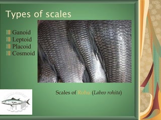
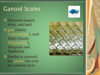
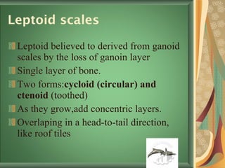
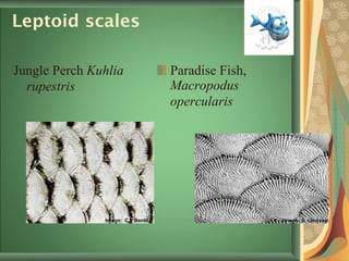
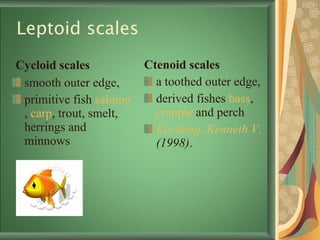
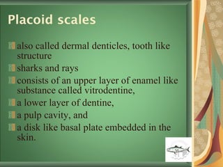
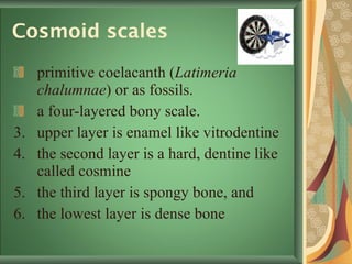
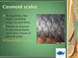
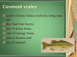
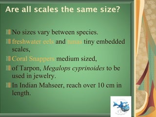
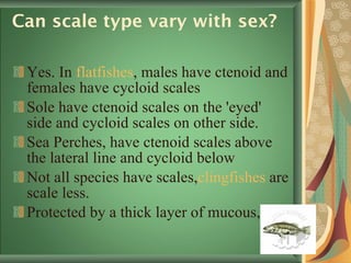
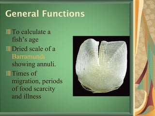
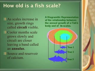
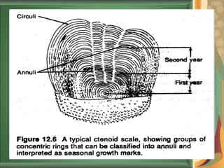
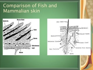
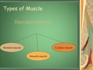
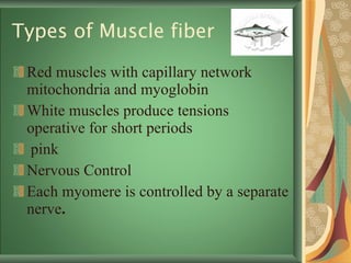
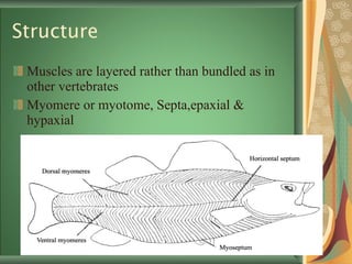
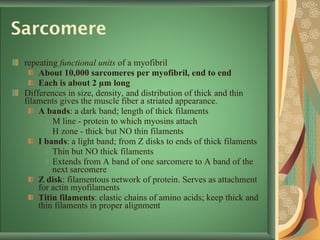
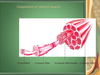
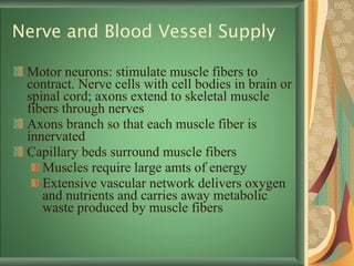
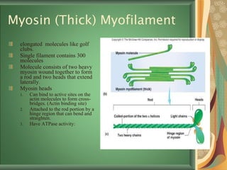
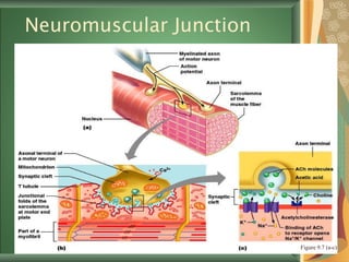
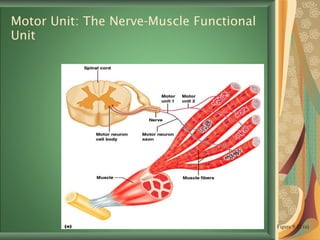
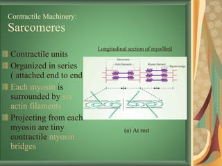
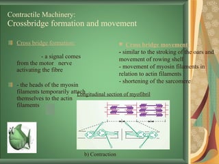
Skin Scales and Skeletal Muscles Contents: Scales Types of Scales:Are all scales the same size?  Can scale type vary with sex?  Slime and Spots  Functions of Scales How old is a fish scale Fish skin Function Skin StructureFish and Mammalian skin Skin Properties Hormonal control of Skin   Skeletal Muscles Types of Muscle Skeletal Muscles function Properties of Muscle Structure Myosin (Thick) Myofilament Actin (Thin) Myofilaments Sliding Filament Model of Contraction Neuromuscular Junction Motor Unit: Contractile Machinery: Sarcomeres Crossbridge formation and movemen
Scales A rigid plate out of an  animal 's  skin  to provide protection.  E volved multiple times  with varying structure and function. Classified as an organism's  integumentary system .  scales usually not eaten.
Types of scales Ganoid  Leptoid  Placoid Cosmoid   Scales of  Rohu  ( Labeo rohita )
Ganoid Scales Diamond-shaped, shiny, and hard.  gars  (family  Lepisosteidae ), reed fishes (family  Polypteridae ), Sturgeons and Paddlefish Similar to cosmoid but  ganoin  lies over the cosmine layer
Leptoid scales Leptoid believed to derived from ganoid scales by the loss of ganoin layer Single layer of bone.  Two forms: cycloid (circular) and ctenoid  (toothed) As they grow,add concentric layers. Overlaping in a head-to-tail direction, like roof tiles
Leptoid scales Jungle Perch  Kuhlia rupestris   Paradise Fish,  Macropodus opercularis
Leptoid scales  Cycloid scales   smooth outer edge, primitive fish  salmon ,  carp , trout, smelt, herrings and minnows Ctenoid scales   a toothed outer edge, derived fishes  bass ,  crappie  and perch Kardong, Kenneth V.  (1998) .
Placoid scales also called dermal denticles, tooth like structure  sharks and rays consists of an upper layer of enamel like substance called vitrodentine,  a lower layer of dentine,  a pulp cavity, and  a disk like basal plate embedded in the skin.
Placoid scales Do not increase in size, new scales must be added as a shark grows Homologous with teeth in all vertebrates. Broadnose Sevengill Shark
Cosmoid scales   primitive coelacanth ( Latimeria chalumnae ) or as fossils. a four-layered bony scale.  upper layer is enamel like vitrodentine the second layer is a hard, dentine like called cosmine the third layer is spongy bone, and  the lowest layer is dense bone
Cosmoid scales In lungfishes, but highly modified, single-layered form.  Similar in structure to placoid probably arose from fusion of placoid scales  Queensland Lungfish
Cosmoid scales scales of bony fishes evolved a long time ago  they had four layers,  one of dense bone, one of spongy bone,  one of dentine and  one of enamel.
Are all scales the same size?  No sizes vary between species. freshwater eels  and  tunas  tiny embedded scales, Coral Snappers  medium sized,  of Tarpon,  Megalops cyprinoides  to be used in jewelry.  In Indian Mahseer, reach over 10 cm in length.
Can scale type vary with sex?  Yes. In  flatfishes , males have ctenoid and females have cycloid scales Sole have ctenoid scales on the 'eyed' side and cycloid scales on other side.  Sea Perches, have ctenoid scales above the lateral line and cycloid below Not all species have scales, clingfishes  are scale less.  Protected by a thick layer of mucous,
Function of Slime and Spots  Hydrodynamic function to reduce water friction and resistance to forward motion Enzymes and antibodies as a defensive system Spots size fluctuate according to nervous stimuli.  The arrangement imbricate or mosaic
General Functions To calculate a fish’s age Dried scale of a  Barramundi  showing annuli. Times of migration, periods of food scarcity and illness
How old is a fish scale?  As scales increase in size, growth rings called  circuli  visible.  Cooler months scale grows slowly and circuli are closer leaving a band called an  annulus .  Source and reservoir   of calcium.
 
Fish skin Barrier, strong, tough and watertight, Ion- and water balance The scales lie in the  Dermis of skin
Skin Structure Two layers, the  Epidermis (outer) layer and the Dermis .  Epidermis is made up of  epithelial cells Inter-spaced between epithelial cells are  slime  cells produce mucous secretions  Dermis  consists of two layers: stratum spongiosum &   stratum compactum. Hypodermis
Comparison of Fish and Mammalian skin
Skin Properties Mucus ->fatty acid+phospholipids Pigment cells (chromatophores) under hormonal or nervous system control  Effector organ  Respond to hormones & neurotransmitters via receptors  Xanthophores , erythrophores  and  melanophores
Hormonal control of Skin   Melanin-concentrating hormone (MCH) Neurotransmitter or neuromodulator  Sensory organs  Nerve endings and receives tactile, thermal, and pain stimuli  e.g. teleost fish Blood vessels  Collagen fibers
Types of Muscle Three types of muscle: Skeletal muscle Smooth muscle Cardiac muscle
 
Skeletal Muscles function Two groups – the  axial  and  appendicular  muscles.  Main function is to support the body viscera. Without them animal could neither move or eat or bear young.  Body movement (Locomotion) Maintenance of posture Constriction of organs and vessel
Types of Muscle fiber Red muscles with capillary network mitochondria and myoglobin  White muscles produce tensions operative for short periods  pink Nervous Control Each myomere is controlled by a separate nerve .
Properties of Muscle Excitability : capacity of muscle to respond to a stimulus Contractility : ability of a muscle to shorten with force Extensibility : muscle can be stretched to its normal resting length and beyond to a limited degree Elasticity : ability of muscle to recoil to original resting length after stretched
Structure Muscles are layered rather than bundled as in other vertebrates  Myomere or myotome, Septa,epaxial & hypaxial
Structure Muscle cells long and cylindrical and have many nuclei.  Actin and myosin with controlling proteins In other vertebrates muscles connected to bone through a tendon.  In fish, arranged vertically throughout whole length of fish.
Muscle Fiber Anatomy Sarcolemma  - cell membrane Surrounds the  sarcoplasm   (cytoplasm of fiber) Contains oxygen-binding protein  myoglobin Punctuated by openings called the  transverse tubules (T-tubules) Narrow tubes that extend into the sarcoplasm  Filled with extracellular fluid Myofibrils  -cylindrical structures within muscle fiber Are bundles of protein filaments (= myofilaments ) When myofibril shortens, muscle shortens (contracts)
Sarcomere repeating  functional units  of a myofibril About 10,000 sarcomeres per myofibril, end to end Each is about 2 µm long Differences in size, density, and distribution of thick and thin filaments gives the muscle fiber a striated appearance. A bands : a dark band; length of thick filaments M line - protein to which myosins attach H zone - thick but NO thin filaments I bands : a light band; from Z disks to ends of thick filaments Thin but NO thick filaments Extends from A band of one sarcomere to A band of the next sarcomere Z disk : filamentous network of protein. Serves as attachment for actin myofilaments Titin filaments : elastic chains of amino acids; keep thick and thin filaments in proper alignment
d) myofibril  c) muscle fibre  b) muscle fibre bundle  a) Muscle belly  Components of skeletal muscle
Nerve and Blood Vessel Supply Motor neurons: stimulate muscle fibers to contract. Nerve cells with cell bodies in brain or spinal cord; axons extend to skeletal muscle fibers through nerves Axons branch so that each muscle fiber is innervated Capillary beds surround muscle fibers Muscles require large amts of energy Extensive vascular network delivers oxygen and nutrients and carries away metabolic waste produced by muscle fibers
Myosin (Thick) Myofilament elongated  molecules like golf clubs.  Single filament contains 300 molecules Molecule consists of two heavy myosin wound together to form a rod and two heads that extend laterally.  Myosin heads Can bind to active sites on the actin molecules to form cross-bridges. (Actin binding site) Attached to the rod portion by a hinge region that can bend and straighten.  Have ATPase activity:
Actin (Thin) Myofilaments Thin Filament : 3 major proteins F (fibrous) actin Tropomyosin Troponin Two strands of fibrous (F) actin form a double helix extending the length of the myofilament; attached at either end at sarcomere. Composed of G actin monomers each of which has a  myosin-binding site  Actin site can bind myosin during muscle contraction.  Tropomyosin Troponin  three subunits:  Tn-A : binds to actin Tn-T :binds to tropomyosin, Tn-C :binds to calcium ions.
Sliding Filament Model of Contraction Thin filaments slide past the thick ones so that the actin and myosin filaments overlap to a greater degree In the relaxed state, thin and thick filaments overlap only slightly Upon stimulation, myosin heads bind to actin and sliding begins
Sliding Filament Model of Contraction Each myosin head binds and detaches several times during contraction, acting like a ratchet to generate tension and propel the thin filaments to the center of the sarcomere As this event occurs throughout the sarcomeres, the muscle shortens
Neuromuscular Junction The neuromuscular junction is formed from: Axonal endings, which have small membranous sacs (synaptic vesicles) that contain the neurotransmitter acetylcholine   (ACh) The motor end plate of a muscle, a specific part of sarcolemma that contains ACh receptors and helps to form the neuromuscular junction Axonal ends and muscle fibers are always separated by a space called the  synaptic cleft
Neuromuscular Junction Figure 9.7 (a-c)
Motor Unit: The Nerve-Muscle Functional Unit A motor unit is a motor neuron and all the muscle fibers it supplies The number of muscle fibers per motor unit can vary from a few (4-6) to hundreds (1200-1500)
Motor Unit: The Nerve-Muscle Functional Unit Figure 9.12 (a)
Contractile Machinery: Sarcomeres Contractile units Organized in series ( attached end to end) Each myosin  is surrounded by  six actin filaments Projecting from each myosin are tiny contractile  myosin bridges Longitudinal section of myofibril (a) At rest
Contractile Machinery: Crossbridge formation and movement Cross bridge formation:   - a signal comes from the motor  nerve activating the fibre  - the heads of the myosin filaments temporarily attach themselves to the actin filaments Cross bridge movement:   -  similar to the stroking of the oars and movement of rowing shell - movement of myosin filaments in relation to actin filaments - shortening of the sarcomere b) Contraction Longitudinal section of myofibril
Contractile Machinery: Optimal Crossbridge formation Sarcomeres should be  optimal distance apart optimal distance is (0.0019-0.0022 mm) At this an optimal number of cross bridges is formed If stretched farther   apart :  - fewer cross bridges can form    less force produced If the sarcomeres are too close together:  - cross bridges interfere with one another    less force produced Longitudinal section of myofibril c) Powerful stretching d) Powerful contraction
Any questions …….. Don’t shy

Scales

  • 1.
  • 2.
    Skin Scales andSkeletal Muscles Contents: Scales Types of Scales:Are all scales the same size? Can scale type vary with sex? Slime and Spots Functions of Scales How old is a fish scale Fish skin Function Skin StructureFish and Mammalian skin Skin Properties Hormonal control of Skin Skeletal Muscles Types of Muscle Skeletal Muscles function Properties of Muscle Structure Myosin (Thick) Myofilament Actin (Thin) Myofilaments Sliding Filament Model of Contraction Neuromuscular Junction Motor Unit: Contractile Machinery: Sarcomeres Crossbridge formation and movemen
  • 3.
    Scales A rigidplate out of an animal 's skin to provide protection. E volved multiple times with varying structure and function. Classified as an organism's integumentary system . scales usually not eaten.
  • 4.
    Types of scalesGanoid Leptoid Placoid Cosmoid Scales of Rohu ( Labeo rohita )
  • 5.
    Ganoid Scales Diamond-shaped,shiny, and hard. gars (family Lepisosteidae ), reed fishes (family Polypteridae ), Sturgeons and Paddlefish Similar to cosmoid but ganoin lies over the cosmine layer
  • 6.
    Leptoid scales Leptoidbelieved to derived from ganoid scales by the loss of ganoin layer Single layer of bone. Two forms: cycloid (circular) and ctenoid (toothed) As they grow,add concentric layers. Overlaping in a head-to-tail direction, like roof tiles
  • 7.
    Leptoid scales JunglePerch Kuhlia rupestris Paradise Fish, Macropodus opercularis
  • 8.
    Leptoid scales Cycloid scales smooth outer edge, primitive fish salmon , carp , trout, smelt, herrings and minnows Ctenoid scales a toothed outer edge, derived fishes bass , crappie and perch Kardong, Kenneth V. (1998) .
  • 9.
    Placoid scales alsocalled dermal denticles, tooth like structure sharks and rays consists of an upper layer of enamel like substance called vitrodentine, a lower layer of dentine, a pulp cavity, and a disk like basal plate embedded in the skin.
  • 10.
    Placoid scales Donot increase in size, new scales must be added as a shark grows Homologous with teeth in all vertebrates. Broadnose Sevengill Shark
  • 11.
    Cosmoid scales primitive coelacanth ( Latimeria chalumnae ) or as fossils. a four-layered bony scale. upper layer is enamel like vitrodentine the second layer is a hard, dentine like called cosmine the third layer is spongy bone, and the lowest layer is dense bone
  • 12.
    Cosmoid scales Inlungfishes, but highly modified, single-layered form. Similar in structure to placoid probably arose from fusion of placoid scales Queensland Lungfish
  • 13.
    Cosmoid scales scalesof bony fishes evolved a long time ago they had four layers, one of dense bone, one of spongy bone, one of dentine and one of enamel.
  • 14.
    Are all scalesthe same size? No sizes vary between species. freshwater eels and tunas tiny embedded scales, Coral Snappers medium sized, of Tarpon, Megalops cyprinoides to be used in jewelry. In Indian Mahseer, reach over 10 cm in length.
  • 15.
    Can scale typevary with sex? Yes. In flatfishes , males have ctenoid and females have cycloid scales Sole have ctenoid scales on the 'eyed' side and cycloid scales on other side. Sea Perches, have ctenoid scales above the lateral line and cycloid below Not all species have scales, clingfishes are scale less. Protected by a thick layer of mucous,
  • 16.
    Function of Slimeand Spots Hydrodynamic function to reduce water friction and resistance to forward motion Enzymes and antibodies as a defensive system Spots size fluctuate according to nervous stimuli. The arrangement imbricate or mosaic
  • 17.
    General Functions Tocalculate a fish’s age Dried scale of a Barramundi showing annuli. Times of migration, periods of food scarcity and illness
  • 18.
    How old isa fish scale? As scales increase in size, growth rings called circuli visible. Cooler months scale grows slowly and circuli are closer leaving a band called an annulus . Source and reservoir of calcium.
  • 19.
  • 20.
    Fish skin Barrier,strong, tough and watertight, Ion- and water balance The scales lie in the Dermis of skin
  • 21.
    Skin Structure Twolayers, the Epidermis (outer) layer and the Dermis . Epidermis is made up of epithelial cells Inter-spaced between epithelial cells are slime cells produce mucous secretions Dermis consists of two layers: stratum spongiosum & stratum compactum. Hypodermis
  • 22.
    Comparison of Fishand Mammalian skin
  • 23.
    Skin Properties Mucus->fatty acid+phospholipids Pigment cells (chromatophores) under hormonal or nervous system control Effector organ Respond to hormones & neurotransmitters via receptors Xanthophores , erythrophores and melanophores
  • 24.
    Hormonal control ofSkin Melanin-concentrating hormone (MCH) Neurotransmitter or neuromodulator Sensory organs Nerve endings and receives tactile, thermal, and pain stimuli e.g. teleost fish Blood vessels Collagen fibers
  • 25.
    Types of MuscleThree types of muscle: Skeletal muscle Smooth muscle Cardiac muscle
  • 26.
  • 27.
    Skeletal Muscles functionTwo groups – the axial and appendicular muscles. Main function is to support the body viscera. Without them animal could neither move or eat or bear young. Body movement (Locomotion) Maintenance of posture Constriction of organs and vessel
  • 28.
    Types of Musclefiber Red muscles with capillary network mitochondria and myoglobin White muscles produce tensions operative for short periods pink Nervous Control Each myomere is controlled by a separate nerve .
  • 29.
    Properties of MuscleExcitability : capacity of muscle to respond to a stimulus Contractility : ability of a muscle to shorten with force Extensibility : muscle can be stretched to its normal resting length and beyond to a limited degree Elasticity : ability of muscle to recoil to original resting length after stretched
  • 30.
    Structure Muscles arelayered rather than bundled as in other vertebrates Myomere or myotome, Septa,epaxial & hypaxial
  • 31.
    Structure Muscle cellslong and cylindrical and have many nuclei. Actin and myosin with controlling proteins In other vertebrates muscles connected to bone through a tendon. In fish, arranged vertically throughout whole length of fish.
  • 32.
    Muscle Fiber AnatomySarcolemma - cell membrane Surrounds the sarcoplasm (cytoplasm of fiber) Contains oxygen-binding protein myoglobin Punctuated by openings called the transverse tubules (T-tubules) Narrow tubes that extend into the sarcoplasm Filled with extracellular fluid Myofibrils -cylindrical structures within muscle fiber Are bundles of protein filaments (= myofilaments ) When myofibril shortens, muscle shortens (contracts)
  • 33.
    Sarcomere repeating functional units of a myofibril About 10,000 sarcomeres per myofibril, end to end Each is about 2 µm long Differences in size, density, and distribution of thick and thin filaments gives the muscle fiber a striated appearance. A bands : a dark band; length of thick filaments M line - protein to which myosins attach H zone - thick but NO thin filaments I bands : a light band; from Z disks to ends of thick filaments Thin but NO thick filaments Extends from A band of one sarcomere to A band of the next sarcomere Z disk : filamentous network of protein. Serves as attachment for actin myofilaments Titin filaments : elastic chains of amino acids; keep thick and thin filaments in proper alignment
  • 34.
    d) myofibril c) muscle fibre b) muscle fibre bundle a) Muscle belly Components of skeletal muscle
  • 35.
    Nerve and BloodVessel Supply Motor neurons: stimulate muscle fibers to contract. Nerve cells with cell bodies in brain or spinal cord; axons extend to skeletal muscle fibers through nerves Axons branch so that each muscle fiber is innervated Capillary beds surround muscle fibers Muscles require large amts of energy Extensive vascular network delivers oxygen and nutrients and carries away metabolic waste produced by muscle fibers
  • 36.
    Myosin (Thick) Myofilamentelongated molecules like golf clubs. Single filament contains 300 molecules Molecule consists of two heavy myosin wound together to form a rod and two heads that extend laterally. Myosin heads Can bind to active sites on the actin molecules to form cross-bridges. (Actin binding site) Attached to the rod portion by a hinge region that can bend and straighten. Have ATPase activity:
  • 37.
    Actin (Thin) MyofilamentsThin Filament : 3 major proteins F (fibrous) actin Tropomyosin Troponin Two strands of fibrous (F) actin form a double helix extending the length of the myofilament; attached at either end at sarcomere. Composed of G actin monomers each of which has a myosin-binding site Actin site can bind myosin during muscle contraction. Tropomyosin Troponin three subunits: Tn-A : binds to actin Tn-T :binds to tropomyosin, Tn-C :binds to calcium ions.
  • 38.
    Sliding Filament Modelof Contraction Thin filaments slide past the thick ones so that the actin and myosin filaments overlap to a greater degree In the relaxed state, thin and thick filaments overlap only slightly Upon stimulation, myosin heads bind to actin and sliding begins
  • 39.
    Sliding Filament Modelof Contraction Each myosin head binds and detaches several times during contraction, acting like a ratchet to generate tension and propel the thin filaments to the center of the sarcomere As this event occurs throughout the sarcomeres, the muscle shortens
  • 40.
    Neuromuscular Junction Theneuromuscular junction is formed from: Axonal endings, which have small membranous sacs (synaptic vesicles) that contain the neurotransmitter acetylcholine (ACh) The motor end plate of a muscle, a specific part of sarcolemma that contains ACh receptors and helps to form the neuromuscular junction Axonal ends and muscle fibers are always separated by a space called the synaptic cleft
  • 41.
  • 42.
    Motor Unit: TheNerve-Muscle Functional Unit A motor unit is a motor neuron and all the muscle fibers it supplies The number of muscle fibers per motor unit can vary from a few (4-6) to hundreds (1200-1500)
  • 43.
    Motor Unit: TheNerve-Muscle Functional Unit Figure 9.12 (a)
  • 44.
    Contractile Machinery: SarcomeresContractile units Organized in series ( attached end to end) Each myosin is surrounded by six actin filaments Projecting from each myosin are tiny contractile myosin bridges Longitudinal section of myofibril (a) At rest
  • 45.
    Contractile Machinery: Crossbridgeformation and movement Cross bridge formation: - a signal comes from the motor nerve activating the fibre - the heads of the myosin filaments temporarily attach themselves to the actin filaments Cross bridge movement: - similar to the stroking of the oars and movement of rowing shell - movement of myosin filaments in relation to actin filaments - shortening of the sarcomere b) Contraction Longitudinal section of myofibril
  • 46.
    Contractile Machinery: OptimalCrossbridge formation Sarcomeres should be optimal distance apart optimal distance is (0.0019-0.0022 mm) At this an optimal number of cross bridges is formed If stretched farther apart : - fewer cross bridges can form  less force produced If the sarcomeres are too close together: - cross bridges interfere with one another  less force produced Longitudinal section of myofibril c) Powerful stretching d) Powerful contraction
  • 47.