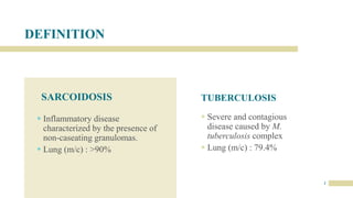
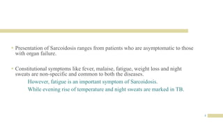
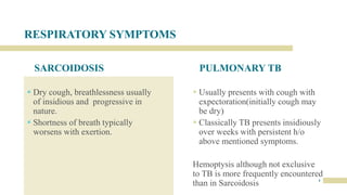
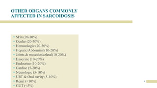
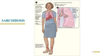
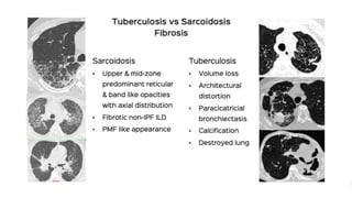
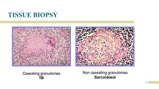
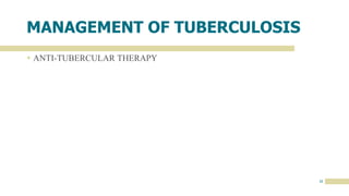
SARCOIDOSIS
V/S
PULMONARY TUBERCULOSIS
“A DIANOSTIC DILEMMA”
By Dr. Gouri Mohan | JR1-Respiratory Medicine
DEFINITION
SARCOIDOSIS
 Inflammatory disease
characterized by the presence of
non-caseating granulomas.
 Lung (m/c) : >90%
TUBERCULOSIS
 Severe and contagious
disease caused by M.
tuberculosis complex
 Lung (m/c) : 79.4%
2
3
 Presentation of Sarcoidosis ranges from patients who are asymptomatic to those
with organ failure.
 Constitutional symptoms like fever, malaise, fatigue, weight loss and night
sweats are non-specific and common to both the diseases.
However, fatigue is an important symptom of Sarcoidosis.
While evening rise of temperature and night sweats are marked in TB.
RESPIRATORY SYMPTOMS
SARCOIDOSIS
 Dry cough, breathlessness usually
of insidious and progressive in
nature.
 Shortness of breath typically
worsens with exertion.
PULMONARY TB
 Usually presents with cough with
expectoration(initially cough may
be dry)
 Classically TB presents insidiously
over weeks with persistent h/o
above mentioned symptoms.
Hemoptysis although not exclusive
to TB is more frequently encountered
than in Sarcoidosis 4
OTHER ORGANS COMMONLY
AFFECTED IN SARCOIDOSIS
 Skin (20-30%)
 Ocular (20-30%)
 Hematologic (20-30%)
 Hepatic/Abdominal(10-20%)
 Joints & musculoskeletal(10-20%)
 Exocrine (10-20%)
 Endocrine (10-20%)
 Cardiac (5-20%)
 Neurologic (5-10%)
 URT & Oral cavity (5-10%)
 Renal (<10%)
 GUT (<5%)
5
SARCOIDOSIS
6
MM.DD.20XX
ADD A FOOTER
7
SKIN MANIFESTATION
LUPUS PERNIO
8
OCULAR INVOLVEMENT
 Anterior uveitis (m/c)
 Lacrimal gland involvement
 Scleral nodules
 Chorioretinitis
 Conjunctivitis
 Optic neuritis
9
 LIVER INVOLVEMENT : Elevation of ALP, Transaminase, Bilirubin(marker of
more advanced liver disease)
Portal hypertension – Ascites and Esophageal varices.
 CARDIAC INVOLVEMENT : Cardiomyopathy (Dilated >Restrictive),
BBB/Arrhythmias
 NERVOUS SYSTEM INVOLVEMENT : Aseptic meningitis/encephalitis, Cranial
neuropathy, Peripheral neuropathy, Central DI, Optic neuritis.
 HEMATOLOGICAL : Mediastinal LN Involvement, Hypersplenism leading to
Anemia, Thrombocytopenia
10
There are certain conditions where the clinicoradiological
manifestations are typical for Sarcoidosis – LOFGREN
SYNDROME, HEERFORDT SYNDROME.
11
TUBERCULOSIS
TB is predominantly a respiratory disease affecting Lungs in approx. 80% of cases.
 About 30% of TB cases involve an extrapulmonary site, occurring with or without
concomitant Lung involvement.
12
ORGANS AFFECTED IN EXTRAPULMONARY
TB : 20.6%
Lymph node (7.6%)
Pleural (4%)
Bone and joint(1.8%)
Peritoneal (1.3%)
Meningeal (0.9%)
GUT (0.9%)
Others (5.3%)
EXTRAPULMONARY
TB
13
Common Forms
PHYSICAL EXAMINATION
SARCOIDOSIS
 Physical findings are infrequent, with
lung crackles heard in <20% of
patients.
 Clubbing is rare.
PULMONARY
TUBERCULOSIS
 Even when relatively extensive
disease is present, Pul TB most
often produces no detectable
abnormality on physical
examination.
 Tachypnea and Hypoxia are
relatively rare, unless there is
extensive lung destruction or miliary
disease 14
DIAGONOSTIC APPROACH
SARCOIDOSIS
 Is almost always a diagnosis of
exclusion.
 There is no reliable, non-invasive
screening test to help confirm a
diagnosis.
 Diagnosis is based on compatible clinical
and radiological manifestations together
with a tissue biopsy.
PULMONARY
TUBERCULOSIS
 The key to early diagnosis is a high index of
suspicion, considering epidemiological risk
factors and suggestive clinic-radiological
features.
 Ultimately the definitive diagnosis requires a
culture confirmation
 In case of high clinical suspicion due to
radiological findings , Culture –ve d/s is the
diagnosis if radiological findings improves
with treatment
15
LABORATORY FINDINGS
SARCOIDOSIS
 Lymphopenia(m/c), Anemia (20%),
Thrombocytopenia – Pancytopenia in
some patients.
 Hypercalcemia and / or
hypercalciuria occur in about 10% of
Sarcoidosis patients
 Elevated ACE Levels
 TST –ve
PULMONARY
TUBERCULOSIS
 AFB Microscopy
 Gene Xpert for MTB which also detects drug
resistance (Rif)
 Mycobacterial Culture remains the gold std.
 Anemia
 TST can be positive but with low specificity
and sensitivity
16
CHEST X RAY
IN
SARCOIDOSIS
17
CHEST X RAY
IN
SARCOIDOSIS
18
CHEST X RAY
IN
TUBERCULOSIS
19
Although “classic
picture” is that of
upper lobe
infiltration &
cavities, virtually
any radiographic
pattern from a
normal film or a
solitary pulmonary
nodule to diffuse
alveolar infiltrates
may be seen
20
21
22
MM.DD.20XX
ADD A FOOTER
23
MM.DD.20XX
ADD A FOOTER
24
MM.DD.20XX
ADD A FOOTER
25
26
TISSUE BIOPSY
27
29
SARCOIDOSIS : TREATMENT
30
31
 PREDNISOLONE 20-40 mg/day - - - - taper to 5-10mg/day
Treatment is usually continued for 6-24 months
 Inhaled steroids appeared to have limited effectiveness in Chronic Pulmonary
Sarcoidosis and are not recommended as sole therapy
 ALTERNATIVE AGENTS : Methotrexate, Chloroquine, Azathioprine, Infliximab
32
MANAGEMENT OF TUBERCULOSIS
 ANTI-TUBERCULAR THERAPY
Thank you

SARCOIDOSIS vs TB.pptx

  • 1.
    SARCOIDOSIS V/S PULMONARY TUBERCULOSIS “A DIANOSTICDILEMMA” By Dr. Gouri Mohan | JR1-Respiratory Medicine
  • 2.
    DEFINITION SARCOIDOSIS  Inflammatory disease characterizedby the presence of non-caseating granulomas.  Lung (m/c) : >90% TUBERCULOSIS  Severe and contagious disease caused by M. tuberculosis complex  Lung (m/c) : 79.4% 2
  • 3.
    3  Presentation ofSarcoidosis ranges from patients who are asymptomatic to those with organ failure.  Constitutional symptoms like fever, malaise, fatigue, weight loss and night sweats are non-specific and common to both the diseases. However, fatigue is an important symptom of Sarcoidosis. While evening rise of temperature and night sweats are marked in TB.
  • 4.
    RESPIRATORY SYMPTOMS SARCOIDOSIS  Drycough, breathlessness usually of insidious and progressive in nature.  Shortness of breath typically worsens with exertion. PULMONARY TB  Usually presents with cough with expectoration(initially cough may be dry)  Classically TB presents insidiously over weeks with persistent h/o above mentioned symptoms. Hemoptysis although not exclusive to TB is more frequently encountered than in Sarcoidosis 4
  • 5.
    OTHER ORGANS COMMONLY AFFECTEDIN SARCOIDOSIS  Skin (20-30%)  Ocular (20-30%)  Hematologic (20-30%)  Hepatic/Abdominal(10-20%)  Joints & musculoskeletal(10-20%)  Exocrine (10-20%)  Endocrine (10-20%)  Cardiac (5-20%)  Neurologic (5-10%)  URT & Oral cavity (5-10%)  Renal (<10%)  GUT (<5%) 5
  • 6.
  • 7.
    MM.DD.20XX ADD A FOOTER 7 SKINMANIFESTATION LUPUS PERNIO
  • 8.
    8 OCULAR INVOLVEMENT  Anterioruveitis (m/c)  Lacrimal gland involvement  Scleral nodules  Chorioretinitis  Conjunctivitis  Optic neuritis
  • 9.
    9  LIVER INVOLVEMENT: Elevation of ALP, Transaminase, Bilirubin(marker of more advanced liver disease) Portal hypertension – Ascites and Esophageal varices.  CARDIAC INVOLVEMENT : Cardiomyopathy (Dilated >Restrictive), BBB/Arrhythmias  NERVOUS SYSTEM INVOLVEMENT : Aseptic meningitis/encephalitis, Cranial neuropathy, Peripheral neuropathy, Central DI, Optic neuritis.  HEMATOLOGICAL : Mediastinal LN Involvement, Hypersplenism leading to Anemia, Thrombocytopenia
  • 10.
    10 There are certainconditions where the clinicoradiological manifestations are typical for Sarcoidosis – LOFGREN SYNDROME, HEERFORDT SYNDROME.
  • 11.
    11 TUBERCULOSIS TB is predominantlya respiratory disease affecting Lungs in approx. 80% of cases.  About 30% of TB cases involve an extrapulmonary site, occurring with or without concomitant Lung involvement.
  • 12.
    12 ORGANS AFFECTED INEXTRAPULMONARY TB : 20.6% Lymph node (7.6%) Pleural (4%) Bone and joint(1.8%) Peritoneal (1.3%) Meningeal (0.9%) GUT (0.9%) Others (5.3%)
  • 13.
  • 14.
    PHYSICAL EXAMINATION SARCOIDOSIS  Physicalfindings are infrequent, with lung crackles heard in <20% of patients.  Clubbing is rare. PULMONARY TUBERCULOSIS  Even when relatively extensive disease is present, Pul TB most often produces no detectable abnormality on physical examination.  Tachypnea and Hypoxia are relatively rare, unless there is extensive lung destruction or miliary disease 14
  • 15.
    DIAGONOSTIC APPROACH SARCOIDOSIS  Isalmost always a diagnosis of exclusion.  There is no reliable, non-invasive screening test to help confirm a diagnosis.  Diagnosis is based on compatible clinical and radiological manifestations together with a tissue biopsy. PULMONARY TUBERCULOSIS  The key to early diagnosis is a high index of suspicion, considering epidemiological risk factors and suggestive clinic-radiological features.  Ultimately the definitive diagnosis requires a culture confirmation  In case of high clinical suspicion due to radiological findings , Culture –ve d/s is the diagnosis if radiological findings improves with treatment 15
  • 16.
    LABORATORY FINDINGS SARCOIDOSIS  Lymphopenia(m/c),Anemia (20%), Thrombocytopenia – Pancytopenia in some patients.  Hypercalcemia and / or hypercalciuria occur in about 10% of Sarcoidosis patients  Elevated ACE Levels  TST –ve PULMONARY TUBERCULOSIS  AFB Microscopy  Gene Xpert for MTB which also detects drug resistance (Rif)  Mycobacterial Culture remains the gold std.  Anemia  TST can be positive but with low specificity and sensitivity 16
  • 17.
  • 18.
  • 19.
    CHEST X RAY IN TUBERCULOSIS 19 Although“classic picture” is that of upper lobe infiltration & cavities, virtually any radiographic pattern from a normal film or a solitary pulmonary nodule to diffuse alveolar infiltrates may be seen
  • 20.
  • 21.
  • 22.
  • 23.
  • 24.
  • 25.
  • 26.
  • 27.
  • 29.
  • 30.
  • 31.
    31  PREDNISOLONE 20-40mg/day - - - - taper to 5-10mg/day Treatment is usually continued for 6-24 months  Inhaled steroids appeared to have limited effectiveness in Chronic Pulmonary Sarcoidosis and are not recommended as sole therapy  ALTERNATIVE AGENTS : Methotrexate, Chloroquine, Azathioprine, Infliximab
  • 32.
    32 MANAGEMENT OF TUBERCULOSIS ANTI-TUBERCULAR THERAPY
  • 33.