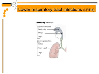
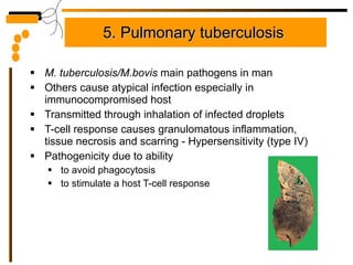
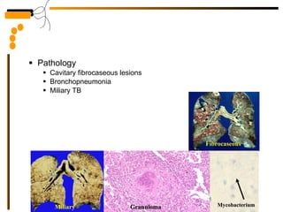
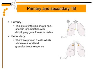
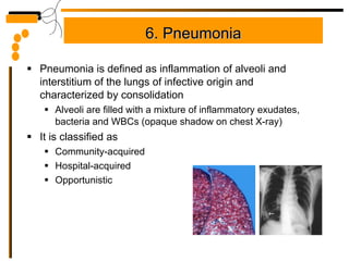
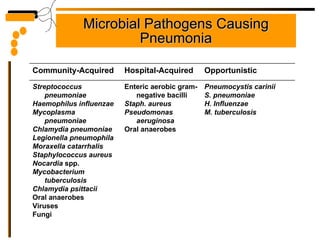
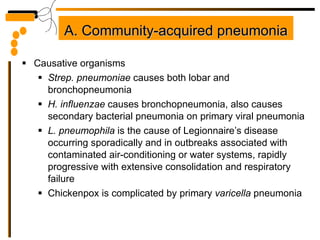
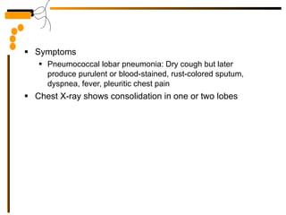
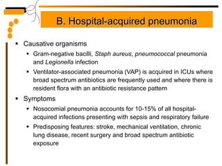
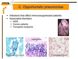
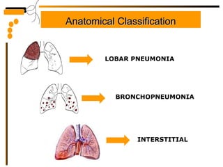
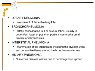
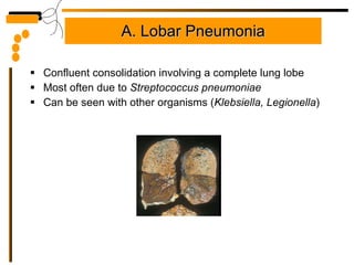
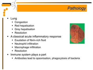
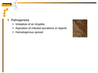
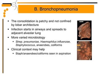
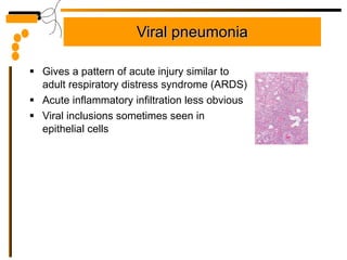
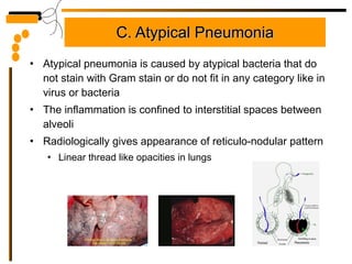
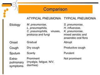
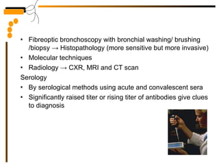
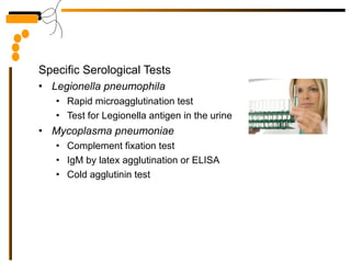
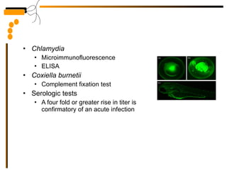
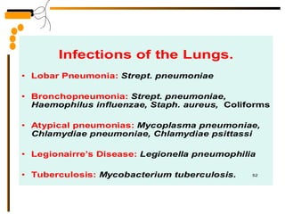
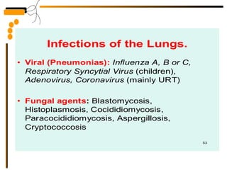
Respiratory Tract Infections Professor Mark Pallen Dr Kamran Afzal Classified Microbiologist
Throat/Nasal Swab specimens Taken under direct visualization with good lighting Areas of exudation, membrane formation or inflammation are choice sites, otherwise rub the tonsillar crypts For  Bordetella pertussis , collect nasopharyngeal or per nasal swab Inform lab in advance to prepare fresh culture media for this organism
For viral agents, instruct patient to gargle with nutrient broth For  M. leprae , nasal swab or nasal washing is used TRANSPORTATION Streptococcal pharyngitis or Diphtheria Stuart tpt medium, silica gel Whooping cough Bordetella tpt medium Viral infection VTM, but if delay, keep at -70 0 C
Collection of Sputum specimens Specimen obtained before start of antibiotic therapy If started, inform lab about antibiotics Instruct patient appropriately Proper labeling Patient’s name, test type, date, clinical info Preferably collect in working hours Collect specimen as early as possible in acute phase, these agents tend to disappear rapidly after the onset of symptoms All specimens for viral culture to be frozen and stored at -70 o C
Give the patient a dry, wide mouthed, leak proof container Collect sufficient quantity, sterile container Early morning specimen is preferred Patient should cough deeply to produce a sputum specimen For MTB culture, collect 3 x fresh, early morning specimens, keep in a refrigerator to be pooled or processed individually In un-cooperative patients, MTB may be recovered from gastric aspiration in the ward
Respiratory tract defences Ventilatory flow Cough Mucociliary clearance mechanisms Mucosal immune system
Predisposing factors for RTIs Low immunity Environmental pollution Smoking Bad nutrition Tumors Alcohol Decreased cough reflex Injury to cilia Decreased function of alveolar macrophages Edema or congestion Retention of secretions
Upper respiratory tract infections  (URTIs)
1. Cold and Flu Causative organisms Rhino, corona, adeno  and  parainfluenza  viruses A novel respiratory pathogen (hMPV) Causes respiratory illness in elderly, young children and immunocompromised patients Symptoms Coryzal symptoms, rhinitis, pharyngitis, laryngitis Management Symptomatic and consists of rest, adequate hydration, simple analgesics and antipyretics Antibacterials are not effective and cause adverse consequences of its overuse in treatment of viral infections
2. Influenza Causative organisms Influenza viruses (A, B and C) Symptoms Severe malaise and myalgia complicated by life-threatening secondary bacterial infections as staphylococcal pneumonia Management Neuraminidase inhibitors (Zanamivir and oseltamivir)
3. Sore throat (pharyngitis) Causative organism Streptococcus pyogenes Symptoms Ranging from fever and symptoms of common cold to inflammation of pharynx with whitish exudate on the tonsils plus enlarged tender cervical lymph nodes Diagnosis Is a must to differentiate between viral or streptococcal infections by taking a throat swab for culture Treatment Wait for results of culture before starting antibiotics Penicillins, cephalosporins and macrolides
3. Laryngitis  Most commonly upper respiratory viruses Diphtheria  C. diphtheriae  produces a cytotoxic exotoxin causing tissue necrosis at site of infection with associated acute inflammation Membrane may narrow airway and/or slough off (asphyxiation)
4. Acute epiglottitis H. influenzae  type B Another cause of acute severe airway compromise in childhood
5. Otitis media Causative organisms Inflammation of middle ear seen most frequently in children under 3 years Streptococcus pneumoniae , H. influenzae, Strep. pyogenes Symptoms Ear pain, if the drum perforates, the pain is relieved and a purulent discharge follows Treatment It should be effective against the 3 main bacterial pathogens Co-amoxiclav or the newer cefixime having high activity against  H. influenzae
6. Acute sinusitis Causative organisms Caused by similar organisms as otitis media Viral upper respiratory tract infections Sometimes associated with dental disease Symptoms Facial pain and tenderness accompanied by headache and purulent nasal discharge Treatment As otitis media Addition of metronidazole (dental disease)
Lower respiratory tract infections  (LRTIs)
1. Acute bronchitis Acute bronchitis is usually infective Chronic bronchitis is a chronic inflammatory condition characterized by thickened, edematous bronchial mucosa with mucus gland hypertrophy and usually caused by smoking Symptoms Yellow or green sputum (pus cells), wheezing Treatment Airflow optimization Physiotherapy to aid expectoration, oxygen and bronchodilatation Antibiotic therapy First-line agents: Doxycycline, Amoxicillin Second-line agents: Co-amoxiclav,  clarithromycin, cefixime Others: Moxifloxacin, a quinolone effective against gram +ve and -ve
2. Bronchiolitis It is characterized by inflammatory changes in the small bronchi and bronchioles but not by consolidation It attacks infants in the 1 st  year of life (airway narrowing) Causative organism Respiratory syncytial virus (RSV) Symptoms Fever, coryzal symptoms which progresses to wheezing, respiratory distress and hypoxia Diagnosis Viral culture of respiratory secretion or IF Treatment  Supportive: oxygen, adequate hydration and vent. assistance Severe cases: Ribavirin
3. Lung abscess Localized suppurative necrosis Organisms commonly cultured Staphylococci Streptococci Gram-negative Anaerobes Frequent mixed infections Pathogenesis Aspiration Pneumonia Septic emboli Tumors  Bronchiectasis
4. Bronchiectasis Dilatation of bronchi and bronchioles secondary to chronic inflammation Usually due to fibrous scarring following infection (pneumonia, tuberculosis, cystic fibrosis) Dilated airways accumulate purulent secretions Associated conditions Obstruction Cystic fibrosis Immotile cilia syndromes Necrotizing pneumonia
5.  Pulmonary tuberculosis   M. tuberculosis/M.bovis  main pathogens in man Others cause atypical infection especially in immunocompromised host Transmitted through inhalation of infected droplets T-cell response causes granulomatous inflammation, tissue necrosis and scarring - Hypersensitivity (type IV) Pathogenicity due to ability to avoid phagocytosis to stimulate a host T-cell response
Pathology Cavitary fibrocaseous lesions Bronchopneumonia Miliary TB Miliary Granuloma Fibrocaseous Mycobacterium
Primary and secondary TB Primary The site of infection shows non-specific inflammation with developing granulomas in nodes Secondary There are primed T cells which stimulate a localised granulomatous response
6. Pneumonia Pneumonia is defined as inflammation of alveoli and interstitium of the lungs of infective origin and characterized by consolidation Alveoli are filled with a mixture of inflammatory exudates, bacteria and WBCs (opaque shadow on chest X-ray) It is classified as Community-acquired Hospital-acquired Opportunistic
Microbial Pathogens Causing Pneumonia             Pneumocystis carinii S. pneumoniae H. Influenzae M. tuberculosis Enteric aerobic gram-negative bacilli Staph. aureus Pseudomonas aeruginosa Oral anaerobes Streptococcus pneumoniae Haemophilus influenzae Mycoplasma pneumoniae Chlamydia pneumoniae Legionella pneumophila Moraxella catarrhalis Staphylococcus aureus Nocardia  spp. Mycobacterium tuberculosis Chlamydia psittacii Oral anaerobes   Viruses Fungi   Opportunistic Hospital-Acquired Community-Acquired
A. Community-acquired pneumonia Causative organisms  Strep. pneumoniae  causes both lobar and bronchopneumonia H. influenzae  causes bronchopneumonia, also causes secondary bacterial pneumonia on primary viral pneumonia L. pneumophila  is the cause of Legionnaire’s disease occurring sporadically and in outbreaks associated with contaminated air-conditioning or water systems, rapidly progressive with extensive consolidation and respiratory failure Chickenpox is complicated by primary  varicella  pneumonia
Symptoms Pneumococcal lobar pneumonia: Dry cough but later produce purulent or blood-stained, rust-colored sputum, dyspnea, fever, pleuritic chest pain Chest X-ray shows consolidation in one or two lobes
B. Hospital-acquired pneumonia Causative organisms Gram-negative bacilli,  Staph aureus ,  pneumococcal  pneumonia and  Legionella  infection Ventilator-associated pneumonia (VAP) is acquired in ICUs where broad spectrum antibiotics are frequently used and where there is resident flora with an antibiotic resistance pattern Symptoms Nosocomial pneumonia accounts for 10-15% of all hospital-acquired infections presenting with sepsis and respiratory failure Predisposing features: stroke, mechanical ventilation, chronic lung disease, recent surgery and broad spectrum antibiotic exposure
Aspiration pneumonia It can be seen in community or hospital Caused by inhalation of stomach contents contaminated by bacteria from the mouth (when the patient vomits while unconscious) Anaerobic bacteria are implicated often accompanied by aerobic organisms Gastric acid is very destructive to lung tissue causing severe tissue necrosis and infection with abscess Risk factors Alcohol, hypnotic drugs and GA Treatment Metronidazole plus amoxicillin or metronidazole plus cefotaxime if you suspect gram-negative infection
C. Opportunistic pneumonias Infections that affect immunosuppressed patients Associated disorders AIDS Cancer patients Transplant recipients Pneumocystis carinii Aspergillus Cytomegalovirus
Anatomical Classification LOBAR PNEUMONIA BRONCHOPNEUMONIA INTERSTITIAL
LOBAR PNEUMONIA Involvement of the entire lung lobe BRONCHOPNEUMONIA Patchy consolidation in 1 or several lobes, usually in dependent lower or posterior portions centered around bronchi and bronchioles INTERSTITIAL PNEUMONIA Inflammation of the interstitium, including the alveolar walls and connective tissue around the bronchovascular tree MILIARY PNEUMONIA Numerous discrete lesions due to hematogenous spread
A. Lobar Pneumonia Confluent consolidation involving a complete lung lobe Most often due to  Streptococcus pneumoniae   Can be seen with other organisms ( Klebsiella, Legionella )
Pathology Lung Congestion  Red hepatisation Grey hepatisation Resolution A classical acute inflammatory response Exudation of fibrin-rich fluid Neutrophil infiltration Macrophage infiltration Resolution Immune system plays a part  Antibodies lead to opsonisation, phagocytosis of bacteria
Pathogenesis Inhalation of air droplets Aspiration of infected secretions or objects Hematogenous spread
B. Bronchopneumonia The consolidation is patchy and not confined by lobar architecture Infection starts in airways and spreads to adjacent alveolar lung More varied microbiology  Strep. pneumoniae, Haemophilus influenzae, Staphylococcus , anaerobes, coliforms Clinical context may help  Staph/anaerobes/coliforms seen in aspiration
Viral pneumonia Gives a pattern of acute injury similar to adult respiratory distress syndrome (ARDS) Acute inflammatory infiltration less obvious Viral inclusions sometimes seen in epithelial cells
C. Atypical Pneumonia Atypical pneumonia is caused by atypical bacteria that do not stain with Gram stain or do not fit in any category like in virus or bacteria The inflammation is confined to interstitial spaces between alveoli  Radiologically gives appearance of reticulo-nodular pattern Linear thread like opacities in lungs
Comparison Not prominent Prominent  (myalgia, fatigue, N/V, diarrhea) Extra-pulmonary symptoms Purulent Scanty  Sputum Productive cough Dry cough Cough Abrupt Gradual Onset S. pneumoniae ,  H. influenzae ,  K. pneumoniae ,  mixed aerobic and anaerobic oral flora M. pneumoniae ,  L.   pneumophilia,   C. pneumophilla,   viruses, protozoa and fungi Etiology TYPICAL PNEUMONIA ATYPICAL PNEUMONIA
Investigations Microscopy is unreliable due to oropharyngeal contamination Gram stain ZN stain for AFB  Sputum culture is the mainstay of diagnosis for pneumonia caused by  pneumococci  and  H. influenzae Total and differential blood count Blood, urine, sputum  ->  Culture/sensitivity Serological investigations Antigen detection in sputum or urine by  Fluorescent methods, Immunoelectrophoresis, Latex agglutination, ELISA
Fibreoptic bronchoscopy with bronchial washing/ brushing /biopsy -> Histopathology (more sensitive but more invasive) Molecular techniques Radiology -> CXR, MRI and CT scan Serology  By serological methods using acute and convalescent sera Significantly raised titer or rising titer of antibodies give clues to diagnosis
Specific Serological Tests Legionella pneumophila   Rapid microagglutination test Test for Legionella antigen in the urine Mycoplasma pneumoniae  Complement fixation test IgM by latex agglutination or ELISA Cold agglutinin test
Chlamydia  Microimmunofluorescence ELISA  Coxiella burnetii  Complement fixation test Serologic tests A four fold or greater rise in titer is confirmatory of an acute infection
Treatment Targeted treatment Pneumococcal  pneumonia Benzyl penicillin or Amoxicillin  Erythromycin (in penicillin-allergic patients)  Combined  β - lactam + macrolide when pneumonia is complicated by  pneumococcal bacteremia H. influenzae Co-amoxiclav, parenteral cefuroxime, cefixime Clarithromycin and azithromycin
Morexella  pneumonia Doesn’t possess cell wall so not susceptible to  β -lactams Tetracyclins and quinolones Staphylococcal  pneumonia Flucloxacillin + a second agent as rifampicin, fusidic acid or gentamycin Legionnaire’s  disease Erythromycin or azithromycin


LAW OF CONSERVATION OF KNOWLEDGE “ No matter how long the lecture may be, the knowledge before and after the lecture remains constant”

RTIs

  • 1.
    Respiratory Tract InfectionsProfessor Mark Pallen Dr Kamran Afzal Classified Microbiologist
  • 2.
    Throat/Nasal Swab specimensTaken under direct visualization with good lighting Areas of exudation, membrane formation or inflammation are choice sites, otherwise rub the tonsillar crypts For Bordetella pertussis , collect nasopharyngeal or per nasal swab Inform lab in advance to prepare fresh culture media for this organism
  • 3.
    For viral agents,instruct patient to gargle with nutrient broth For M. leprae , nasal swab or nasal washing is used TRANSPORTATION Streptococcal pharyngitis or Diphtheria Stuart tpt medium, silica gel Whooping cough Bordetella tpt medium Viral infection VTM, but if delay, keep at -70 0 C
  • 4.
    Collection of Sputumspecimens Specimen obtained before start of antibiotic therapy If started, inform lab about antibiotics Instruct patient appropriately Proper labeling Patient’s name, test type, date, clinical info Preferably collect in working hours Collect specimen as early as possible in acute phase, these agents tend to disappear rapidly after the onset of symptoms All specimens for viral culture to be frozen and stored at -70 o C
  • 5.
    Give the patienta dry, wide mouthed, leak proof container Collect sufficient quantity, sterile container Early morning specimen is preferred Patient should cough deeply to produce a sputum specimen For MTB culture, collect 3 x fresh, early morning specimens, keep in a refrigerator to be pooled or processed individually In un-cooperative patients, MTB may be recovered from gastric aspiration in the ward
  • 6.
    Respiratory tract defencesVentilatory flow Cough Mucociliary clearance mechanisms Mucosal immune system
  • 7.
    Predisposing factors forRTIs Low immunity Environmental pollution Smoking Bad nutrition Tumors Alcohol Decreased cough reflex Injury to cilia Decreased function of alveolar macrophages Edema or congestion Retention of secretions
  • 8.
    Upper respiratory tractinfections (URTIs)
  • 9.
    1. Cold andFlu Causative organisms Rhino, corona, adeno and parainfluenza viruses A novel respiratory pathogen (hMPV) Causes respiratory illness in elderly, young children and immunocompromised patients Symptoms Coryzal symptoms, rhinitis, pharyngitis, laryngitis Management Symptomatic and consists of rest, adequate hydration, simple analgesics and antipyretics Antibacterials are not effective and cause adverse consequences of its overuse in treatment of viral infections
  • 10.
    2. Influenza Causativeorganisms Influenza viruses (A, B and C) Symptoms Severe malaise and myalgia complicated by life-threatening secondary bacterial infections as staphylococcal pneumonia Management Neuraminidase inhibitors (Zanamivir and oseltamivir)
  • 11.
    3. Sore throat(pharyngitis) Causative organism Streptococcus pyogenes Symptoms Ranging from fever and symptoms of common cold to inflammation of pharynx with whitish exudate on the tonsils plus enlarged tender cervical lymph nodes Diagnosis Is a must to differentiate between viral or streptococcal infections by taking a throat swab for culture Treatment Wait for results of culture before starting antibiotics Penicillins, cephalosporins and macrolides
  • 12.
    3. Laryngitis Most commonly upper respiratory viruses Diphtheria C. diphtheriae produces a cytotoxic exotoxin causing tissue necrosis at site of infection with associated acute inflammation Membrane may narrow airway and/or slough off (asphyxiation)
  • 13.
    4. Acute epiglottitisH. influenzae type B Another cause of acute severe airway compromise in childhood
  • 14.
    5. Otitis mediaCausative organisms Inflammation of middle ear seen most frequently in children under 3 years Streptococcus pneumoniae , H. influenzae, Strep. pyogenes Symptoms Ear pain, if the drum perforates, the pain is relieved and a purulent discharge follows Treatment It should be effective against the 3 main bacterial pathogens Co-amoxiclav or the newer cefixime having high activity against H. influenzae
  • 15.
    6. Acute sinusitisCausative organisms Caused by similar organisms as otitis media Viral upper respiratory tract infections Sometimes associated with dental disease Symptoms Facial pain and tenderness accompanied by headache and purulent nasal discharge Treatment As otitis media Addition of metronidazole (dental disease)
  • 16.
    Lower respiratory tractinfections (LRTIs)
  • 17.
    1. Acute bronchitisAcute bronchitis is usually infective Chronic bronchitis is a chronic inflammatory condition characterized by thickened, edematous bronchial mucosa with mucus gland hypertrophy and usually caused by smoking Symptoms Yellow or green sputum (pus cells), wheezing Treatment Airflow optimization Physiotherapy to aid expectoration, oxygen and bronchodilatation Antibiotic therapy First-line agents: Doxycycline, Amoxicillin Second-line agents: Co-amoxiclav, clarithromycin, cefixime Others: Moxifloxacin, a quinolone effective against gram +ve and -ve
  • 18.
    2. Bronchiolitis Itis characterized by inflammatory changes in the small bronchi and bronchioles but not by consolidation It attacks infants in the 1 st year of life (airway narrowing) Causative organism Respiratory syncytial virus (RSV) Symptoms Fever, coryzal symptoms which progresses to wheezing, respiratory distress and hypoxia Diagnosis Viral culture of respiratory secretion or IF Treatment Supportive: oxygen, adequate hydration and vent. assistance Severe cases: Ribavirin
  • 19.
    3. Lung abscessLocalized suppurative necrosis Organisms commonly cultured Staphylococci Streptococci Gram-negative Anaerobes Frequent mixed infections Pathogenesis Aspiration Pneumonia Septic emboli Tumors Bronchiectasis
  • 20.
    4. Bronchiectasis Dilatationof bronchi and bronchioles secondary to chronic inflammation Usually due to fibrous scarring following infection (pneumonia, tuberculosis, cystic fibrosis) Dilated airways accumulate purulent secretions Associated conditions Obstruction Cystic fibrosis Immotile cilia syndromes Necrotizing pneumonia
  • 21.
    5. Pulmonarytuberculosis M. tuberculosis/M.bovis main pathogens in man Others cause atypical infection especially in immunocompromised host Transmitted through inhalation of infected droplets T-cell response causes granulomatous inflammation, tissue necrosis and scarring - Hypersensitivity (type IV) Pathogenicity due to ability to avoid phagocytosis to stimulate a host T-cell response
  • 22.
    Pathology Cavitary fibrocaseouslesions Bronchopneumonia Miliary TB Miliary Granuloma Fibrocaseous Mycobacterium
  • 23.
    Primary and secondaryTB Primary The site of infection shows non-specific inflammation with developing granulomas in nodes Secondary There are primed T cells which stimulate a localised granulomatous response
  • 24.
    6. Pneumonia Pneumoniais defined as inflammation of alveoli and interstitium of the lungs of infective origin and characterized by consolidation Alveoli are filled with a mixture of inflammatory exudates, bacteria and WBCs (opaque shadow on chest X-ray) It is classified as Community-acquired Hospital-acquired Opportunistic
  • 25.
    Microbial Pathogens CausingPneumonia             Pneumocystis carinii S. pneumoniae H. Influenzae M. tuberculosis Enteric aerobic gram-negative bacilli Staph. aureus Pseudomonas aeruginosa Oral anaerobes Streptococcus pneumoniae Haemophilus influenzae Mycoplasma pneumoniae Chlamydia pneumoniae Legionella pneumophila Moraxella catarrhalis Staphylococcus aureus Nocardia spp. Mycobacterium tuberculosis Chlamydia psittacii Oral anaerobes Viruses Fungi   Opportunistic Hospital-Acquired Community-Acquired
  • 26.
    A. Community-acquired pneumoniaCausative organisms Strep. pneumoniae causes both lobar and bronchopneumonia H. influenzae causes bronchopneumonia, also causes secondary bacterial pneumonia on primary viral pneumonia L. pneumophila is the cause of Legionnaire’s disease occurring sporadically and in outbreaks associated with contaminated air-conditioning or water systems, rapidly progressive with extensive consolidation and respiratory failure Chickenpox is complicated by primary varicella pneumonia
  • 27.
    Symptoms Pneumococcal lobarpneumonia: Dry cough but later produce purulent or blood-stained, rust-colored sputum, dyspnea, fever, pleuritic chest pain Chest X-ray shows consolidation in one or two lobes
  • 28.
    B. Hospital-acquired pneumoniaCausative organisms Gram-negative bacilli, Staph aureus , pneumococcal pneumonia and Legionella infection Ventilator-associated pneumonia (VAP) is acquired in ICUs where broad spectrum antibiotics are frequently used and where there is resident flora with an antibiotic resistance pattern Symptoms Nosocomial pneumonia accounts for 10-15% of all hospital-acquired infections presenting with sepsis and respiratory failure Predisposing features: stroke, mechanical ventilation, chronic lung disease, recent surgery and broad spectrum antibiotic exposure
  • 29.
    Aspiration pneumonia Itcan be seen in community or hospital Caused by inhalation of stomach contents contaminated by bacteria from the mouth (when the patient vomits while unconscious) Anaerobic bacteria are implicated often accompanied by aerobic organisms Gastric acid is very destructive to lung tissue causing severe tissue necrosis and infection with abscess Risk factors Alcohol, hypnotic drugs and GA Treatment Metronidazole plus amoxicillin or metronidazole plus cefotaxime if you suspect gram-negative infection
  • 30.
    C. Opportunistic pneumoniasInfections that affect immunosuppressed patients Associated disorders AIDS Cancer patients Transplant recipients Pneumocystis carinii Aspergillus Cytomegalovirus
  • 31.
    Anatomical Classification LOBARPNEUMONIA BRONCHOPNEUMONIA INTERSTITIAL
  • 32.
    LOBAR PNEUMONIA Involvementof the entire lung lobe BRONCHOPNEUMONIA Patchy consolidation in 1 or several lobes, usually in dependent lower or posterior portions centered around bronchi and bronchioles INTERSTITIAL PNEUMONIA Inflammation of the interstitium, including the alveolar walls and connective tissue around the bronchovascular tree MILIARY PNEUMONIA Numerous discrete lesions due to hematogenous spread
  • 33.
    A. Lobar PneumoniaConfluent consolidation involving a complete lung lobe Most often due to Streptococcus pneumoniae Can be seen with other organisms ( Klebsiella, Legionella )
  • 34.
    Pathology Lung Congestion Red hepatisation Grey hepatisation Resolution A classical acute inflammatory response Exudation of fibrin-rich fluid Neutrophil infiltration Macrophage infiltration Resolution Immune system plays a part Antibodies lead to opsonisation, phagocytosis of bacteria
  • 35.
    Pathogenesis Inhalation ofair droplets Aspiration of infected secretions or objects Hematogenous spread
  • 36.
    B. Bronchopneumonia Theconsolidation is patchy and not confined by lobar architecture Infection starts in airways and spreads to adjacent alveolar lung More varied microbiology Strep. pneumoniae, Haemophilus influenzae, Staphylococcus , anaerobes, coliforms Clinical context may help Staph/anaerobes/coliforms seen in aspiration
  • 37.
    Viral pneumonia Givesa pattern of acute injury similar to adult respiratory distress syndrome (ARDS) Acute inflammatory infiltration less obvious Viral inclusions sometimes seen in epithelial cells
  • 38.
    C. Atypical PneumoniaAtypical pneumonia is caused by atypical bacteria that do not stain with Gram stain or do not fit in any category like in virus or bacteria The inflammation is confined to interstitial spaces between alveoli Radiologically gives appearance of reticulo-nodular pattern Linear thread like opacities in lungs
  • 39.
    Comparison Not prominentProminent (myalgia, fatigue, N/V, diarrhea) Extra-pulmonary symptoms Purulent Scanty Sputum Productive cough Dry cough Cough Abrupt Gradual Onset S. pneumoniae , H. influenzae , K. pneumoniae , mixed aerobic and anaerobic oral flora M. pneumoniae , L. pneumophilia, C. pneumophilla, viruses, protozoa and fungi Etiology TYPICAL PNEUMONIA ATYPICAL PNEUMONIA
  • 40.
    Investigations Microscopy isunreliable due to oropharyngeal contamination Gram stain ZN stain for AFB Sputum culture is the mainstay of diagnosis for pneumonia caused by pneumococci and H. influenzae Total and differential blood count Blood, urine, sputum -> Culture/sensitivity Serological investigations Antigen detection in sputum or urine by Fluorescent methods, Immunoelectrophoresis, Latex agglutination, ELISA
  • 41.
    Fibreoptic bronchoscopy withbronchial washing/ brushing /biopsy -> Histopathology (more sensitive but more invasive) Molecular techniques Radiology -> CXR, MRI and CT scan Serology By serological methods using acute and convalescent sera Significantly raised titer or rising titer of antibodies give clues to diagnosis
  • 42.
    Specific Serological TestsLegionella pneumophila Rapid microagglutination test Test for Legionella antigen in the urine Mycoplasma pneumoniae Complement fixation test IgM by latex agglutination or ELISA Cold agglutinin test
  • 43.
    Chlamydia MicroimmunofluorescenceELISA Coxiella burnetii Complement fixation test Serologic tests A four fold or greater rise in titer is confirmatory of an acute infection
  • 44.
    Treatment Targeted treatmentPneumococcal pneumonia Benzyl penicillin or Amoxicillin Erythromycin (in penicillin-allergic patients) Combined β - lactam + macrolide when pneumonia is complicated by pneumococcal bacteremia H. influenzae Co-amoxiclav, parenteral cefuroxime, cefixime Clarithromycin and azithromycin
  • 45.
    Morexella pneumoniaDoesn’t possess cell wall so not susceptible to β -lactams Tetracyclins and quinolones Staphylococcal pneumonia Flucloxacillin + a second agent as rifampicin, fusidic acid or gentamycin Legionnaire’s disease Erythromycin or azithromycin
  • 46.
  • 47.
  • 48.
  • 49.
  • 50.
  • 51.
    LAW OF CONSERVATIONOF KNOWLEDGE “ No matter how long the lecture may be, the knowledge before and after the lecture remains constant”