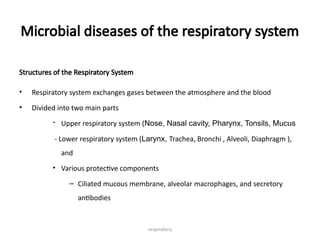
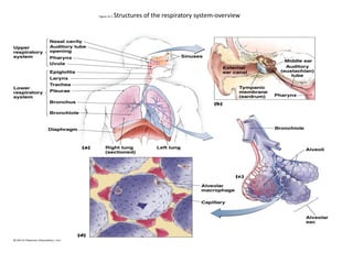
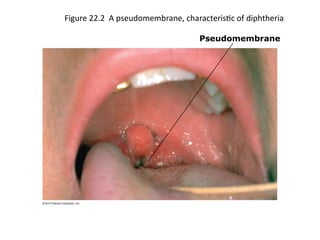
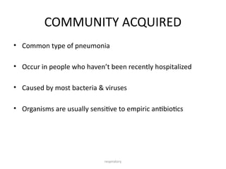
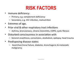
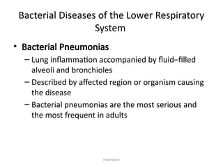
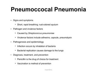
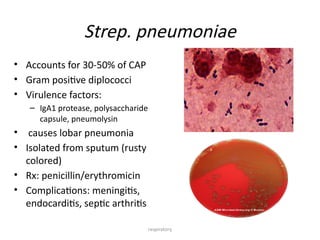
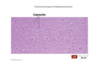
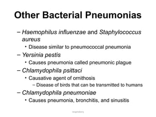
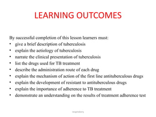
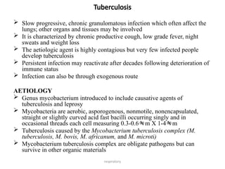
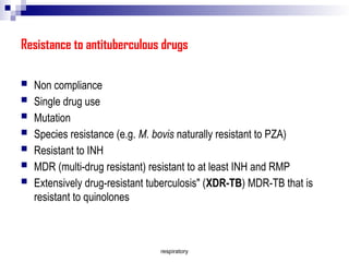
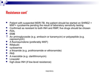
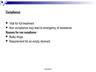
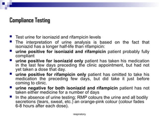
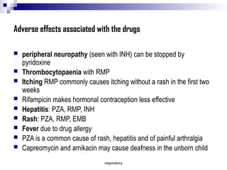
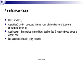
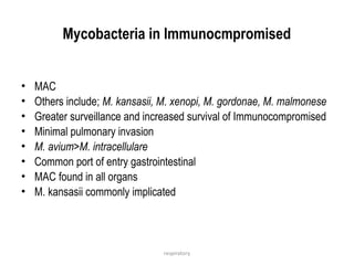
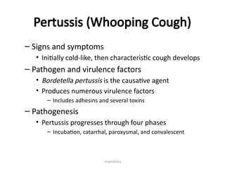
Diseases of the Respiratory
System
Dr R.S. MKAKOSYA
microbiology
respiratory
Intended learning outcomes
• Recall anatomy
• The Normal flora
• Aetiology
• Pathogenesis
• Diagnosis
• Management
• Prevention
respiratory
Microbial diseases of the respiratory system
Structures of the Respiratory System
• Respiratory system exchanges gases between the atmosphere and the blood
• Divided into two main parts
- Upper respiratory system (Nose, Nasal cavity, Pharynx, Tonsils, Mucus
- Lower respiratory system (Larynx, Trachea, Bronchi , Alveoli, Diaphragm ),
and
• Various protective components
– Ciliated mucous membrane, alveolar macrophages, and secretory
antibodies
Figure 22.1 Structures of the respiratory system-overview
respiratory
Normal Microbiota of the Respiratory System
– Lower respiratory system
• Typically microorganisms are not present
– Upper respiratory system
• Colonized by many microorganisms
• Normal microbiota limit growth of pathogens
• Normal microbiota may be opportunistic pathogens
respiratory
Bacterial Diseases of the Upper Respiratory
System, Sinuses, and Ears
• Streptococcal Respiratory Diseases
– Signs and symptoms
• Sore throat and difficulty swallowing
• May progress to scarlet or rheumatic fever
– Pathogen and virulence factors
• Caused by group A streptococci (S. pyogenes)
• Variety of virulence factors
– M proteins, hyaluronic acid capsule, streptokinases, C5a peptidase, pyrogenic
toxins, streptolysins
respiratory
Streptococcal Respiratory Diseases
– Pathogenesis
• Occurs when normal microbiota are depleted, large inoculum is
introduced, or adaptive immunity is impaired
– Epidemiology
• Spread via respiratory droplets
• Occurs most often in cold weather
– Diagnosis, treatment, and prevention
• Often confused with viral pharyngitis
• Penicillin is an effective treatment
respiratory
Diphtheria
– Signs and symptoms
• Presence of a pseudomembrane that can obstruct airways
– Pathogen and virulence factors
• Caused by Corynebacterium diphtheriae
• Virulence factors
– C. diptheriae produces diphtheria toxin
» Prevents polypeptide synthesis and causes cell death
Figure 22.2 A pseudomembrane, characteristic of diphtheria
Pseudomembrane
respiratory
Diphtheria
– Pathogenesis and epidemiology
• Spread via respiratory droplets or skin contact
• Symptomatic in immunocompromised or nonimmune individuals
– Diagnosis, treatment, and prevention
• Diagnosis based on presence of a pseudomembrane
• Treat with antitoxin and antibiotics
• Immunization is an effective prevention
respiratory
Sinusitis and Otitis Media
– Signs and symptoms
• Sinusitis causes pain and pressure of the affected sinus
accompanied by malaise
• Otitis media results in severe pain in the ears
– Pathogen and virulence factors
• Caused by various respiratory microbiota
– May be due to upper respiratory system and auditory tube
damage
respiratory
Sinusitis and Otitis Media
– Pathogenesis and epidemiology
• Bacteria in the pharynx spread to the sinuses via the throat
• Sinusitis is more common in adults
• Otitis media is more common in children
– Diagnosis, treatment, and prevention
• Symptoms often diagnostic
• No known way to prevent sinusitis
respiratory
Infections of the lower respiratory system
(pneumonia)
• Definition of pneumonia
• Classifications of pneumonia
• Risk factors for pneumonia
• Management of pneumonia
• Assessment for severity of pneumonia
• Complications
• Prevention
respiratory
DEFINITION
• PATHOLOGICAL:
– Inflammation of the lung parenchyma
– Polymorphonuclear leukocyte exudate in and around alveoli, terminal
bronchioles
– Develops in 3 stages:
• Engorgement: lung is wet, edematous & congested
• Red hepatization: lung is red, dry, friable & solid
• Grey hepatization: softened lung & exudation of yellow purulent
fluid
respiratory
Clinical presentation
• CLINICAL:
– Acute illness consisting of a syndrome of
• Fever, cough, sputum production and
• Clinical signs of consolidation and
• Typical chest radiograph (CXR) changes
respiratory
pathogenesis
• Microbes can access the lower respiratory tract by:
– Inhalation of aerosolized material
– Aspiration of normal flora of the upper respiratory tract
– Seeding from other infected sites via bloodstream
• Only microbes of <5µm diameter reach the alveoli to cause
inflammation
respiratory
CLASSIFICATION
• SITE
– Lobar vs. bronchopneumonia
• AETIOLOGY
– Community acquired vs. nosocomial pneumnia
• MICROBIOLOGY
– Causative organism: bacteria, virus, fungi
• TYPICAL VS. ATYPICAL
respiratory
• Involvement of a distinct
region of the lung
• Polymorph exudate clots in
the alveoli rendering them
solid
• Classically seen in
previously healthy young
people
• Usually caused by S.
pneumoniae
1. SITE: lobar/segmental
respiratory
• Usually bilateral
• Consolidation is scattered
throughout the lung fields
• Mainly seen in
– Elderly
– Debilitating or chronic
respiratory disease, e.g.
chronic bronchitis
2. SITE: bronchopneumonia
respiratory
• INTERSTITIAL
– Involves invasion of the
lung interstitium
– Characteristic of viral
infections of the lungs
• LUNG ABSCESS
– a.k.a. necrotizing
pneumonia
– Cavitation & destruction
of the lung parenchyma
3. SITE: interstitial vs. abscess
respiratory
COMMUNITY ACQUIRED
• Common type of pneumonia
• Occur in people who haven’t been recently hospitalized
• Caused by most bacteria & viruses
• Organisms are usually sensitive to empiric antibiotics
respiratory
RISK FACTORS
• Immune deficiency:
– Primary, e.g. complement deficiency
– Secondary, e.g. HIV infection, malnutrition
• Extremes of age,
• Prior viral & other respiratory tract infections
– Asthma, bronciectasis, chronic bronchitis, COPD, cystic fibrosis
• Disturbed consciousness in association with
– General anaethesia, convulsions, alcoholism, epilepsy, head trauma
• Predisposing disease states:
– heart/liver/renal failure, diabetes, bronchogenic & metastatic
malignancy
respiratory
Bacterial Diseases of the Lower Respiratory
System
• Bacterial Pneumonias
– Lung inflammation accompanied by fluid–filled
alveoli and bronchioles
– Described by affected region or organism causing
the disease
– Bacterial pneumonias are the most serious and
the most frequent in adults
respiratory
Pneumoccocal Pneumonia
– Signs and symptoms
• Short, rapid breathing; rust-colored sputum
– Pathogen and virulence factors
• Caused by Streptococcus pneumoniae
• Virulence factors include adhesins, capsule, pneumolysin
– Pathogenesis and epidemiology
• Infection occurs by inhalation of bacteria
• Bacterial replication causes damage to the lungs
– Diagnosis, treatment, and prevention
• Penicillin is the drug of choice for treatment
• Vaccination is method of prevention
• Accounts for 30-50% of CAP
• Gram positive diplococci
• Virulence factors:
– IgA1 protease, polysaccharide
capsule, pneumolysin
• causes lobar pneumonia
• Isolated from sputum (rusty
colored)
• Rx: penicillin/erythromicin
• Complications: meningitis,
endocarditis, septic arthritis
Strep. pneumoniae
respiratory
respiratory
Primary Atypical (Mycoplasmal) Pneumonia
– Signs and symptoms
• Include fever, malaise, sore throat, excessive sweating
– Pathogen and virulence factors
• Caused by Mycoplasma pneumoniae
• Virulence factors include an adhesion protein
– Epidemiology
• Bacteria spread by nasal secretions
– Diagnosis, treatment, and prevention
• Treated with tetracycline and erythromycin
• Prevention difficult since infected individuals may be
asymptomatic
respiratory
Klebsiella Pneumonia
– Signs and symptoms
• Pneumonia symptoms combined with a thick, bloody
sputum
– Pathogen and virulence factors
• Caused by Klebsiella pneumoniae
• Virulence factors include a capsule
– Pathogenesis and epidemiology
• Immunocompromised individuals at greatest risk for
infection
– Diagnosis, treatment, and prevention
• Treated with antimicrobials
• Prevention involves good aseptic technique by health care
workers
The prominent capsule of Klebsiella pneumoniae
Capsules
respiratory
Other Bacterial Pneumonias
– Haemophilus influenzae and Staphylococcus
aureus
• Disease similar to pneumococcal pneumonia
– Yersinia pestis
• Causes pneumonia called pneumonic plague
– Chlamydophila psittaci
• Causative agent of ornithosis
– Disease of birds that can be transmitted to humans
– Chlamydophila pneumoniae
• Causes pneumonia, bronchitis, and sinusitis
TUBERCULOSIS
R.S. Mkakosya
respiratory
LEARNING OUTCOMES
By successful completion of this lesson learners must:
• give a brief description of tuberculosis
• explain the aetiology of tuberculosis
• narrate the clinical presentation of tuberculosis
• list the drugs used for TB treatment
• describe the administration route of each drug
• explain the mechanism of action of the first line antituberculous drugs
• explain the development of resistant to antituberculous drugs
• explain the importance of adherence to TB treatment
• demonstrate an understanding on the results of treatment adherence test
respiratory
Tuberculosis
 Slow progressive, chronic granulomatous infection which often affect the
lungs; other organs and tissues may be involved
 It is characterized by chronic productive cough, low grade fever, night
sweats and weight loss
 The aetiologic agent is highly contagious but very few infected people
develop tuberculosis
 Persistent infection may reactivate after decades following deterioration of
immune status
 Infection can also be through exogenous route
AETIOLOGY
 Genus mycobacterium introduced to include causative agents of
tuberculosis and leprosy
 Mycobacteria are aerobic, asporogenous, nonmotile, nonencapsulated,
straight or slightly curved acid fast bacilli occurring singly and in
occasional threads each cell measuring 0.3-0.6m X 1-4m
 Tuberculosis caused by the Mycobacterium tuberculosis complex (M.
tuberculosis, M. bovis, M. africanum, and M. microti)
 Mycobacterium tuberculosis complex are obligate pathogens but can
survive in other organic materials
respiratory
 Can survive in sputum for 3 months, 2-6 months in the soil, 6
months in the laboratory
 Sensitive to UV light, heat (destroyed during pasteurization)
 Susceptible to alcohol, formaldehyde and gluteraldehyde
 Resistant to acids, alkali and quaternary ammonium
compounds
 Rabbits are more sensitive to M. bovis than to M. tuberculosis
 Guinea pigs sensitive to both M. tuberculosis and M. bovis
respiratory
PATHOGENESIS
 Inhalation of droplets with viable bacilli or ingestion of contaminated
food/drink
 Bacilli deposited into the alveolar space where they are engulfed by
alveolar macrophages
 Infectious inoculum resists intracellular destruction and persists,
eventually multiplying and killing the macrophages
 Virulence due to the ability of surviving in macrophages
 Accumulating mycobacteria stimulate an inflammatory focus which
matures into a granulomatous lesion characterized by a mononuclear cell
infiltrate surrounding a core of degenerating epitheloid and
multinucleated giant cells
 Lesions become enveloped in fibrin and the center progresses to caseous
necrosis
 Erosion of caseous tubercles into adjacent airways result in cavitation and
release of bacilli into sputum
respiratory
CLINICAL MANIFESTATIONS
Primary TB
 Generally subclinical but positive to PPD; Fever, non-productive cough, shortness of breath
 X-ray of the chest may show patchy or lobular infiltrate in the anterior segment of the upper,
middle and lower lobes
 Pleurisy without parenchyma infiltrate, pleuritic chest pain, fever, chills, sweats and dyspnea
Progressive TB
 Low grade fever, night sweats, fatigability, loss of appetite, weight loss, cough and occasional
haemoptysis
 Sputum smear positive
Post Primary Reactivation
 Night sweats, chills, fatigue, fever, haemoptysis, physical examination reveals dullness and
rales in the upper lung fields. PPD and culture positive
Extra-pulmonary TB
 Clinical manifestation general e.g. fever or typical of the organ involved e.g. dysuria in
genitourinary TB, back pain in vertebral TB (1) Lymphatic TB (2) genitourinary TB (3) vertebral
and articular TB (4) meningeal TB (5) Peritoneal TB (6) perirdial TB (7) milliary TB
respiratory
CLINICAL MANIFESTATIONS
Diagnosis of tuberculosis-overview
respiratory
TREATMENT
 1ST
Line drugs: Isoniazid (INH; isonicotinic acid hydrazide), rifampicin, pyrazinamide
(PZA)and/or ethambutol (EMB) for the first 2 months then continue with isoniazid
and rifampicin
Antituberculous drugs
Isoniazid: Most active against M. tuberculosis where it exerts bactericidal effect. Well
absorbed when given orally and eliminated through urine. May cause peripheral
neuropathy and mental disturbance which can be prevented by giving pyridoxine
(vit. B6)
Rifampicin: One of the rifamycins. Broad spectrum antibiotic active against M. leprae,
M. marinum, M.kansasii and M. haemophilum. Together with rifabutin are
semisynthetic derivative of the naturally occurring antibiotic rifamycin B. Rifabutin
is active against M. avium complex. Rifamycin act by binding to the  subunit of
the DNA dependent RNA polymerase and prevent initiation of protein synthesis.
Causes flu-like syndrome especially when given intermittently. Contraindicated in
administration of steroids, oral contraceptives and antiepleptic drugs
Pyrazinamide: Synthetic derivative of nicotinamide. Bactericidal only in acidic
environment and after intracellular conversion by a bacterial amidase to pyrazinoic
acid
All the three are hepatotoxic
Ethambutol: Generally mycobacteristatic but has a wider spectra activity over the
mycobacteria. Causes ocular damage
respiratory
Second-line drugs (SLDs) used for the treatment of TB
 Aminoglycosides e.g. kanamycin, amikacin
 Polypeptides e.g. capreomycin, viomycin, enviomycin
 Fluoroquinolones e.g. ciprofloxacin, levofloxacin, moxilofloxacin
 Thioamides e.g. prothionamide, ethionamide
 Cycloserine and
 P-aminosalicylic acid
Other drugs in the second line treatment or Third line
 Rifabutin, linezolid, thioacetazone, macrolides (e.g. clarithromycin),
thiolidazine, vitamin D and arginine
respiratory
Acceptable abbreviations: Antituberculous drugs can abbreviated
as below:
First line tuberculosis drugs
Isoniazid INH H
Rifampicin RMP R
Pyrazinamide PZA Z
Ethambutol EMB E
Streptomycin STM S
Second line tuberculosis drugs
Ciprofloxacin CIP
P-aminosalicylic acid PAS P
Moxifloxacin MXF
respiratory
Resistance to antituberculous drugs
 Non compliance
 Single drug use
 Mutation
 Species resistance (e.g. M. bovis naturally resistant to PZA)
 Resistant to INH
 MDR (multi-drug resistant) resistant to at least INH and RMP
 Extensively drug-resistant tuberculosis" (XDR-TB) MDR-TB that is
resistant to quinolones
respiratory
Resistance cont’
 Patient with suspected MDR-TB, the patient should be started on SHREZ +
MXF+ cycolserine pending the result of laboratory sensitivity testing
 Confirmed as resistant to both INH and RMP, five drugs should be chosen
 PZA,
 EMB
 An aminoglycoside (e.g. amikacin or kanamycin) or polypeptide (e.g.
capreomycin)
 A fluoroquinolone (preferably MXF)
 Rifabutin
 cycloserine
 a thioanide (e.g. prothionamide or ethionamide)
 PAS
 A macrolide (e.g. clarithhromycin)
 Linezolid
 high-dose INH (if low-level resistance)
respiratory
Compliance
 Vital for full treatment
 Non compliance may lead to emergency of resistance
Reasons for non compliance
 Bulky drugs
 Requirement for an empty stomach
respiratory
Compliance Testing
 Test urine for isoniazid and rifampicin levels
 The interpretation of urine analysis is based on the fact that
isoniazid has a longer half-life than rifampicin:
 urine positive for isoniazid and rifampicin patient probably fully
compliant
 urine positive for isoniazid only patient has taken his medication
in the last few days preceding the clinic appointment, but had not
yet taken a dose that day.
 urine positive for rifampicin only patient has omitted to take his
medication the preceding few days, but did take it just before
coming to clinic.
 urine negative for both isoniazid and rifampicin patient has not
taken either medicine for a number of days
 In the absence of urine testing; RMP colours the urine and all bodily
secretions (tears, sweat, etc.) an orange-pink colour (colour fades
6-8 hours after each dose).
respiratory
Adverse effects associated with the drugs
 peripheral neuropathy (seen with INH) can be stopped by
pyridoxine
 Thrombocytopaenia with RMP
 Itching RMP commonly causes itching without a rash in the first two
weeks
 Rifampicin makes hormonal contraception less effective
 Hepatitis: PZA, RMP, INH
 Rash: PZA, RMP, EMB
 Fever due to drug allergy
 PZA is a common cause of rash, hepatitis and of painful arthralgia
 Capreomycin and amikacin may cause deafness in the unborn child
respiratory
A model prescription
 2HREZ/4HR3
 A prefix (2 and 4) denotes the number of months the treatment
should be given for
 A subscript (3) denotes intermittent dosing (so 3 means three times a
week) and
 No subscript means daily dosing
respiratory
Mycobacteria in Immunocmpromised
• MAC
• Others include; M. kansasii, M. xenopi, M. gordonae, M. malmonese
• Greater surveillance and increased survival of Immunocompromised
• Minimal pulmonary invasion
• M. avium>M. intracellulare
• Common port of entry gastrointestinal
• MAC found in all organs
• M. kansasii commonly implicated
respiratory
Pertussis (Whooping Cough)
– Signs and symptoms
• Initially cold-like, then characteristic cough develops
– Pathogen and virulence factors
• Bordetella pertussis is the causative agent
• Produces numerous virulence factors
– Includes adhesins and several toxins
– Pathogenesis
• Pertussis progresses through four phases
– Incubation, catarrhal, paroxysmal, and convalescent
respiratory
Pertussis (Whooping Cough)
– Epidemiology
• Highly contagious
• Bacteria spread through the air in airborne droplets
– Diagnosis, treatment, and prevention
• Symptoms are usually diagnostic
• Treatment is primarily supportive
• Prevention is with the DTaP vaccine
respiratory
Inhalational Anthrax
– Signs and symptoms
• Initially resembles a cold or flu
• Progresses to severe coughing, lethargy, shock, and
death
– Pathogen and virulence factors
• Bacillus anthracis is the causative agent
• Virulence factors include a capsule and anthrax toxin
– Pathogenesis and epidemiology
• Anthrax not spread from person to person
• Acquired by contact or inhalation of endospores
respiratory
Inhalation Anthrax
– Diagnosis, treatment, and prevention
• Diagnosis based on identification of bacteria in sputum
• Early and aggressive antimicrobial treatment necessary
• Anthrax vaccine available to selected individuals
respiratory
Mycoses of the Lower Respiratory System
• Coccidioidomycosis
– Signs and symptoms
• Resembles pneumonia or tuberculosis
• Can become systemic in immunocompromised persons
– Pathogen and virulence factors
• Caused by Coccidioides immitis
• Pathogen assumes yeast form at human body
temperature
– Pathogenesis
• Arthroconidia from the soil enter the body through
inhalation
Coccidioidomycosis lesions in subcutaneous tissue
Spherules of Coccidioides immitis
Spherule Spores
respiratory
• Coccidioidomycosis
– Epidemiology
• Almost exclusively in southwestern U.S. and northern
Mexico
– Diagnosis, treatment, and prevention
• Diagnosed by presence of spherules in clinical
specimens
• Treat with amphotericin B
• Protective masks can prevent exposure to arthroconidia
respiratory
Mycoses of the Lower Respiratory System cont’
• Blastomycosis
– Signs and symptoms
• Flulike symptoms
• Systemic infections can produce lesions on the face
and upper body or purulent lesions on various organs
– Pathogen
• Caused by Blastomyces dermatitidis
• Pathogenic yeast form at human body temperature
Cutaneous blastomycosis in an American woman
respiratory
• Blastomycosis
– Pathogenesis and epidemiology
• Enters body through inhalation of dust carrying
fungal spores
• Incidence of human infection is increasing
– Diagnosis, treatment, and prevention
• Diagnosis based on fungus identification in
clinical samples
• Treated with amphotericin B
• Relapse common in AIDS patients
respiratory
Mycoses of the Lower Respiratory System cont’
• Histoplasmosis
– Signs and symptoms
• Asymptomatic in most cases
• Symptomatic infection causes coughing with bloody
sputum or skin lesions
– Pathogen
• Caused by Histoplasmosis capsulatum
• Pathogenic yeast form at human body temperature
respiratory
• Histoplasmosis
– Pathogenesis and epidemiology
• Humans inhale airborne spores from the soil
• Prevalent in the eastern U.S.
– Diagnosis, treatment, and prevention
• Diagnosis based on fungus identification in clinical
samples
• Infections in immunocompetent individuals typically
resolve without treatment
respiratory
Mycoses of the Lower Respiratory System
• Pneumocystis Pneumonia (PCP)
– Signs and symptoms
• Difficulty breathing, anemia, hypoxia, and fever
– Pathogen
• Caused by Pneumocystis jirovecii
– Pathogenesis and epidemiology
• Transmitted by inhalation of droplets containing the fungus
• Common disease in AIDS patients
– Diagnosis, treatment, and prevention
• Diagnosis based on clinical and microscopic findings
• Treat with trimethoprim and sulfamethoxazole
• Impossible to prevent infection with P. jirovecii
respiratory
Viral Diseases of the Upper Respiratory
System
• Common Cold
– Signs and symptoms
• Sneezing, runny nose, congestion, sore throat, malaise, and
cough
– Pathogens and virulence factors
• Enteroviruses (rhinoviruses) are the most common cause
• Numerous other viruses cause colds
– Pathogenesis
• Cold viruses replicate in and then kill infected cells
Rhinoviruses, the most common cause of colds
respiratory
Common Cold
– Epidemiology
• Rhinoviruses are highly infective
• Spread by coughing/sneezing, fomites, or person-to-
person contact
– Diagnosis, treatment, and prevention
• Signs and symptoms are usually diagnostic
• Pleconaril can reduce duration of symptoms
• Hand antisepsis is important preventive measure
respiratory
Influenza
– Signs and symptoms
• Sudden fever, pharyngitis, congestion, cough, myalgia
– Pathogens and virulence factors
• Influenza virus types A and B are the causative agents
• Mutations in hemagglutinin and neuraminidase
produce new strains
respiratory
• Influenza
– Pathogenesis
• Symptoms produced by the immune response to the
virus
• Flu patients are susceptible to secondary bacterial
infections
– Virus causes damage to the lung epithelium
– Epidemiology
• Transmitted via inhalation of viruses or by self-
inoculation
• Complications occur most often in the elderly, children,
and individuals with chronic diseases
respiratory
Viral Diseases of the Lower Respiratory System
• Influenza
– Diagnosis, treatment, and prevention
• Signs and symptoms during a community-wide
outbreak are often diagnostic
• Treatment involves supportive care to relieve
symptoms
• Oseltamivir and zanamivir can be administered early in
infection
• Prevent by immunization with a multivalent vaccine
© 2012 Pearson Education Inc.
respiratory
Viral Diseases of the Lower Respiratory System
• Severe Acute Respiratory Syndrome (SARS)
– Signs and symptoms
• High fever, shortness of breath, and difficulty breathing
• Later develop dry cough and pneumonia
– Pathogen and virulence factors
• Caused by a coronavirus called SARS virus
– Pathogenesis and epidemiology
• SARS virus spreads via respiratory droplets
– Diagnosis, treatment, and prevention
• Diagnosis based on signs and symptoms of SARS
• Treatment is supportive
respiratory
Viral Diseases of the Lower Respiratory System
• Other Viral Respiratory Diseases
– Other viruses cause respiratory disease in children,
the elderly, or immunocompromised individuals
• Cytomegalovirus
• Metapneumovirus
– Estimated to be the second most common cause of viral
respiratory disease
• Parainfluenza viruses
– Three strains cause croup and viral pneumonia
– Occur primarily in young children

Microbial Diseases Respiratory System.pptx

  • 1.
    Diseases of theRespiratory System Dr R.S. MKAKOSYA microbiology
  • 2.
    respiratory Intended learning outcomes •Recall anatomy • The Normal flora • Aetiology • Pathogenesis • Diagnosis • Management • Prevention
  • 3.
    respiratory Microbial diseases ofthe respiratory system Structures of the Respiratory System • Respiratory system exchanges gases between the atmosphere and the blood • Divided into two main parts - Upper respiratory system (Nose, Nasal cavity, Pharynx, Tonsils, Mucus - Lower respiratory system (Larynx, Trachea, Bronchi , Alveoli, Diaphragm ), and • Various protective components – Ciliated mucous membrane, alveolar macrophages, and secretory antibodies
  • 4.
    Figure 22.1 Structuresof the respiratory system-overview
  • 5.
    respiratory Normal Microbiota ofthe Respiratory System – Lower respiratory system • Typically microorganisms are not present – Upper respiratory system • Colonized by many microorganisms • Normal microbiota limit growth of pathogens • Normal microbiota may be opportunistic pathogens
  • 6.
    respiratory Bacterial Diseases ofthe Upper Respiratory System, Sinuses, and Ears • Streptococcal Respiratory Diseases – Signs and symptoms • Sore throat and difficulty swallowing • May progress to scarlet or rheumatic fever – Pathogen and virulence factors • Caused by group A streptococci (S. pyogenes) • Variety of virulence factors – M proteins, hyaluronic acid capsule, streptokinases, C5a peptidase, pyrogenic toxins, streptolysins
  • 7.
    respiratory Streptococcal Respiratory Diseases –Pathogenesis • Occurs when normal microbiota are depleted, large inoculum is introduced, or adaptive immunity is impaired – Epidemiology • Spread via respiratory droplets • Occurs most often in cold weather – Diagnosis, treatment, and prevention • Often confused with viral pharyngitis • Penicillin is an effective treatment
  • 8.
    respiratory Diphtheria – Signs andsymptoms • Presence of a pseudomembrane that can obstruct airways – Pathogen and virulence factors • Caused by Corynebacterium diphtheriae • Virulence factors – C. diptheriae produces diphtheria toxin » Prevents polypeptide synthesis and causes cell death
  • 9.
    Figure 22.2 Apseudomembrane, characteristic of diphtheria Pseudomembrane
  • 10.
    respiratory Diphtheria – Pathogenesis andepidemiology • Spread via respiratory droplets or skin contact • Symptomatic in immunocompromised or nonimmune individuals – Diagnosis, treatment, and prevention • Diagnosis based on presence of a pseudomembrane • Treat with antitoxin and antibiotics • Immunization is an effective prevention
  • 11.
    respiratory Sinusitis and OtitisMedia – Signs and symptoms • Sinusitis causes pain and pressure of the affected sinus accompanied by malaise • Otitis media results in severe pain in the ears – Pathogen and virulence factors • Caused by various respiratory microbiota – May be due to upper respiratory system and auditory tube damage
  • 12.
    respiratory Sinusitis and OtitisMedia – Pathogenesis and epidemiology • Bacteria in the pharynx spread to the sinuses via the throat • Sinusitis is more common in adults • Otitis media is more common in children – Diagnosis, treatment, and prevention • Symptoms often diagnostic • No known way to prevent sinusitis
  • 13.
    respiratory Infections of thelower respiratory system (pneumonia) • Definition of pneumonia • Classifications of pneumonia • Risk factors for pneumonia • Management of pneumonia • Assessment for severity of pneumonia • Complications • Prevention
  • 14.
    respiratory DEFINITION • PATHOLOGICAL: – Inflammationof the lung parenchyma – Polymorphonuclear leukocyte exudate in and around alveoli, terminal bronchioles – Develops in 3 stages: • Engorgement: lung is wet, edematous & congested • Red hepatization: lung is red, dry, friable & solid • Grey hepatization: softened lung & exudation of yellow purulent fluid
  • 15.
    respiratory Clinical presentation • CLINICAL: –Acute illness consisting of a syndrome of • Fever, cough, sputum production and • Clinical signs of consolidation and • Typical chest radiograph (CXR) changes
  • 16.
    respiratory pathogenesis • Microbes canaccess the lower respiratory tract by: – Inhalation of aerosolized material – Aspiration of normal flora of the upper respiratory tract – Seeding from other infected sites via bloodstream • Only microbes of <5µm diameter reach the alveoli to cause inflammation
  • 17.
    respiratory CLASSIFICATION • SITE – Lobarvs. bronchopneumonia • AETIOLOGY – Community acquired vs. nosocomial pneumnia • MICROBIOLOGY – Causative organism: bacteria, virus, fungi • TYPICAL VS. ATYPICAL
  • 18.
    respiratory • Involvement ofa distinct region of the lung • Polymorph exudate clots in the alveoli rendering them solid • Classically seen in previously healthy young people • Usually caused by S. pneumoniae 1. SITE: lobar/segmental
  • 19.
    respiratory • Usually bilateral •Consolidation is scattered throughout the lung fields • Mainly seen in – Elderly – Debilitating or chronic respiratory disease, e.g. chronic bronchitis 2. SITE: bronchopneumonia
  • 20.
    respiratory • INTERSTITIAL – Involvesinvasion of the lung interstitium – Characteristic of viral infections of the lungs • LUNG ABSCESS – a.k.a. necrotizing pneumonia – Cavitation & destruction of the lung parenchyma 3. SITE: interstitial vs. abscess
  • 21.
    respiratory COMMUNITY ACQUIRED • Commontype of pneumonia • Occur in people who haven’t been recently hospitalized • Caused by most bacteria & viruses • Organisms are usually sensitive to empiric antibiotics
  • 22.
    respiratory RISK FACTORS • Immunedeficiency: – Primary, e.g. complement deficiency – Secondary, e.g. HIV infection, malnutrition • Extremes of age, • Prior viral & other respiratory tract infections – Asthma, bronciectasis, chronic bronchitis, COPD, cystic fibrosis • Disturbed consciousness in association with – General anaethesia, convulsions, alcoholism, epilepsy, head trauma • Predisposing disease states: – heart/liver/renal failure, diabetes, bronchogenic & metastatic malignancy
  • 23.
    respiratory Bacterial Diseases ofthe Lower Respiratory System • Bacterial Pneumonias – Lung inflammation accompanied by fluid–filled alveoli and bronchioles – Described by affected region or organism causing the disease – Bacterial pneumonias are the most serious and the most frequent in adults
  • 24.
    respiratory Pneumoccocal Pneumonia – Signsand symptoms • Short, rapid breathing; rust-colored sputum – Pathogen and virulence factors • Caused by Streptococcus pneumoniae • Virulence factors include adhesins, capsule, pneumolysin – Pathogenesis and epidemiology • Infection occurs by inhalation of bacteria • Bacterial replication causes damage to the lungs – Diagnosis, treatment, and prevention • Penicillin is the drug of choice for treatment • Vaccination is method of prevention
  • 25.
    • Accounts for30-50% of CAP • Gram positive diplococci • Virulence factors: – IgA1 protease, polysaccharide capsule, pneumolysin • causes lobar pneumonia • Isolated from sputum (rusty colored) • Rx: penicillin/erythromicin • Complications: meningitis, endocarditis, septic arthritis Strep. pneumoniae respiratory
  • 26.
    respiratory Primary Atypical (Mycoplasmal)Pneumonia – Signs and symptoms • Include fever, malaise, sore throat, excessive sweating – Pathogen and virulence factors • Caused by Mycoplasma pneumoniae • Virulence factors include an adhesion protein – Epidemiology • Bacteria spread by nasal secretions – Diagnosis, treatment, and prevention • Treated with tetracycline and erythromycin • Prevention difficult since infected individuals may be asymptomatic
  • 27.
    respiratory Klebsiella Pneumonia – Signsand symptoms • Pneumonia symptoms combined with a thick, bloody sputum – Pathogen and virulence factors • Caused by Klebsiella pneumoniae • Virulence factors include a capsule – Pathogenesis and epidemiology • Immunocompromised individuals at greatest risk for infection – Diagnosis, treatment, and prevention • Treated with antimicrobials • Prevention involves good aseptic technique by health care workers
  • 28.
    The prominent capsuleof Klebsiella pneumoniae Capsules
  • 29.
    respiratory Other Bacterial Pneumonias –Haemophilus influenzae and Staphylococcus aureus • Disease similar to pneumococcal pneumonia – Yersinia pestis • Causes pneumonia called pneumonic plague – Chlamydophila psittaci • Causative agent of ornithosis – Disease of birds that can be transmitted to humans – Chlamydophila pneumoniae • Causes pneumonia, bronchitis, and sinusitis
  • 30.
  • 31.
    respiratory LEARNING OUTCOMES By successfulcompletion of this lesson learners must: • give a brief description of tuberculosis • explain the aetiology of tuberculosis • narrate the clinical presentation of tuberculosis • list the drugs used for TB treatment • describe the administration route of each drug • explain the mechanism of action of the first line antituberculous drugs • explain the development of resistant to antituberculous drugs • explain the importance of adherence to TB treatment • demonstrate an understanding on the results of treatment adherence test
  • 32.
    respiratory Tuberculosis  Slow progressive,chronic granulomatous infection which often affect the lungs; other organs and tissues may be involved  It is characterized by chronic productive cough, low grade fever, night sweats and weight loss  The aetiologic agent is highly contagious but very few infected people develop tuberculosis  Persistent infection may reactivate after decades following deterioration of immune status  Infection can also be through exogenous route AETIOLOGY  Genus mycobacterium introduced to include causative agents of tuberculosis and leprosy  Mycobacteria are aerobic, asporogenous, nonmotile, nonencapsulated, straight or slightly curved acid fast bacilli occurring singly and in occasional threads each cell measuring 0.3-0.6m X 1-4m  Tuberculosis caused by the Mycobacterium tuberculosis complex (M. tuberculosis, M. bovis, M. africanum, and M. microti)  Mycobacterium tuberculosis complex are obligate pathogens but can survive in other organic materials
  • 33.
    respiratory  Can survivein sputum for 3 months, 2-6 months in the soil, 6 months in the laboratory  Sensitive to UV light, heat (destroyed during pasteurization)  Susceptible to alcohol, formaldehyde and gluteraldehyde  Resistant to acids, alkali and quaternary ammonium compounds  Rabbits are more sensitive to M. bovis than to M. tuberculosis  Guinea pigs sensitive to both M. tuberculosis and M. bovis
  • 34.
    respiratory PATHOGENESIS  Inhalation ofdroplets with viable bacilli or ingestion of contaminated food/drink  Bacilli deposited into the alveolar space where they are engulfed by alveolar macrophages  Infectious inoculum resists intracellular destruction and persists, eventually multiplying and killing the macrophages  Virulence due to the ability of surviving in macrophages  Accumulating mycobacteria stimulate an inflammatory focus which matures into a granulomatous lesion characterized by a mononuclear cell infiltrate surrounding a core of degenerating epitheloid and multinucleated giant cells  Lesions become enveloped in fibrin and the center progresses to caseous necrosis  Erosion of caseous tubercles into adjacent airways result in cavitation and release of bacilli into sputum
  • 35.
    respiratory CLINICAL MANIFESTATIONS Primary TB Generally subclinical but positive to PPD; Fever, non-productive cough, shortness of breath  X-ray of the chest may show patchy or lobular infiltrate in the anterior segment of the upper, middle and lower lobes  Pleurisy without parenchyma infiltrate, pleuritic chest pain, fever, chills, sweats and dyspnea Progressive TB  Low grade fever, night sweats, fatigability, loss of appetite, weight loss, cough and occasional haemoptysis  Sputum smear positive Post Primary Reactivation  Night sweats, chills, fatigue, fever, haemoptysis, physical examination reveals dullness and rales in the upper lung fields. PPD and culture positive Extra-pulmonary TB  Clinical manifestation general e.g. fever or typical of the organ involved e.g. dysuria in genitourinary TB, back pain in vertebral TB (1) Lymphatic TB (2) genitourinary TB (3) vertebral and articular TB (4) meningeal TB (5) Peritoneal TB (6) perirdial TB (7) milliary TB
  • 36.
  • 37.
  • 38.
    respiratory TREATMENT  1ST Line drugs:Isoniazid (INH; isonicotinic acid hydrazide), rifampicin, pyrazinamide (PZA)and/or ethambutol (EMB) for the first 2 months then continue with isoniazid and rifampicin Antituberculous drugs Isoniazid: Most active against M. tuberculosis where it exerts bactericidal effect. Well absorbed when given orally and eliminated through urine. May cause peripheral neuropathy and mental disturbance which can be prevented by giving pyridoxine (vit. B6) Rifampicin: One of the rifamycins. Broad spectrum antibiotic active against M. leprae, M. marinum, M.kansasii and M. haemophilum. Together with rifabutin are semisynthetic derivative of the naturally occurring antibiotic rifamycin B. Rifabutin is active against M. avium complex. Rifamycin act by binding to the  subunit of the DNA dependent RNA polymerase and prevent initiation of protein synthesis. Causes flu-like syndrome especially when given intermittently. Contraindicated in administration of steroids, oral contraceptives and antiepleptic drugs Pyrazinamide: Synthetic derivative of nicotinamide. Bactericidal only in acidic environment and after intracellular conversion by a bacterial amidase to pyrazinoic acid All the three are hepatotoxic Ethambutol: Generally mycobacteristatic but has a wider spectra activity over the mycobacteria. Causes ocular damage
  • 39.
    respiratory Second-line drugs (SLDs)used for the treatment of TB  Aminoglycosides e.g. kanamycin, amikacin  Polypeptides e.g. capreomycin, viomycin, enviomycin  Fluoroquinolones e.g. ciprofloxacin, levofloxacin, moxilofloxacin  Thioamides e.g. prothionamide, ethionamide  Cycloserine and  P-aminosalicylic acid Other drugs in the second line treatment or Third line  Rifabutin, linezolid, thioacetazone, macrolides (e.g. clarithromycin), thiolidazine, vitamin D and arginine
  • 40.
    respiratory Acceptable abbreviations: Antituberculousdrugs can abbreviated as below: First line tuberculosis drugs Isoniazid INH H Rifampicin RMP R Pyrazinamide PZA Z Ethambutol EMB E Streptomycin STM S Second line tuberculosis drugs Ciprofloxacin CIP P-aminosalicylic acid PAS P Moxifloxacin MXF
  • 41.
    respiratory Resistance to antituberculousdrugs  Non compliance  Single drug use  Mutation  Species resistance (e.g. M. bovis naturally resistant to PZA)  Resistant to INH  MDR (multi-drug resistant) resistant to at least INH and RMP  Extensively drug-resistant tuberculosis" (XDR-TB) MDR-TB that is resistant to quinolones
  • 42.
    respiratory Resistance cont’  Patientwith suspected MDR-TB, the patient should be started on SHREZ + MXF+ cycolserine pending the result of laboratory sensitivity testing  Confirmed as resistant to both INH and RMP, five drugs should be chosen  PZA,  EMB  An aminoglycoside (e.g. amikacin or kanamycin) or polypeptide (e.g. capreomycin)  A fluoroquinolone (preferably MXF)  Rifabutin  cycloserine  a thioanide (e.g. prothionamide or ethionamide)  PAS  A macrolide (e.g. clarithhromycin)  Linezolid  high-dose INH (if low-level resistance)
  • 43.
    respiratory Compliance  Vital forfull treatment  Non compliance may lead to emergency of resistance Reasons for non compliance  Bulky drugs  Requirement for an empty stomach
  • 44.
    respiratory Compliance Testing  Testurine for isoniazid and rifampicin levels  The interpretation of urine analysis is based on the fact that isoniazid has a longer half-life than rifampicin:  urine positive for isoniazid and rifampicin patient probably fully compliant  urine positive for isoniazid only patient has taken his medication in the last few days preceding the clinic appointment, but had not yet taken a dose that day.  urine positive for rifampicin only patient has omitted to take his medication the preceding few days, but did take it just before coming to clinic.  urine negative for both isoniazid and rifampicin patient has not taken either medicine for a number of days  In the absence of urine testing; RMP colours the urine and all bodily secretions (tears, sweat, etc.) an orange-pink colour (colour fades 6-8 hours after each dose).
  • 45.
    respiratory Adverse effects associatedwith the drugs  peripheral neuropathy (seen with INH) can be stopped by pyridoxine  Thrombocytopaenia with RMP  Itching RMP commonly causes itching without a rash in the first two weeks  Rifampicin makes hormonal contraception less effective  Hepatitis: PZA, RMP, INH  Rash: PZA, RMP, EMB  Fever due to drug allergy  PZA is a common cause of rash, hepatitis and of painful arthralgia  Capreomycin and amikacin may cause deafness in the unborn child
  • 46.
    respiratory A model prescription 2HREZ/4HR3  A prefix (2 and 4) denotes the number of months the treatment should be given for  A subscript (3) denotes intermittent dosing (so 3 means three times a week) and  No subscript means daily dosing
  • 47.
    respiratory Mycobacteria in Immunocmpromised •MAC • Others include; M. kansasii, M. xenopi, M. gordonae, M. malmonese • Greater surveillance and increased survival of Immunocompromised • Minimal pulmonary invasion • M. avium>M. intracellulare • Common port of entry gastrointestinal • MAC found in all organs • M. kansasii commonly implicated
  • 48.
    respiratory Pertussis (Whooping Cough) –Signs and symptoms • Initially cold-like, then characteristic cough develops – Pathogen and virulence factors • Bordetella pertussis is the causative agent • Produces numerous virulence factors – Includes adhesins and several toxins – Pathogenesis • Pertussis progresses through four phases – Incubation, catarrhal, paroxysmal, and convalescent
  • 49.
    respiratory Pertussis (Whooping Cough) –Epidemiology • Highly contagious • Bacteria spread through the air in airborne droplets – Diagnosis, treatment, and prevention • Symptoms are usually diagnostic • Treatment is primarily supportive • Prevention is with the DTaP vaccine
  • 50.
    respiratory Inhalational Anthrax – Signsand symptoms • Initially resembles a cold or flu • Progresses to severe coughing, lethargy, shock, and death – Pathogen and virulence factors • Bacillus anthracis is the causative agent • Virulence factors include a capsule and anthrax toxin – Pathogenesis and epidemiology • Anthrax not spread from person to person • Acquired by contact or inhalation of endospores
  • 51.
    respiratory Inhalation Anthrax – Diagnosis,treatment, and prevention • Diagnosis based on identification of bacteria in sputum • Early and aggressive antimicrobial treatment necessary • Anthrax vaccine available to selected individuals
  • 52.
    respiratory Mycoses of theLower Respiratory System • Coccidioidomycosis – Signs and symptoms • Resembles pneumonia or tuberculosis • Can become systemic in immunocompromised persons – Pathogen and virulence factors • Caused by Coccidioides immitis • Pathogen assumes yeast form at human body temperature – Pathogenesis • Arthroconidia from the soil enter the body through inhalation
  • 53.
    Coccidioidomycosis lesions insubcutaneous tissue
  • 54.
    Spherules of Coccidioidesimmitis Spherule Spores
  • 55.
    respiratory • Coccidioidomycosis – Epidemiology •Almost exclusively in southwestern U.S. and northern Mexico – Diagnosis, treatment, and prevention • Diagnosed by presence of spherules in clinical specimens • Treat with amphotericin B • Protective masks can prevent exposure to arthroconidia
  • 56.
    respiratory Mycoses of theLower Respiratory System cont’ • Blastomycosis – Signs and symptoms • Flulike symptoms • Systemic infections can produce lesions on the face and upper body or purulent lesions on various organs – Pathogen • Caused by Blastomyces dermatitidis • Pathogenic yeast form at human body temperature
  • 57.
    Cutaneous blastomycosis inan American woman
  • 58.
    respiratory • Blastomycosis – Pathogenesisand epidemiology • Enters body through inhalation of dust carrying fungal spores • Incidence of human infection is increasing – Diagnosis, treatment, and prevention • Diagnosis based on fungus identification in clinical samples • Treated with amphotericin B • Relapse common in AIDS patients
  • 59.
    respiratory Mycoses of theLower Respiratory System cont’ • Histoplasmosis – Signs and symptoms • Asymptomatic in most cases • Symptomatic infection causes coughing with bloody sputum or skin lesions – Pathogen • Caused by Histoplasmosis capsulatum • Pathogenic yeast form at human body temperature
  • 60.
    respiratory • Histoplasmosis – Pathogenesisand epidemiology • Humans inhale airborne spores from the soil • Prevalent in the eastern U.S. – Diagnosis, treatment, and prevention • Diagnosis based on fungus identification in clinical samples • Infections in immunocompetent individuals typically resolve without treatment
  • 61.
    respiratory Mycoses of theLower Respiratory System • Pneumocystis Pneumonia (PCP) – Signs and symptoms • Difficulty breathing, anemia, hypoxia, and fever – Pathogen • Caused by Pneumocystis jirovecii – Pathogenesis and epidemiology • Transmitted by inhalation of droplets containing the fungus • Common disease in AIDS patients – Diagnosis, treatment, and prevention • Diagnosis based on clinical and microscopic findings • Treat with trimethoprim and sulfamethoxazole • Impossible to prevent infection with P. jirovecii
  • 62.
    respiratory Viral Diseases ofthe Upper Respiratory System • Common Cold – Signs and symptoms • Sneezing, runny nose, congestion, sore throat, malaise, and cough – Pathogens and virulence factors • Enteroviruses (rhinoviruses) are the most common cause • Numerous other viruses cause colds – Pathogenesis • Cold viruses replicate in and then kill infected cells
  • 63.
    Rhinoviruses, the mostcommon cause of colds
  • 64.
    respiratory Common Cold – Epidemiology •Rhinoviruses are highly infective • Spread by coughing/sneezing, fomites, or person-to- person contact – Diagnosis, treatment, and prevention • Signs and symptoms are usually diagnostic • Pleconaril can reduce duration of symptoms • Hand antisepsis is important preventive measure
  • 65.
    respiratory Influenza – Signs andsymptoms • Sudden fever, pharyngitis, congestion, cough, myalgia – Pathogens and virulence factors • Influenza virus types A and B are the causative agents • Mutations in hemagglutinin and neuraminidase produce new strains
  • 66.
    respiratory • Influenza – Pathogenesis •Symptoms produced by the immune response to the virus • Flu patients are susceptible to secondary bacterial infections – Virus causes damage to the lung epithelium – Epidemiology • Transmitted via inhalation of viruses or by self- inoculation • Complications occur most often in the elderly, children, and individuals with chronic diseases
  • 67.
    respiratory Viral Diseases ofthe Lower Respiratory System • Influenza – Diagnosis, treatment, and prevention • Signs and symptoms during a community-wide outbreak are often diagnostic • Treatment involves supportive care to relieve symptoms • Oseltamivir and zanamivir can be administered early in infection • Prevent by immunization with a multivalent vaccine © 2012 Pearson Education Inc.
  • 68.
    respiratory Viral Diseases ofthe Lower Respiratory System • Severe Acute Respiratory Syndrome (SARS) – Signs and symptoms • High fever, shortness of breath, and difficulty breathing • Later develop dry cough and pneumonia – Pathogen and virulence factors • Caused by a coronavirus called SARS virus – Pathogenesis and epidemiology • SARS virus spreads via respiratory droplets – Diagnosis, treatment, and prevention • Diagnosis based on signs and symptoms of SARS • Treatment is supportive
  • 69.
    respiratory Viral Diseases ofthe Lower Respiratory System • Other Viral Respiratory Diseases – Other viruses cause respiratory disease in children, the elderly, or immunocompromised individuals • Cytomegalovirus • Metapneumovirus – Estimated to be the second most common cause of viral respiratory disease • Parainfluenza viruses – Three strains cause croup and viral pneumonia – Occur primarily in young children

Editor's Notes

  • #25 Capsule is antiphagocytic, inhibiting complement deposition and phagocytosis IgA1 protease is an extracellular protease that specifically cleaves human IgA1 in the hinge region Pneumolysin is an intracellular membrane-damaging toxin which is released by autolysis. Inhibits neutrophil chemotaxis & phagocytosis