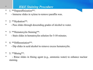
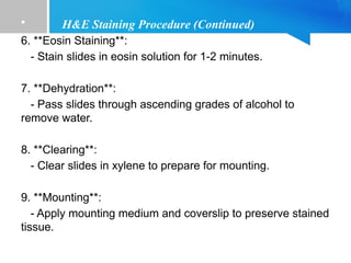
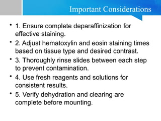
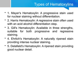
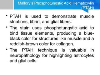
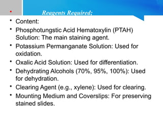
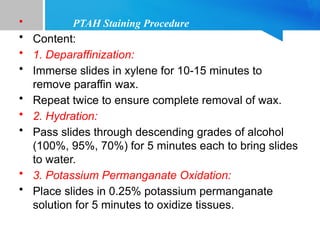
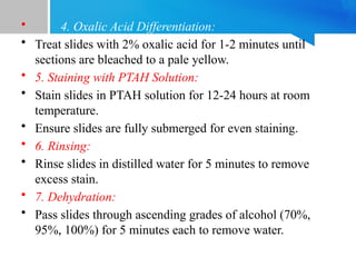
Routine Staining Procedures
Hematoxylin and Eosin Staining, Various
Types of Hematoxylins, and Mallory’s
Phosphotungstic Acid Hematoxylin (PTAH)
Author; Jagroop Singh
PhD Biochemistry
Introduction
• Routine staining procedures are essential for
visualizing tissue structure and morphology in
histological studies.
• Hematoxylin and Eosin (H&E) staining is the most
widely used technique, providing excellent
contrast between the nucleus and cytoplasm.
• Mallory’s Phosphotungstic Acid Hematoxylin
(PTAH) is used for demonstrating specific tissue
components, such as muscle striations and fibrin.
Hematoxylin and Eosin (H&E) Staining
• Hematoxylin stains the nuclei of cells blue
or purple, providing excellent contrast.
• Eosin stains the cytoplasm and
extracellular matrix pink, complementing
the nuclear stain.
• The H&E staining procedure involves
several steps: fixation, dehydration,
clearing, embedding, sectioning, staining,
and mounting.
• H&E Staining Procedure
• 1. **Deparaffinization**:
• - Immerse slides in xylene to remove paraffin wax.
• 2. **Hydration**:
• - Pass slides through descending grades of alcohol to water.
• 3. **Hematoxylin Staining**:
• - Stain slides in hematoxylin solution for 5-10 minutes.
• 4. **Differentiation**:
• - Dip slides in acid alcohol to remove excess hematoxylin.
• 5. **Bluing**:
• - Rinse slides in bluing agent (e.g., ammonia water) to enhance nuclear
staining.
• H&E Staining Procedure (Continued)
6. **Eosin Staining**:
- Stain slides in eosin solution for 1-2 minutes.
7. **Dehydration**:
- Pass slides through ascending grades of alcohol to
remove water.
8. **Clearing**:
- Clear slides in xylene to prepare for mounting.
9. **Mounting**:
- Apply mounting medium and coverslip to preserve stained
tissue.
Important Considerations
• 1. Ensure complete deparaffinization for
effective staining.
• 2. Adjust hematoxylin and eosin staining times
based on tissue type and desired contrast.
• 3. Thoroughly rinse slides between each step
to prevent contamination.
• 4. Use fresh reagents and solutions for
consistent results.
• 5. Verify dehydration and clearing are
complete before mounting.
Types of Hematoxylins
• 1. Mayer's Hematoxylin: A progressive stain used
for nuclear staining without differentiation.
• 2. Harris Hematoxylin: A regressive stain often used
with an acid alcohol differentiation step.
• 3. Gill's Hematoxylin: Available in three strengths,
suitable for both progressive and regressive
staining.
• 4. Ehrlich's Hematoxylin: A naturally ripened stain
providing intense nuclear staining.
• 5. Delafield's Hematoxylin: A ripened stain providing
good nuclear detail.
Mallory’s Phosphotungstic Acid Hematoxylin
(PTAH)
• PTAH is used to demonstrate muscle
striations, fibrin, and glial fibers.
• The stain uses phosphotungstic acid to
bind tissue elements, producing a blue-
black color for structures like muscle and a
reddish-brown color for collagen.
• The PTAH technique is valuable in
neuropathology for highlighting astrocytes
and glial cells.
• Reagents Required;
• Content:
• Phosphotungstic Acid Hematoxylin (PTAH)
Solution: The main staining agent.
• Potassium Permanganate Solution: Used for
oxidation.
• Oxalic Acid Solution: Used for differentiation.
• Dehydrating Alcohols (70%, 95%, 100%): Used
for dehydration.
• Clearing Agent (e.g., xylene): Used for clearing.
• Mounting Medium and Coverslips: For preserving
stained slides.
• PTAH Staining Procedure
• Content:
• 1. Deparaffinization:
• Immerse slides in xylene for 10-15 minutes to
remove paraffin wax.
• Repeat twice to ensure complete removal of wax.
• 2. Hydration:
• Pass slides through descending grades of alcohol
(100%, 95%, 70%) for 5 minutes each to bring slides
to water.
• 3. Potassium Permanganate Oxidation:
• Place slides in 0.25% potassium permanganate
solution for 5 minutes to oxidize tissues.
• 4. Oxalic Acid Differentiation:
• Treat slides with 2% oxalic acid for 1-2 minutes until
sections are bleached to a pale yellow.
• 5. Staining with PTAH Solution:
• Stain slides in PTAH solution for 12-24 hours at room
temperature.
• Ensure slides are fully submerged for even staining.
• 6. Rinsing:
• Rinse slides in distilled water for 5 minutes to remove
excess stain.
• 7. Dehydration:
• Pass slides through ascending grades of alcohol (70%,
95%, 100%) for 5 minutes each to remove water.
• 8. Clearing:
• Clear slides in xylene for 5 minutes to
prepare for mounting.
• Repeat clearing step twice to ensure
transparency.
• 9. Mounting:
• Apply mounting medium and coverslip to
preserve stained tissue.
Applications of PTAH Staining
• 1. Muscle Pathology: Demonstrates cross-
striations and changes in muscle fibers.
• 2. Connective Tissue: Highlights fibrin and
collagen fibers.
• 3. Neuropathology: Identifies glial cells
and structures in the central nervous
system.
• 4. Tumor Pathology: Differentiates
between various tissue components in
tumors.
• Troubleshooting Common Issues
• Content:
• Faint Staining:
• Increase staining time or concentration of
PTAH solution.
• Overstaining:
• Reduce PTAH staining time or increase
differentiation time with oxalic acid.
• Incomplete Deparaffinization:
• Ensure sufficient xylene treatment and
extend deparaffinization time if necessary.
Conclusion
• Hematoxylin and Eosin (H&E) staining is
fundamental in histology, providing essential
contrast for tissue examination.
• Mallory’s Phosphotungstic Acid Hematoxylin
(PTAH) offers specialized staining for specific
tissue components.
• Understanding the applications and techniques
of these stains is crucial for accurate
histopathological analysis.
THANK YOU

Routine_Staining_Procedures_H&E_and_PTAH_v2.pptx

  • 1.
    Routine Staining Procedures Hematoxylinand Eosin Staining, Various Types of Hematoxylins, and Mallory’s Phosphotungstic Acid Hematoxylin (PTAH) Author; Jagroop Singh PhD Biochemistry
  • 2.
    Introduction • Routine stainingprocedures are essential for visualizing tissue structure and morphology in histological studies. • Hematoxylin and Eosin (H&E) staining is the most widely used technique, providing excellent contrast between the nucleus and cytoplasm. • Mallory’s Phosphotungstic Acid Hematoxylin (PTAH) is used for demonstrating specific tissue components, such as muscle striations and fibrin.
  • 3.
    Hematoxylin and Eosin(H&E) Staining • Hematoxylin stains the nuclei of cells blue or purple, providing excellent contrast. • Eosin stains the cytoplasm and extracellular matrix pink, complementing the nuclear stain. • The H&E staining procedure involves several steps: fixation, dehydration, clearing, embedding, sectioning, staining, and mounting.
  • 4.
    • H&E StainingProcedure • 1. **Deparaffinization**: • - Immerse slides in xylene to remove paraffin wax. • 2. **Hydration**: • - Pass slides through descending grades of alcohol to water. • 3. **Hematoxylin Staining**: • - Stain slides in hematoxylin solution for 5-10 minutes. • 4. **Differentiation**: • - Dip slides in acid alcohol to remove excess hematoxylin. • 5. **Bluing**: • - Rinse slides in bluing agent (e.g., ammonia water) to enhance nuclear staining.
  • 5.
    • H&E StainingProcedure (Continued) 6. **Eosin Staining**: - Stain slides in eosin solution for 1-2 minutes. 7. **Dehydration**: - Pass slides through ascending grades of alcohol to remove water. 8. **Clearing**: - Clear slides in xylene to prepare for mounting. 9. **Mounting**: - Apply mounting medium and coverslip to preserve stained tissue.
  • 6.
    Important Considerations • 1.Ensure complete deparaffinization for effective staining. • 2. Adjust hematoxylin and eosin staining times based on tissue type and desired contrast. • 3. Thoroughly rinse slides between each step to prevent contamination. • 4. Use fresh reagents and solutions for consistent results. • 5. Verify dehydration and clearing are complete before mounting.
  • 7.
    Types of Hematoxylins •1. Mayer's Hematoxylin: A progressive stain used for nuclear staining without differentiation. • 2. Harris Hematoxylin: A regressive stain often used with an acid alcohol differentiation step. • 3. Gill's Hematoxylin: Available in three strengths, suitable for both progressive and regressive staining. • 4. Ehrlich's Hematoxylin: A naturally ripened stain providing intense nuclear staining. • 5. Delafield's Hematoxylin: A ripened stain providing good nuclear detail.
  • 8.
    Mallory’s Phosphotungstic AcidHematoxylin (PTAH) • PTAH is used to demonstrate muscle striations, fibrin, and glial fibers. • The stain uses phosphotungstic acid to bind tissue elements, producing a blue- black color for structures like muscle and a reddish-brown color for collagen. • The PTAH technique is valuable in neuropathology for highlighting astrocytes and glial cells.
  • 9.
    • Reagents Required; •Content: • Phosphotungstic Acid Hematoxylin (PTAH) Solution: The main staining agent. • Potassium Permanganate Solution: Used for oxidation. • Oxalic Acid Solution: Used for differentiation. • Dehydrating Alcohols (70%, 95%, 100%): Used for dehydration. • Clearing Agent (e.g., xylene): Used for clearing. • Mounting Medium and Coverslips: For preserving stained slides.
  • 10.
    • PTAH StainingProcedure • Content: • 1. Deparaffinization: • Immerse slides in xylene for 10-15 minutes to remove paraffin wax. • Repeat twice to ensure complete removal of wax. • 2. Hydration: • Pass slides through descending grades of alcohol (100%, 95%, 70%) for 5 minutes each to bring slides to water. • 3. Potassium Permanganate Oxidation: • Place slides in 0.25% potassium permanganate solution for 5 minutes to oxidize tissues.
  • 11.
    • 4. OxalicAcid Differentiation: • Treat slides with 2% oxalic acid for 1-2 minutes until sections are bleached to a pale yellow. • 5. Staining with PTAH Solution: • Stain slides in PTAH solution for 12-24 hours at room temperature. • Ensure slides are fully submerged for even staining. • 6. Rinsing: • Rinse slides in distilled water for 5 minutes to remove excess stain. • 7. Dehydration: • Pass slides through ascending grades of alcohol (70%, 95%, 100%) for 5 minutes each to remove water.
  • 12.
    • 8. Clearing: •Clear slides in xylene for 5 minutes to prepare for mounting. • Repeat clearing step twice to ensure transparency. • 9. Mounting: • Apply mounting medium and coverslip to preserve stained tissue.
  • 13.
    Applications of PTAHStaining • 1. Muscle Pathology: Demonstrates cross- striations and changes in muscle fibers. • 2. Connective Tissue: Highlights fibrin and collagen fibers. • 3. Neuropathology: Identifies glial cells and structures in the central nervous system. • 4. Tumor Pathology: Differentiates between various tissue components in tumors.
  • 14.
    • Troubleshooting CommonIssues • Content: • Faint Staining: • Increase staining time or concentration of PTAH solution. • Overstaining: • Reduce PTAH staining time or increase differentiation time with oxalic acid. • Incomplete Deparaffinization: • Ensure sufficient xylene treatment and extend deparaffinization time if necessary.
  • 15.
    Conclusion • Hematoxylin andEosin (H&E) staining is fundamental in histology, providing essential contrast for tissue examination. • Mallory’s Phosphotungstic Acid Hematoxylin (PTAH) offers specialized staining for specific tissue components. • Understanding the applications and techniques of these stains is crucial for accurate histopathological analysis.
  • 16.