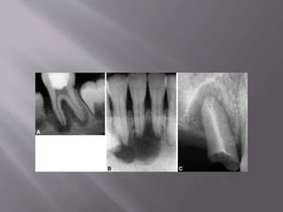
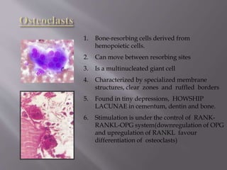
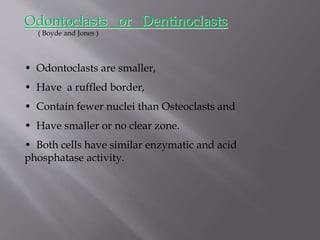
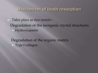
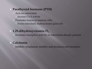
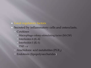
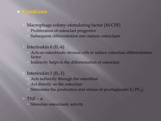
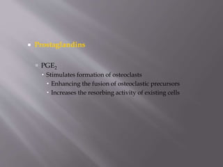
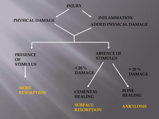
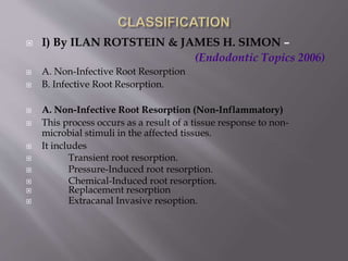
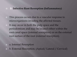
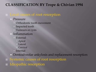
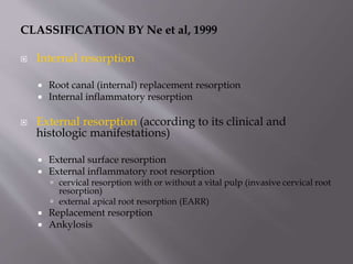
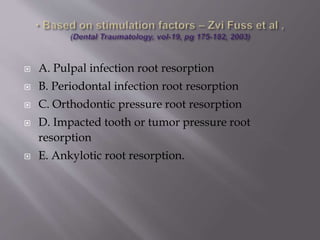
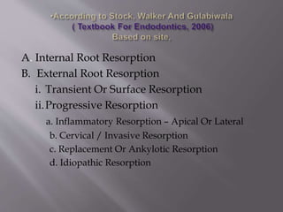
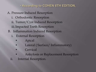
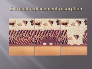
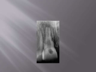
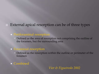
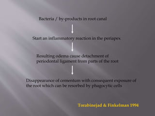
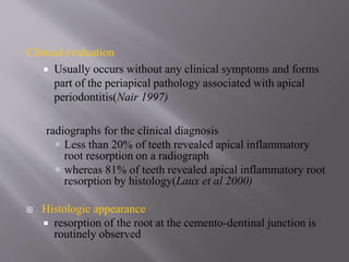
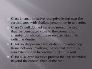
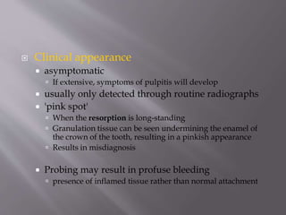
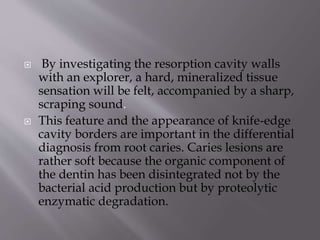
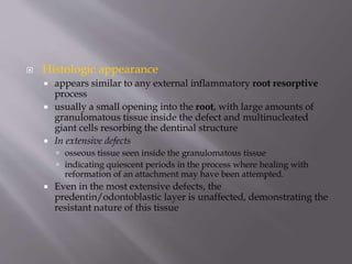
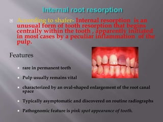
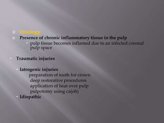
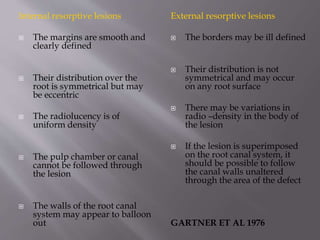
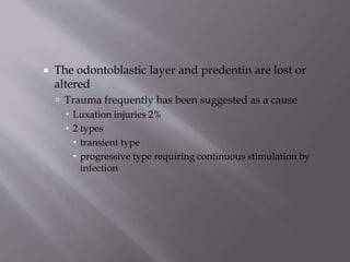
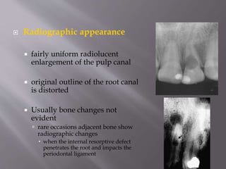
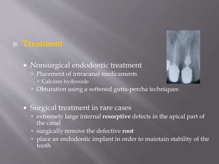
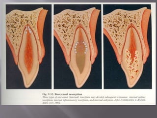
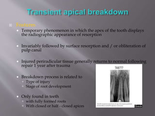
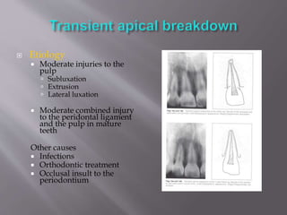
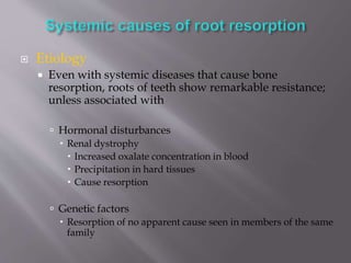
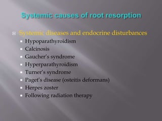
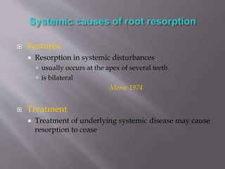
SlideShare a Scribd company logo
1 of 183
PRESENTED BY
RENJITH RAJ
III MDS
 Introduction
 Definitions
 Physiological mechanisms inhibiting root
resorption
 Key calls involved
 Mechanism of resorption
 Requirements for the presence of resorption
 Classification
 External resorption and its various types
 Internal resorption and types
 Journal review
 Conclusion
 Tooth resorption is considered as a reactive mechanism
of pulpal and periodontal tissues in response to
various injuries .
 Unlike bone, roots of permanent teeth are not resorbed
normally.
 If occurs, it is because of some pathological reasons-
considered an unfortunate and unpredictable
phenomenon.
 Decidous teeth show physiologic resorption before
they are shed off.
Resorption : (AAE,1944 ) : Resorption is a condition associated
with either physiologic or pathologic process resulting
in a loss of dentin, cementum or bone.
Root resorption : (C.W.Barclay) : It is a pathological process, of
internal or external origin, which occurs when the natural
protection of predentin and odontoblasts in root canal or
precementum and cementoblasts on the root surface are damaged
or removed.
Internal resorption : ( Grossman ): Internal resorption is an
idiopathic, slow or fast progressive resorptive process, occurring
in the dentin of the pulp chamber or root canals of teeth.
External resorption : ( Grossman ): External resorption is a lytic
process, occurring in the cementum or cementum and dentin of
the roots of teeth.
 Physiological process
 Necessary precursor to the eruption
of permanent teeth.
Permanent teeth root resorption
Pathological inflammatory processs
Results from injury to or irritation of pdl and or pulp
 Unlike deciduous teeth, permanent teeth rarely
undergo root resorption
 Even in the presence of peri-radicular inflammation,
resorption will occur primarily on the bone side of the
attachment apparatus and the root will be resistant to
it.
 Al-though many theories have been put forward, the
reason for the resistance of the root to resorption is not
fully understood.
 One theory maintains that remnants of the epithelial root
sheath surround the root like a net, therefore imparting a
resistance to resorption and subsequent ankylosis
 inhibitory effects of organic precementum and predentin
(hypothesis by Andreason). This theory is based on the
premise that the cementum and predentin covering on
dentin are essential elements in the resistance of the dental
root to resorption.
 Another function of the cemental layer is related to its
ability to inhibit the movement of toxins if present in
the root canal space into the surrounding periodontal
tissues
 another hypothesis for the relative resistance of tooth
to resorption maintains that intrinsic factors found in
predentin and cementum act as inhibitors of resorptive
cells.
 One such factor is Tumor Necrosis Factor
osteoprotegerin (OPG), a novel member of the tumor
necrosis factor(TNF) superfamily that has the ability to
inhibit osteoclast- mediated bone loss.
 Cementoblasts, periodontal ligament cells, and
human pulpal cells possess the ability to produce OPG
 KEY CELLS INVOLVED:-
Tooth resorption involves an elaborate interaction among
 Inflammatory cells
 Hard tissue cells
 Resorbing cells
 Monocytes
 Macrophages
 Osteoclasts
 Odontoclasts
(According to Pierce AM et al.)
1. Bone-resorbing cells derived from
hemopoietic cells.
2. Can move between resorbing sites
3. Is a multinucleated giant cell
4. Characterized by specialized membrane
structures, clear zones and ruffled borders
5. Found in tiny depressions, HOWSHIP
LACUNAE in cementum, dentin and bone.
6. Stimulation is under the control of RANK-
RANKL-OPG system(downregulation of OPG
and upregulation of RANKL favour
differentiation of osteoclasts)
• Odontoclasts are smaller,
• Have a ruffled border,
• Contain fewer nuclei than Osteoclasts and
• Have smaller or no clear zone.
• Both cells have similar enzymatic and acid
phosphatase activity.
Odontoclasts or Dentinoclasts
( Boyde and Jones )
 Takes place as two events:-
Degradation of the inorganic crystal structures
 Hydroxyapatite
Degradation of the organic matrix
 Type I collagen
 By producing an acidic (ph-3 to 4.5)
 Highly active polarized proton pump
 Within the ruffled border of clastic cells
 Carbonic anhydrase II
 Present intracellularly
 Catalyses CO2 H2CO3
 Readily available source of H+ ion
 Acid phosphatase enzyme also favors resorption process
 Degradation of the organic structure
 By three groups of proteinase enzymes
 Act at / just below neutral pH of 7.4
 Collagenase
 Matrix metalloproteinase (MMP)
 Act at acidic pH
 Cysteine proteinase
 Regulatory factors
 Systemic regulatory factors
 Local regulatory factors
 Systemic regulatory factors
 Promote resorption
 Parathyroid hormone (PTH)
 1,25-dihydroxyvitamin D3
 Inhibit resorption
 Calcitonin
 Parathyroid hormone (PTH)
 Acts on osteoclasts
 Increase CA II activity
 Promotes fusion of marrow cells
 Forms osteoclastic multinucleated giant cells
 1,25-dihydroxyvitamin D3
 Increases resorptive activity of osteoclasts already present
 Calcitonin
 Inhibits cytoplasmic motility and produces cell retraction
 Local regulatory factors
 Secreted by inflammatory cells and osteoclasts.
 Cytokines
 Macrophage colony-stimulating factor (M-CSF)
 Interleukin 6 (IL-6)
 Interleukin 1 (IL-1)
 TNF – α
 Arachidonic acid metabolites (PGE2)
 Endotoxin (lipopolysaccharides)
 Cytokines
 Macrophage colony-stimulating factor (M-CSF)
 Proliferation of osteoclast progenitor
 Subsequent differentiation into mature osteoclasts
 Interleukin 6 (IL-6)
 Acts on osteoblastic stromal cells to induce osteoclast differentiation
factor
 Indirectly helps in the differentiation of osteoclast
 Interleukin 1 (IL-1)
 Acts indirectly through the osteoblast
 Act directly on the osteoclast
 Stimulates the production and release of prostaglandin E2
(PG2)
 TNF – α
 Stimulate osteoclastic activity
 Prostaglandins
 PGE2
 Stimulates formation of osteoclasts
 Enhancing the fusion of osteoclastic precursors
 Increases the resorbing activity of existing cells
 Bacterial endotoxin / LPS
(lipopolysaccharide)
 Induction of osteolytic factors
 Lysosomal enzyme release
 Collagenase release from macrophages
 Osteoblastic secretion of osteolytic factors
 IL-1
 IL-6
 M-CSF
 PGE2
 The loss or alteration of the protective layer
 pre-cementum
 pre-dentin
 Sustained inflammation must occur to the
unprotected root surface
 Directly
 due to the trauma of a dental injury
 Especially intrusive injury
 Indirectly
 Inflammation in reaction to the traumatic injury
 varies according to the stimulus it is exposed to after
the injury
 has the potential to cause extensive damage to the
protective layer
 The inflammatory response caused by the
dental injury can be divided into two critical
phases
 Destructive phase
 Healing phase
Destructive phase
 where active resorption between the dried-out cells
with multinucleated giant cells takes place
 This destruction will continue as long as there is
stimulus present to allow the inflammation to develop
 stimulus only exist for a short period of time
 Healing will take place without intervention by the dentist
 If the inflammatory stimulus is long-standing
 the destructive root resorption will continue until either no root
structure remains or the stimulus is removed by the
intervention of the dentist.
 Identified by radiolucent appearance of the tooth and bone on
x-rays
 Healing phase
 The critical factor in determining the outcome after a
dental traumatic injury has occurred is the type of
cells that repopulate the root surface during the
healing phase
 Cementoblasts
 type of healing termed
 cemental healing or surface resorption will occur
 the outcome will be favorable
 Osteoblasts
 the conditions for healing will be unfavorable
 Ankylosis, replacement resorption or osseous replacement
 The type of tissue that will cover the root surface is
dependent on
 the surface area of the root damage
 destruction of over 20% of the root surface is required
for osseous replacement to occur
 the relative proximity of the cells to the root; i.e.
how far and how fast the cells can travel in order to
cover the damaged root surface
 surface area of root damage is dependent on
 the scale of the initial injury
 which cannot be reversed.
 the extent of the destructive inflammatory
response.
 opportunity here for the initial inflammation to be
minimized by actions taken both immediately after the injury
 where the way in which the tooth is handled is of crucial
importance
 by the pharmacological manipulation of the inflammatory
response
PRESENCE
OF
STIMULUS
ABSENCE OF
STIMULUS
CEMENTAL
HEALING
BONE
HEALING
SURFACE
RESORPTION
ANKYLOSIS
MORE
RESORPTION
< 20 %
DAMAGE
> 20 %
DAMAGE
INJURY
PHYSICAL DAMAGE
INFLAMMATION
ADDED PHYSICAL DAMAGE
 I) By ILAN ROTSTEIN & JAMES H. SIMON –
(Endodontic Topics 2006)
 A. Non-Infective Root Resorption
 B. Infective Root Resorption.
 A. Non-Infective Root Resorption (Non-Inflammatory)
 This process occurs as a result of a tissue response to non-
microbial stimuli in the affected tissues.
 It includes
 Transient root resorption.
 Pressure-Induced root resorption.
 Chemical-Induced root resorption.
 Replacement resorption
 Extracanal Invasive resoption.
 B. Infective Root Resorption (Inflammatory)
 This process occurs due to a vascular response to
microorganisms invading the affected tissues.
 It may occur in both the pulp space and the
periodontium and may be located either within the
root canal space (internal resorption) or on the external
root surface of the root (external resorption).
 a. Internal Resorption
 b. External Resorption. (Apical/ Lateral / Cervical)
CLASSIFICATION BY Trope & Chivian 1994
 Local causes of root resorption
 Pressure
 Orthodontic tooth movement
 Impacted tooth
 Tumours or cysts
 Inflammation
 External
 Apical
 Lateral
 Cervical
 Internal
 Dentoalveolar ankylosis and replacement resorption
 Systemic causes of root resorption
 Idiopathic resorption
CLASSIFICATION BY Ne et al, 1999
 Internal resorption
 Root canal (internal) replacement resorption
 Internal inflammatory resorption
 External resorption (according to its clinical and
histologic manifestations)
 External surface resorption
 External inflammatory root resorption
 cervical resorption with or without a vital pulp (invasive cervical root
resorption)
 external apical root resorption (EARR)
 Replacement resorption
 Ankylosis
 A. Pulpal infection root resorption
 B. Periodontal infection root resorption
 C. Orthodontic pressure root resorption
 D. Impacted tooth or tumor pressure root
resorption
 E. Ankylotic root resorption.
A. Internal Root Resorption
B. External Root Resorption
i. Transient Or Surface Resorption
ii.Progressive Resorption
a. Inflammatory Resorption – Apical Or Lateral
b. Cervical / Invasive Resorption
c. Replacement Or Ankylotic Resorption
d. Idiopathic Resorption
A. Pressure Induced Resorption
i. Orthodontic Resorption
ii. Tumor/Cyst Induced Resorption
iii.Impacted Teeth Resorption
B. Inflammation Induced Resorption
i. External Resorption
• Apical
• Lateral ( Surface/ Inflammatory)
• Cervical
• Ankylosis or Replacement Resorption
ii. Internal Resorption.
1. Trauma induced tooth resorption
 Surface resorption
 Transient apical internal resorption
 Pressure resorption
 Orthodontic resorption
 Replacement resorption
2. Infection induced tooth resorption
 Internal inflammatory root resorption
 External inflammatory root resorption
 Communicating internal-external resorption
1. External surface resorption/ Repair related
resorption
1. External infection related resorption /
Inflammatory root resorption
2. External trauma related replacement
resorption / Ankylosis
3. External spontaneous ankylotic resorption
5.External multiple sites of ankylosis /
Infection related resorption
6.Cervical invasive resorption
7. Internal surface resorption
8. Internal infection related root resorption
9. Internal replacement resorption
The root resorption that is initiated in the periodontium and affecting
the external or lateral surfaces of a tooth.
Can be classified into four categories by its clinical and histologic
manifestations. (Rita F.ne, Guttman et al;quintessence international 1999)
External resorption
 Surface resorption
 lnflammatory root resorption
(a) Cervical resorption with or without a vital pulp
(b) External apical root resorption
 Replacement resorption
 Ankylosis
Definition
Small, superficial resorption cavities in the cementum
and the outermost layers of the dentin, without an
inflammatory reaction in the periodontal ligament
Andreason & Hjorting –Hansen (1966)
Etiology
Acute injury:- concussion, subluxation and
lateral luxation.
Chronic injury:-orthodontic treatment, traumatic
occlusion, pressure from cysts, ectopically
erupting teeth.
 Typical feature:-when the trauma or pressure is discontinued,
spontaneous healing takes place.
Pathogenesis
The injured tissue adjacent to the root and surface cementum are
removed by macrophages and osteoclasts.
Takes 2-4 weeks
Repair is by progenitor cells from the pdl, new cementum is formed
with insertion of pdl fibres.
Radiographic features
After 2-4 weeks , localised widening of the pdl space
Slight cavitations may be seen on root surface
Very limited size-cant be recogonized radiographically.
 T
Treatment
 100% repair takes place in almost all cases.
 Endodontic treatment not indicated.
 Combined injury to pulp and pdl and where bacteria
located in the pulp space and dentinal tubules trigger
osteoclastic activity on root surface.
 Can affect all parts of the root.
 Rapidly progressing-result in total resorption of the
root within a few months.
Etiology
Related to acute trauma
Intrusion and replantation of avulsed teeth
 Pathogenesis
 once the initial resorption has penetrated the cementum and
exposed the dentinal tubules, toxins from infected root canal
diffuse through the exposed dentinal tubules to pdl.
 Continuation of osteoclastic process and associated inflammation
in the pdl-resorption of adjacent alveolar bone.
 Process progresses, root dentin is resorbed and root canal becomes
exposed.
 If endodontic therapy is done-resorptive process will be arrested
and the resorption cavity filled with cementum or bone.
Clinical findings
 Increased mobility
 Dull percussion tone
 Sometimes tooth may be extruded
 Sensitivity test gives no response
 May exhibit a sinus tract
Radiographic findings
Progressive cavitations involving root and adjacent alveolar bone.
Result in total loss of tooth structure within a few months.
 Endodontic implications
Requires immediate endodontic treatment to control or remove the
osteoclast promoting factors.
Treatment
Goal is to remove bacteria in the root canal and dentinal
tubules.-allow healing in entire periradicular space.
Mature teeth
Endodontic treatment-prophylactic extirpation of the pulp
in replanted avulsed tooth
Biomechanical preparation with Naocl and Ca(OH).
Calcium hydroxide perform disinfection and canal can be
filled 2-3 weeks after treatment.
Immature teeth
Pulp becomes necrotic before root is fully devoloped- apical opening is
often too large to create a resistance to retain the root canal filling.
Apexification procedures using calcium hydroxide have been
performed with good success – disadvantage: –
takes many months to form an apical barrier to allow placement of a
root canal filling.
Long term use can weaken the dentin,by dissolving its organic
content-result in cervical root fracture on even slight impact.
Root canal filling can be done immediately without waiting for a
biological response if MTA is used as a physical barrier apically.
 Represents a sequel to a defect or injury to pdl cells, including the
cell layer next to the cementum.
Etiology
Acute trauma:- severe luxations
 Lateral luxations
 Intrusions
 Replantation of avulsed teeth
Homeostasis of pdl is lacking.
Healing events take place from adjacent healthy pdl or the bony
alveolus leading to formation of a normal pdl in former and a bony
bridge between socket wall and the root surface.
Moderate sized injuries(1-4mm2)
 An initial ankylosis forms.
 Tooth-allowed functional mobility by the use of a
nonrigid splint, small areas of resorption can be replaced
with new cementum and pdl attatchment. (Transcient ankylosis)
Extensive injuries(›4mm2 )
Progressive ankylosis-tooth becomes an integral part of bone
remodelling system
In children- active process- gradual infraocclusion and arrested
devolopment of the alveolar process.
Combination of resorption –loss of ankylosed teeth within 1-5 years.
Older individuals- replacement resorption is significantly slower and
often allows a tooth to function for much longer periods of time(5-
20 yrs).position of tooth remains the same.
Clinical findings
 Tooth appears very form in its socket.
 High metallic sound on percussion.
 Demonstrated 4-6 weeks post – trauma.
Radiographic findings
 Diagnosed radiographically within 2 months after injury
 Complete disappearance of the periodontal space
 Uneven root surface contour (Moth-eaten appearance)
 Treatment
 No treatment
 Very little can be done to reverse this initial
attachment damage, therefore the main focus of
treatment is to do everything possible to limit the
destructive inflammatory process that occurs
immediately after the injury. If the inflammation is
minimized, the surface area of damaged root surface
will also be kept to a minimum.
 Treatment strategies are therefore directed at
avoiding or minimizing the severity of the
initial inflammatory response
 Prevention of the initial injury
 Minimizing additional damage after the initial injury
 Pharmacological manipulation (shut down) of the
initial inflammatory response
 Possibly stimulating cemental, rather than bone,
healing
 Slowing down the osseous replacement when it is
inevitable
1. Prevention of the initial injury
Preventive measures have been to found to limit
the occurence of tooth injuries
 Athletes-usage of mouth-guards and other protective
devices found to be proven protection against tooth
trauma.
2. Minimizing Additional Damage after the Injury
 Luxations
 Gentle repositioning of the tooth in its original position as
soon as possible.
 Patient should be sent to emergency room or dentist .
 Splinting
 functional splint for 7-10 days
 splint should be constructed to allow adequate cleaning
 Examples of functional splints
 TTS splint (Titanium Trauma
Splint)
 This splint allows space for
adequate cleaning, thereby
minimizing infection due to plaque
and food debris
 It is flexible in a horizontal and
vertical direction
 Resin splint
 whilst allowing functional
movement, is too thick ,
impossible for patients to clean it.
 Avulsions
 replanting the tooth as soon as possible
 extra-oral dry time must be minimized
 appropriate storage medium
 Milk
 Sterile saline
 Saliva
 Hank’s Balanced Salt Solution
 ViaSpan
3. Pharmacological manipulation (shut down) of the initial
inflammatory response
 Drugs that affects osteoclasts present at the site of resorption
 Tetracyclines
 Sustained antimicrobial effect
 Anti-resorptive properties
 direct inhibitory effect on osteoclasts and collagenase
 Significantly more cemental healing
 Drugs that affect the recruitment of osteoclasts to the injury
site –reduce osteoclastic bone resorption.
 Glucocorticoids
 Topical dexamethasone was found to be useful while systemic
usage was not
 Bisphoshonates
 Alendronate
 Amino acids
 Taurine
 Combination of the two types of drugs
 synergistic effect on the inhibition of root resorption
 Ledermix
 a drug combining tetracycline and corticosteroids
 ART (Antiresorptive Regenerative Therapy )
by Pohl et al 2005
 Comprises a combination of different treatment
strategies for a synergistic effect
 Local application of a glucocorticoid
 Systemic and local application of tetracyclines
 Use of Enamel Matrix Derivative (EMD) e.g. Emdogain
4. Stimulate Cemental Healing
 ‘Conditioned Medium'
 supernatant of cultured gingival
fibroblasts, that contain a number
of biologically active factors
 ViaSpan
 Emdogain (Enamel Matrix
Protein; Biora, Malmo, Sweden)
 for teeth with extended extra oral
dry times
 makes the root more resistant to
resorption
 stimulates the formation of new
periodontal ligament from the
socket
Emdogain placed on tooth and
in the socket before replantation
5. Slow down 'inevitable' osseous replacement
 When the periodontal ligament on the root surface is
definitely destroyed
 intrusive injuries
 avulsion injuries with extended extra-oral dry times
 Intrusive injuries
 the tooth is repositioned
 inevitable osseous replacement accepted
 Avulsion injuries with extended dry times
 All remaining periodontal ligament debris is
removed from the root by thorough curettage
 Fluoride
 root is soaked in fluoride for 5 min before replantation
 effectively slow down remodeling of the root to bone
 Bisphosphonates
 slows down the osseous replacement
 More expensive than fluoride
 Emdogain A
 When there is no known etiology
 First reported-mueller & rony(1930)
2 TYPES:-
-APICAL
-CERVICAL
(Lydiatt et al 1989, Yusof & Ghazali 1989)
 Mostly seen in young adults
 Maxillary teeth frequently involved
 Resorption starts apically, progresses coronally
 Gradual shortening and rounding of roots
Cervical type
Starts in cervical region and approaches pulp.
Does not seem to be mediated from pulp space.
 Pathogenesis
Suspected that triggering factors exist for osteoclastic and
odontoclastic activity producing root resorption
 Treatment
Not mediated from pulp space- so interceptive endodontic
procedures are not indicated.
 Appear to begin at mesial or distal CEJ
 Initially presents as a radiolucency with
scalloped margin
 In time found to undermine enamel
Single idiopathic root resorption
Can be confused with root caries
Clinical and radiographic findings are helpful in
establishing correct diagnosis.
Root caries
 Lesion of dentin with
gingival recession
 Clinically soft to the
touch of an explorer
 Radiographically
illdefined, saucer like
and radiolucent.
Sharp edge of cavity
border –diagnostic
finding
Cervical resorption
 Gingival condition
might appear either
normal or inflammed
 Covered by soft tissue
 Almost same –
incidental
radiographic finding
Treatment
 No preventive or therapeutic regimen for this type
of resorption.
 Not mediated from pulp space- endodontic
treatment wont do any good. There is no known
preventive or therapeutic regimen for this
condition, and monitoring accompanied by
periodontal maintenance is recommended
 Usually observation and extraction of teeth.
 Further studies needed to identify the specific
cellular mechanism responsible to determine
therapeutic measures.
 A 27-year-old male was referred to the Endodontic
Department of Shiraz University of Medical Sciences,
School of Dental Medicine by his family dentist for
evaluation of extensive root resorption affecting most
teeth.
 The patient was a truck driver with no history of
medical disorders. A family history revealed no early
tooth loss in parents or grandparents. His two brothers
and three sisters were also examined, and panoramic
radiographs revealed no similar condition.
 Clinical examination revealed normal soft tissues
and normal dentition particularly the morphology
and size of the crowns.
 The periodontal condition was normal with no
abnormal pocketing. Tooth mobility was within
the physiological range.
 A panoramic radiograph and tracing together
with the periapical radiographs revealed extensive
apical root resorption in all teeth except teeth 18,
23, 31–34, 37and 41–44. Eighteen teeth had apical
root resorption, mostly in the maxilla
 On the basis of the history, clinical examination and
radiographic evaluation, a diagnosis of multiple
idiopathic apical root resorption was made.
 The patient was given instructions to maintain proper
oral hygiene and was scheduled for frequent recall
visits in order to prevent periodontal bone loss and
further compromiseof crown : root ratio.
 Nomeclature
 Inflammatory root resorption of endodontic origin
 is usually known as apical inflammatory root
resorption
 External apical root resorption (EARR)
 Definition
External apical root resorption is a pathological
condition, characterized by the resorption of hard
tissues (cementum, dentin) and sustained by a local
inflammatory reaction , in most cases because of the
presence of infected necrotic dental pulp, which is able
to maintain the whole process
Hammarstrom & Lindskog 1985
 Cause
 All causes of apical periodontitis
 Infected necrotic pulp
 caries (predominant cause)
 Overinstrumentation during endodontic therapy
 Trauma
 Features
 intact cementum on the root surface (in most routine
cases)
 communications primarily through the apical
foramina or, occasionally, through accessory canals
 Invariably, slight resorption of the root at the
cemento-dentinal junction
Clinical features
 Early stages-asymptomatic
 As the process progresses, teeth become
symptomatic.
 Periradicular abscess devolop
Radiographically
 Radiolucency observed at external surface of
dentin and adjacent bone.
 External apical resorption can be of three types
 Periforaminal resorption
 Defined as the area of resorption not comprising the outline of
the foramen, but the surrounding area
 Foraminal resorption
 Defined as the resorption within the outline or perimeter of the
foramen
 Combined
Vier & Figueiredo 2002
Bacteria / by-products in root canal
Start an inflammatory reaction in the periapex
Resulting odema cause detachment of
periodontal ligament from parts of the root
Disappearance of cementum with consequent exposure of
the root which can be resorbed by phagocytic cells
Torabinejad & Finkelman 1994
Clinical evaluation
 Usually occurs without any clinical symptoms and forms
part of the periapical pathology associated with apical
periodontitis(Nair 1997)
radiographs for the clinical diagnosis
 Less than 20% of teeth revealed apical inflammatory
root resorption on a radiograph
 whereas 81% of teeth revealed apical inflammatory root
resorption by histology(Laux et al 2000)
 Histologic appearance
 resorption of the root at the cemento-dentinal junction is
routinely observed
 Treatment
 Removing the stimulus for the underlying inflammatory
process
 Working length determination
 Weine’s method
 Electronic method
 Application of intracanal medications
 Ca (OH)2
 Activ point(chlorhexidine 5%)
 Obtaining a good apical seal
 Ca (OH)2 for apexification
 Thermoplasticized gutta percha techniques
 Definition
External root resorption associated with
marginal periodontitis without pulpal
involvement is most commonly referred to as
cervical invasive root resorption (because of the
marginal location of the defect)
Heithersay 1999
 Nomenclature
 Peripheral cervical resorption
 Burrowing resorption
 Pseudo pink spot
 Subepithelial external root resorption
 Extracanal invasive resorption
 May occur after injury to the precementum,
apical to the epithelial attatchment , followed
by bacterial stimulation originating from
periodontal sulcus.
 Injury can be due to
dental trauma, bleaching procedures,
periodonal procedures, orthodontic treatment
 Bacteria from sulcus penetrate patent dentinal tubules
coronal to the epithelial attatchment and exit apical to
epithelial attatchment without penetrating pulp space.
 Damaged root surface –invaded by clast cells ,
penetrate the dentin though a small denuded area-
spread of resorption in root.
 Resorption process reach supragingival area of crown-
vascularised granulation tissue visible through enamel-
pinkish discolouration.
Radiographically,
 Single resorpton lacunae in the dentin usually
at the crestal bone level, especially to the
coronal and apical direction.
 Resorption progresses, radiolucency may be
observed at the alveolar bone adjacent to the
resorption lacunae of the dentin.
 Features
 inflammatory origin
 most often referred to as cervical root resorption
 occurs immediately below the epithelial attachment of the
tooth, usually but not exclusively in the cervical area of the
tooth
 Also called
 Extracanal invasive resorption (Frank AL)
 Invasive cervical resorption (Heithersay GS)
 Subepithelial external root resorption (Trope M)
 The pulp plays no role in cervical root resorption
and is mostly normal in these cases(Frank &
Torabinejad 1998)
 Class 1- small invasive resorptive lesion near the
cervical area with shallow penetration in to dentin.
 Class 2- well defined invasive resorptive lesion
that has penetrated close to the coronal pup
chamber but shows little or no extension in to
radicular dentin
 Class 3 – deeper invasion of dentin by resorbing
tissue not only involving the coronal dentin , but
also extending in to coronal third of the root.
 Class 4 –a large invasive process that has extended
beyond the coronal third of the root
 Clinical appearance
 asymptomatic
 If extensive, symptoms of pulpitis will develop
 usually only detected through routine radiographs
 'pink spot'
 When the resorption is long-standing
 Granulation tissue can be seen undermining the enamel of
the crown of the tooth, resulting in a pinkish appearance
 Results in misdiagnosis
 Probing may result in profuse bleeding
 presence of inflamed tissue rather than normal attachment
 By investigating the resorption cavity walls
with an explorer, a hard, mineralized tissue
sensation will be felt, accompanied by a sharp,
scraping sound.
 This feature and the appearance of knife-edge
cavity borders are important in the differential
diagnosis from root caries. Caries lesions are
rather soft because the organic component of
the dentin has been disintegrated not by the
bacterial acid production but by proteolytic
enzymatic degradation.
 Histologic appearance
 appears similar to any external inflammatory root resorptive
process
 usually a small opening into the root, with large amounts of
granulomatous tissue inside the defect and multinucleated
giant cells resorbing the dentinal structure
 In extensive defects
 osseous tissue seen inside the granulomatous tissue
 indicating quiescent periods in the process where healing with
reformation of an attachment may have been attempted.
 Even in the most extensive defects, the
predentin/odontoblastic layer is unaffected, demonstrating the
resistant nature of this tissue
Tooth immediately after careful
extraction. (Right) Same tooth after exca-
vation of the granulation tissue. Notice
the layer of dentin and predentin that
separated the resorbing tissue from the
dental pulp
 Radiographic appearance
 resorptive process occurs mesially or distally on the root
surface
 Single resorption lacunae in the dentin usually at the crestal bone
level
 The radiolucency expands coronally and apically in the dentin,
and reaches, but usually does not perforate, the root canal
 irregular, diffuse radiolucency of nonuniform radio-density
(Makkes & van Velzen 1975).
 resorptive process is buccal or palatal-lingual
 the radiographic picture depends on the extent to which the
resorptive process has spread in the dentin
 It can be seen as a radiolucency at the attachment level or have
spread a considerable way coronally or apically with a mottled
appearance
 Because the pulp is not involved, root canal outline can
usually be distinguished through the resorptive defect
Pinkish discoloration of the left central incisor
caused by invasion of the cer-vical region of the
tooth by fibrovascular tissue derived from the
periodontal ligament. (Right) The parallel
radiograph shows a rather irregular
radiolucency , involving not only the coronal
dentin but also ex-tending to the coronal third of
the root. The characteristic radiopaque line
separating the lesion from the root canal can be
identified.
 Treatment
 The aim of treatment is two-fold:
 Stop continuation of resorption
 removing all the granulomatous tissue from the root
 In cases where removal of granulomatous tissue would cause
unacceptable damage to supporting structures, an attempt is
made to severe the blood supply to the granulomatous tissue, thus
killing the resorptive cells and inhibiting progression of the
resorptive process
 Replace the unprotected root surface with a foreign material
that clastic cells cannot be attached to or penetrate
 Any well-sealing dental restorative material
 inhibits attachment by clastic cells
 also does not allow attachment by those cells that provide for a normal
periodontal ligament
 root attachment will only occur apical to the filling material, leaving a
periodontal pocket that is unacceptable
 Treatment approaches
 Internal approach
 External approach
 combinations
External Approach
 determine the exact location of the
defect (buccal or lingual-palatal)
 Angled radiographs
 full thickness flap is raised
 granulomatous tissue is removed
from the root and the bone defect
 with a curette or bur
 also remove granulomatous tissue
from sound, healthy bone
 so that revascularization of the
resorbing tissues will not occur
 The opening into the root should be
as conservative as possible
 The root defect is filled with a
restorative material
 flap replaced in a way as to
minimize the periodontal defect
after healing
 Indications
 For the small coronal defect
The flap is raised, meaning the
blood supply to the granulomatous
tissue in the root has been cut. B.
The tissue is removed from the root
and bone and C. the
root is filled with a composite resin
filling.
Internal Approach
 trichloracetic acid (Heithersay)
 The acid will chemically burn the granulomatous
tissue, thus necrosing it and providing space for the
filling material internally
 calcium hydroxide
 but may take multiple applications to achieve the
same results
 Theoretically, a bur could be used but the
chances of pulp exposure and/or extensive
attachment damage are very high with this
approach
External Approach
 Traditional approach
 remove the entire root cementum surface
adjacent to the granulomatous tissue in the
dentin
 Alternate approach
 remove the resorptive tissue from a small
opening at the most apical extent of the
affected root
 leaving coronal root surface onto which a new
attachment might develop
 opening repaired with an acceptable
restorative material
 the defect and denuded root surface is covered
with a spacer (freeze-dried bone), and the
entire area covered with a Gore-Tex
membrane (Gore Tex, W. L. Gore, Flagstatt,
AZ).
 Forced eruption
 If the remaining root apical to the resorption defect is long
enough to maintain the tooth
 The resorption defect is moved to a position coronal to the
adjacent attachment.
 Defect cleaned and restored
 Forced eruption/re-intrusion
 If the crown root ratio is not ideal
 after the repair is complete the tooth can be orthodontically
moved into its original position

 Intentional replantation
 if the practitioner is confident that the resorbed root will not
fracture on extraction
 with or without re-intrusion
External Approach/Internal Approach
 elective endodontic therapy is often the best choice in extensive
lesions
 A flap is raised and the granulomatous tissue is aggressively
removed from the bony defect only
 A barrier membrane is used
 To stop new tissue from growing into the root
 To stop revascularization of the necrotic tissue left inside the root
 After approximately one month, an opening is made externally
above the attachment and the necrotic granulomatous tissue is
removed and replaced with a filling material
 mineral trioxide aggregate (MTA)
 Indication
 Opening of the root defect is small
 If opening into the root is large
 the treatment plan should be re-assessed with extraction an option if a
large periodontal defect is to be expected after surgery
Internal Approach
 Not predictable enough in these extensive cases
 Should not be considered
 offer poor prognosis
 extraction and replacement
with an implant or fixed bridge
is the preferred approach
 A 22-year-old woman entered the clinic with a major
complaint of a discoloring upper right lateral incisor.
The patient had undergone intracoronal bleaching
treatment 3 years previously in her upper right central
incisor
 The intracoronal bleaching procedure was performed
with a mix of sodium perborate and 3% hydrogen
peroxide , resulting in overbleaching to allow better
color matching with time.
 The current clinical examination of the upper lateral
incisor tooth revealed a significant change in the color
of the cervical area of the tooth
 A periapical radiograph showed a significant
cervical radiolucent lesion, which led to the
diagnosis of a class 3 ICR lesion
 Tooth vitality tests produced positive and
normal results and a probe was used to
confirm the presence of a marginal bone defect
(examined under local anesthesia).
 The existence of a large labial-cervical defect
led to the clinical diagnosis of ICR.
Radiographic view presenting a
process of cervical resorption in the
maxillary right lateral incisor.
Frontal view of maxillary right
lateral incisor 3 years after
treatment of
the adjacent central. Note the
cervical discoloration
 A limited labial flap was raised, revealing a large
resorption lesion with soft tissue growing into the
tooth. The lesion borders included the cementoenamel
junction and down to the cervical part of the root.
 The lesion was cleaned with a slow speed handpiece,
exposing sound tooth material on all aspects, while
maintaining the pulp’s integrity and vitality.
 The lesion was sealed with a temporary filling material
(IRM; Dentsply, Tulsa, OK), and the flap was repo-
sitioned, with hidden sutures secured to the palatal
tissue
Flap elevation of the cervical
lesion. Note the granulation tissue.
After removal of the granulation
tissue.
 Following the patient’s request, all efforts were made
to keep the tooth. Therefore, it was decided to try and
pull the root rapidly from within the alveolar socket by
using orthodontic extrusion combined with
fiberotomy.
 The patient received all the necessary information
including the risks and the immediate need for a root
canal treatment before onset of movement .
 The orthodontic movement pulled the tooth from
within the alveolar socket successfully, rapidly
bringing the lesion out of the bone above the crest.
Fiberotomy was performed twice during the
movement, and the temporary restoration material was
replaced with a glass ionomer filling material when the
lesion’s apical borders could be controlled
Orthodontic root extrusion,
onset of movement. After second fiberotomy procedure.
Note exposed gingival lesion
borders
Radiographic view close to end
of movement
 The tooth was then secured to the adjacent teeth
for a retention period of 3 weeks, and after this
stabilization period, a provisional acrylic
restoration was provided.
 Tooth mobility was examined and found to be
acceptable, and after a comprehensive evaluation
,it was decided to continue and prepare a crown
for that tooth.
 A composite post and core restoration was
prepared indirectly on healthy and sound tooth
margins that were cleaned carefully with 90%
trichloroacetic acid.
 After cementing the composite post and core, a second
provisional acrylic restoration was prepared to enable
full tissue maturation after the orthodontic process
Impressions were taken for the fabrication of a
zirconium coping.
 The coping’s accuracy was checked and sent for
porcelain layering. The final result fully met the
patient’s expectations .
 The restored tooth is examined as part of the patient’s
periodic maintenance and shows properly healed
tissues and uneventful function
Occlusal view of root prepared
for a post and core, leaving
sound dentin treated with TCA
Follow-up at 42 months: clinical
and x-ray examination.
Prevention
 Nonvital bleaching
 Protection of the dentinal tubules
 Remove the gutta-percha apical to the cervical line to remove
discolored dentin, but do not extend the preparation into the
root. Use the crestal bone as a guide.
 Place a layer of cement (IRM, Cavit, glass ionomer) to prevent
ingress of the bleaching agent apically and into the cervical
dentinal tubules
 Use of calcium hydroxide beneath the access restoration
 Do not use heat
 30% hydrogen peroxide activated with heat damages the
cementum layer through the dentinal tubules
 Avoid etching the dentin
 Some techniques suggest etching of the dentin before
bleaching; however, a recent study showed similar bleaching
results with and without etching
 Do not use Superoxol as it is caustic
 Some advocate sodium perborate (USP) and water for the
walking bleach and report excellent results with no history of
external resorption
 Others showed in vitro the effectiveness of sodium perborate
and water as a bleaching agent, though it took longer to work
 Use of Carbamide peroxide for intracoronal
bleaching
 Orthodontic therapy
 All orthodontic forces, in particular tipping forces,
should be as light as possible
 in order that they do not crush the attachment apparatus while
the tooth is being uprighted
 Surgical procedures
 Avoid surgical procedures, including excessive use of
surgical elevators
 Surgical procedures that could damage the cervical
margin, for example the canine wire lasso, before the
orthodontic movement of an impacted canine should
not be used; rather, surgical exposure and banding
with acid etch and resin should be used to facilitate
coronal movement
 Periodontal procedures
 Avoid procedures that leave the root surface denuded
 Pressure
 orthodontic tooth movement
 impacted teeth
 tumors
 Orthodontic tooth movement
 In most cases, 'controlled' trauma
 pressure is spread evenly over a root area
 minimizing the inflammatory response
 favors resorption of the bone rather than the root
 In rare cases
 pressure is localized to the apical region
 cause cemental damage and apical root resorption.
 considerable shortening of the root
 Orthodontic root resorption
 'sterile' inflammation
 1st comprehensive study on root resorption
after orthodontic treatment by Ketcham
 2 types of root resorption in connection with
orthodontic treatment
 Small superficial resorptions that undergoes repair
 Resorption in the apical area which leads to
permanent root shortening
 Superficial resorption
 Frequently preceded by the hyalinization of the
periodontal ligament
Clinical features
 Asymptomatic.
 Shortening of roots.
 Pulp is usually vital.
Radiographs
 Resorption seen in apical third.
 no radiolucency observed in bone or root
 Root resorption due to orthodontic pressure has
always been considered to be external root resorption.
However, this is not necessarily true. Damage takes
place at the apex of the tooth near the cemental-
dentinal junction due to the orthodontic pressure.
Therefore, protective damage can be either cementum
or predentin. Because the predentin can also be
affected, it is not unusual to see radiographic evidence
of internal apical resorption during the active stage of
the process. In 1997, Bender et al. suggested the term
periapical replacement resorption (PARR) for
describing this type of resorption
 Pressure resorption due
to orthodontic
treatment. A. Teeth at
the start of orthodontic
treatment. B. Severe
root resorption on the
left central incisor after
orthodontic treatment.
Note that, even though
considerable resorption
has taken place,
favorable healing at the
apex with cementum
can be seen. (Courtesy
Dr IB Bender)
 Healing of Orthodontically Induced Root Resorption by Ultrasound
in Man
 ULTRASOUND
 Root resorption is one of the adverse outcomes of orthodontic tooth
movement
 It compromises the crown-root ratio
 leads to increased malpractice litigation against orthodontists
 Previous researches have shown that Low-intensity pulsed
ultrasound (LIPUS) can enhance healing of traumatized different
types of connective including dental tissues without any adverse
effect
 SEM study showed statistically significant decrease in number and
the area of resorption lacuanae in the LIPUS treated premolars.
 Histological examination revealed healing of the resorped root
surface by deposition of new cellular reparative cementum. Also,
LIPUS application enhances the formation of more cementum
deposition on top of the regular old cementum.
 May provide a valuable and clinically acceptable method for
minimizing orthodontically induced root resorption in human.
(T. EL-BIALY and I. ELSHAMY, 2003)
 For impacted teeth or tumors, the resorption
will occur wherever the pressure from the
impaction/tumor occurs
 Usually slow growing lesions- Cysts,
ameloblastoma, fibro osseous lesions and giant
cell tumours.
 Tooth asymptomatic, pulp is vital
 Treatment is relatively easy, in that removal of
the source of the pressure will result in the
cessation of resorption in the majority of cases
 According to shafer- Internal resorption is an
unusual form of tooth resorption that begins
centrally within the tooth , apparently initiated
in most cases by a peculiar inflammation of the
pulp.
Features
 rare in permanent teeth
 Pulp usually remains vital
 characterized by an oval-shaped enlargement of the root canal
space
 Typically asymptomatic and discovered on routine radiographs
 Pathognomic feature is pink spot appearance of tooth.
 Etiology
 Presence of chronic inflammatory tissue in the pulp
 pulp tissue becomes inflamed due to an infected coronal
pulp space
 Traumatic injuries
 Iatrogenic injuries
preparation of tooth for crown
deep restorative procedures
application of heat over pulp
pulpotomy using ca(oh)
 Idiopathic
Internal resorptive lesions
 The margins are smooth and
clearly defined
 Their distribution over the
root is symmetrical but may
be eccentric
 The radiolucency is of
uniform density
 The pulp chamber or canal
cannot be followed through
the lesion
 The walls of the root canal
system may appear to balloon
out
External resorptive lesions
 The borders may be ill defined
 Their distribution is not
symmetrical and may occur
on any root surface
 There may be variations in
radio –density in the body of
the lesion
 If the lesion is superimposed
on the root canal system, it
should be possible to follow
the canal walls unaltered
through the area of the defect
GARTNER ET AL 1976
 The odontoblastic layer and predentin are lost or
altered
 Trauma frequently has been suggested as a cause
 Luxation injuries 2%
 2 types
 transient type
 progressive type requiring continuous stimulation by
infection
 Radiographic appearance
 fairly uniform radiolucent
enlargement of the pulp canal
 original outline of the root canal
is distorted
 Usually bone changes not
evident
 rare occasions adjacent bone show
radiographic changes
 when the internal resorptive defect
penetrates the root and impacts the
periodontal ligament
 Treatment
 Nonsurgical endodontic treatment
 Placement of intracanal medicaments
 Calcium hydroxide
 Obturation using a softened gutta-percha techniques
 Surgical treatment in rare cases
 extremely large internal resorptive defects in the apical part of
the canal
 surgically remove the defective root
 place an endodontic implant in order to maintain stability of the
tooth
Etiology
 Low grade irritation of pulpal tissue
 Chronic irreversible pulpitis
 Partial necrosis
 Absent or damaged odontoblastic layer and
predentin
 Trauma
 Application of extreme heat to the tooth
 Pathophysiology
 Resorption of dentin
 Subsequent deposition of hard tissue that resembles bone or
cementum but not dentin
 Clinical evaluation
 Typically asymptomatic
 May respond within normal limits to thermal or electric pulp
test
 Radiographic evaluation
 Generally appears as enlargement of canal space
 Discontinuity of the normal canal space
 Engorged with a less radiodense material at later stage.
 Histologic evaluation
 normal pulp tissue replaced by a cancellous type of
hard tissue
 Continuous formation of bone or osteodentin
 Gradual enlargement of the pulp space
 Variations
 Internal tunneling resorption
 Arrest of resorptive process
 Complete pulp canal obliteration
 Treatment
 Nonsurgical root canal therapy
Internal inflammatory resorption
Progresive loss of dentin is present without the deposition of
any form of hard tissue in the resorption cavity
 Etiology
 Chronic inflammation of pulp
 Most commonly found in the cervical region
 Pathophysiology
 Progressive loss of root substance without depositin of hard tissue
in the resorption cavity
 Clinical evaluation
 Generally asymptomatic
 Pain if perforation occurs
 Usually coronal pulp is necrotic
 Can be of 2 types
 Transient
 Progressive
 Transient type
 Frequently in traumatized teeth that has undergone
orthodontic/periodontic treatment
 Only loss of odontoblasts and predentin
 Is very shallow
 Is self limiting
 Repaired with new hard tissue
 Progressive variety
 Ongoing stimulation by bacteria
 Radiographic evaluation
 Appears as a circumscribed ,oval enlargement continuous with
the root canal wall
 Histologic evaluation
 Normal pulpal tissue is present
 Transforms into granulomatous tissue with giant cells that resorb
the predentin of the root canal
 Necrotic zone containig bacteria usually found coronal to
resorbing tissue
 Treatment
 Perforation absent
 Nonsurgical root canal therapy
 Perforation present
 Root canal therapy
 Access for repair
 Periodontal procedure
 Root extrusion
 Internal root resorption has been described as
intraradicular or apical accordingto the location in
which the condition is observed . (shanon patel,
dominico ricucci JOE july 2010)
Intraradicular internal resorption
 is an inflammatory condition that results in
progressive destruction of intraradicular dentin and
dentinal tubules along the middle and apical thirds of
the canal walls.
 The resorptive spaces might be filled by granulation
tissue only or in combination with bone-like or
cementum-like mineralized tissues.
 The condition is more frequently observed in male
than female subjects
 Compared with intraradicular internal resorption,
apical internal resorption is a fairly common
occurrence in teeth with periapical lesions.
Chemomechanical Debridement of the Root Canal
 Ultrasonic activation of irrigants after mechanical
preparation of root canals has been shown to reduce the
number of bacteria.
 Given the inaccessibility of internal root resorption lesions
to chemomechanical debridement, ultrasonic activation of
irrigants should be viewed as an essential step in the
disinfection of the internal resorption defect
 However, even with the use of ultrasonic instruments,
bacteria might still remain in confined areas
 Thus, an intracanal, antibacterial medicament should be
used to improve disinfection of the inaccessible root
resorption defects
 Calcium hydroxide is antibacterial and has been shown to
effectively eradicate bacteria that persist after chemomechanical
instrumentation.
 Calcium hydroxide has also been shown to have a synergistic
effect when used in conjunction with sodium hypochlorite to
remove organic debris from the root canal
 Nevertheless, some case reports demonstrated the inability of
calcium hydroxide to eliminate bacteria in ramifications because
of its low solubility and inactivation by dentin, tissue fluids, and
organic matter.
 Despite these limitations, the use of multiple calcium hydroxide
dressings has been advocated to enhance chemomechanical
debridement of the internal root resorption defects.
Obturation of the Root Canal
 The primary objective of root canal treatment is to
disinfect the root canal system. This is followed by
obturation of the disinfected canal with an appropriate
root-filling material to prevent it from reinfection.
 By their very nature, internal root resorption defects can
be difficult to obturate adequately. To completely seal
the resorptive defect, the obturation material should be
flowable.
 Gutta-percha is the most commonly used filling
material in endodontics
 thermoplastic gutta-percha techniques are significantly
better in filling resorptive cavities.
 In situations when the root wall has been perforated,
mineral trioxide aggregate (MTA) should be considered the
material of choice to seal the perforation.
 MTA is biocompatible and has been shown to be effective in
repairing furcation perforations andlateral root perforations
 The material is well-tolerated by peri-radicular tissues and
has been shown to support almost complete regeneration of
the periodontium.
 In addition, MTA has superior sealing properties when
compared with other materials.
 A hybridtechnique might also be used to obturate
canals; the canal apical to the resorption defect is
obturated with gutta-percha, and then the resorp-tion
defect and associated perforation are sealed with MTA
 When internal resorption has rendered the tooth
untreatable or unrestorable, extraction is the only
treatment option.
 Can occur
simultaneously on
the same tooth
 May appear on
separate or joined
defects
 May eventually
communicate
 Features
 Temporary phenomenon in which the apex of the tooth displays
the radiographic appearance of resorption
 Invariably followed by surface resorption and / or obliteration of
pulp canal
 Injured periradicular tissue generally returns to normal following
repair 1 year after trauma
 Breakdown process is related to
 Type of injury
 Stage of root development
 Only found in teeth
 with fully formed roots
 With closed or half - closed apices
 Etiology
 Moderate injuries to the
pulp
 Subluxation
 Extrusion
 Lateral luxation
 Moderate combined injury
to the peridontal ligament
and the pulp in mature
teeth
Other causes
 Infections
 Orthodontic treatment
 Occlusal insult to the
periodontium
 Radiographic evaluation
 Transient localised change in the size of the apical
periodontal ligament space
 Blunting of the apex from surface resorption
 Pulp canal obliteration may be seen
 Treatment
 No treatment
 Etiology
 Even with systemic diseases that cause bone
resorption, roots of teeth show remarkable resistance;
unless associated with
 Hormonal disturbances
 Renal dystrophy
 Increased oxalate concentration in blood
 Precipitation in hard tissues
 Cause resorption
 Genetic factors
 Resorption of no apparent cause seen in members of the same
family
 Systemic diseases and endocrine disturbances
 Hypoparathyroidism
 Calcinosis
 Gaucher’s syndrome
 Hyperparathyroidism
 Turner’s syndrome
 Paget’s disease (osteitis deformans)
 Herpes zoster
 Following radiation therapy
 Features
 Resorption in systemic disturbances
 usually occurs at the apex of several teeth
 is bilateral
Morse 1974
 Treatment
 Treatment of underlying systemic disease may cause
resorption to cease
Renjith root resorptn

More Related Content

What's hot

Perforation in Endodontics
Perforation in EndodonticsPerforation in Endodontics
Perforation in EndodonticsGurmeen Kaur
 
Root Resorption
Root ResorptionRoot Resorption
Root ResorptionIAU Dent
 
Class III, IV, V Cavity preparations for Composites- SELVI
Class III, IV, V Cavity preparations for Composites- SELVIClass III, IV, V Cavity preparations for Composites- SELVI
Class III, IV, V Cavity preparations for Composites- SELVIPalaniselvi Kamaraj
 
Stainless steel crowns
Stainless steel crownsStainless steel crowns
Stainless steel crownsmahesh kumar
 
Restoration of the endodontically treated tooth
Restoration of the endodontically treated tooth Restoration of the endodontically treated tooth
Restoration of the endodontically treated tooth Azheen Mohamad Kharib
 
Apexification and Apexogenesis
Apexification and ApexogenesisApexification and Apexogenesis
Apexification and Apexogenesisprincesoni3954
 
Labial bow Functions , Constructions , Types & Indications.
Labial bow Functions , Constructions , Types & Indications.Labial bow Functions , Constructions , Types & Indications.
Labial bow Functions , Constructions , Types & Indications.Mohammad Reza Vatankhah
 
Custom made post & Core in endodontics
Custom made post & Core in endodonticsCustom made post & Core in endodontics
Custom made post & Core in endodonticsDr. Arpit Viradiya
 
INTRACANAL MEDICAMENTS IN ENDODONTICS
INTRACANAL MEDICAMENTS IN ENDODONTICSINTRACANAL MEDICAMENTS IN ENDODONTICS
INTRACANAL MEDICAMENTS IN ENDODONTICSSk Aziz Ikbal
 
3 b combination syndrome
3 b  combination syndrome3 b  combination syndrome
3 b combination syndromeAmal Kaddah
 
Pedia exodontia
Pedia exodontiaPedia exodontia
Pedia exodontiaIAU Dent
 
management of non vital open apex roots/ orthodontic course by indian dental...
 management of non vital open apex roots/ orthodontic course by indian dental... management of non vital open apex roots/ orthodontic course by indian dental...
management of non vital open apex roots/ orthodontic course by indian dental...Indian dental academy
 
Obturating materials for primary tooth
Obturating materials for primary toothObturating materials for primary tooth
Obturating materials for primary toothjhansi mutyala
 
Concepts of rpi/ dental implantology courses
Concepts of rpi/ dental implantology coursesConcepts of rpi/ dental implantology courses
Concepts of rpi/ dental implantology coursesIndian dental academy
 
Mandibular Major Connectors
Mandibular Major ConnectorsMandibular Major Connectors
Mandibular Major ConnectorsAamir Godil
 

What's hot (20)

Perforation in Endodontics
Perforation in EndodonticsPerforation in Endodontics
Perforation in Endodontics
 
Root Resorption
Root ResorptionRoot Resorption
Root Resorption
 
Root resorption
Root resorptionRoot resorption
Root resorption
 
Class III, IV, V Cavity preparations for Composites- SELVI
Class III, IV, V Cavity preparations for Composites- SELVIClass III, IV, V Cavity preparations for Composites- SELVI
Class III, IV, V Cavity preparations for Composites- SELVI
 
Stainless steel crowns
Stainless steel crownsStainless steel crowns
Stainless steel crowns
 
telescope presentation.pptx
telescope presentation.pptxtelescope presentation.pptx
telescope presentation.pptx
 
Dental splinting
Dental splintingDental splinting
Dental splinting
 
Restoration of the endodontically treated tooth
Restoration of the endodontically treated tooth Restoration of the endodontically treated tooth
Restoration of the endodontically treated tooth
 
Apexification and Apexogenesis
Apexification and ApexogenesisApexification and Apexogenesis
Apexification and Apexogenesis
 
Labial bow Functions , Constructions , Types & Indications.
Labial bow Functions , Constructions , Types & Indications.Labial bow Functions , Constructions , Types & Indications.
Labial bow Functions , Constructions , Types & Indications.
 
Custom made post & Core in endodontics
Custom made post & Core in endodonticsCustom made post & Core in endodontics
Custom made post & Core in endodontics
 
INTRACANAL MEDICAMENTS IN ENDODONTICS
INTRACANAL MEDICAMENTS IN ENDODONTICSINTRACANAL MEDICAMENTS IN ENDODONTICS
INTRACANAL MEDICAMENTS IN ENDODONTICS
 
3 b combination syndrome
3 b  combination syndrome3 b  combination syndrome
3 b combination syndrome
 
Pedia exodontia
Pedia exodontiaPedia exodontia
Pedia exodontia
 
Deep carious lesions
Deep carious lesionsDeep carious lesions
Deep carious lesions
 
management of non vital open apex roots/ orthodontic course by indian dental...
 management of non vital open apex roots/ orthodontic course by indian dental... management of non vital open apex roots/ orthodontic course by indian dental...
management of non vital open apex roots/ orthodontic course by indian dental...
 
Obturating materials for primary tooth
Obturating materials for primary toothObturating materials for primary tooth
Obturating materials for primary tooth
 
Endo crown
Endo crownEndo crown
Endo crown
 
Concepts of rpi/ dental implantology courses
Concepts of rpi/ dental implantology coursesConcepts of rpi/ dental implantology courses
Concepts of rpi/ dental implantology courses
 
Mandibular Major Connectors
Mandibular Major ConnectorsMandibular Major Connectors
Mandibular Major Connectors
 

Similar to Renjith root resorptn

ROOT RESORPTION IN ORTHODONTICS
ROOT RESORPTION IN ORTHODONTICSROOT RESORPTION IN ORTHODONTICS
ROOT RESORPTION IN ORTHODONTICSPooja Kale
 
Healthy Solution For Maintaining Optimal Oral Health
Healthy Solution For Maintaining Optimal Oral HealthHealthy Solution For Maintaining Optimal Oral Health
Healthy Solution For Maintaining Optimal Oral HealthvelmuruganR20
 
root surface treatment in periodontics
root surface treatment in periodonticsroot surface treatment in periodontics
root surface treatment in periodonticsBhargavi Vedula
 
BONE LOSS AND PATTERNS OF BONE DESTRUCTION ishu.pptx
BONE LOSS AND PATTERNS OF BONE DESTRUCTION ishu.pptxBONE LOSS AND PATTERNS OF BONE DESTRUCTION ishu.pptx
BONE LOSS AND PATTERNS OF BONE DESTRUCTION ishu.pptxDr. Ishu SINGLA
 
7.Bone loss n patterns of bone destruction.pptx
7.Bone loss n patterns of bone destruction.pptx7.Bone loss n patterns of bone destruction.pptx
7.Bone loss n patterns of bone destruction.pptxDrNavyadidla
 
The biology of tooth movement.pdf
The biology of tooth movement.pdfThe biology of tooth movement.pdf
The biology of tooth movement.pdfNay Aung
 
presention - pulpal pathosis
presention - pulpal pathosispresention - pulpal pathosis
presention - pulpal pathosisaidasalimii
 
Pulp capping materials
Pulp capping materialsPulp capping materials
Pulp capping materialsyasmeen ahmed
 
Implants for the aged patient
Implants for the aged patientImplants for the aged patient
Implants for the aged patientDrAtulKoundel
 
Biodegradable implants
Biodegradable implantsBiodegradable implants
Biodegradable implantsDr Imran Jan
 

Similar to Renjith root resorptn (20)

ROOT RESORPTION IN ORTHODONTICS
ROOT RESORPTION IN ORTHODONTICSROOT RESORPTION IN ORTHODONTICS
ROOT RESORPTION IN ORTHODONTICS
 
Root resorption
Root resorptionRoot resorption
Root resorption
 
Healthy Solution For Maintaining Optimal Oral Health
Healthy Solution For Maintaining Optimal Oral HealthHealthy Solution For Maintaining Optimal Oral Health
Healthy Solution For Maintaining Optimal Oral Health
 
Root r
Root rRoot r
Root r
 
TOOTH RESORPTION
TOOTH RESORPTIONTOOTH RESORPTION
TOOTH RESORPTION
 
root surface treatment in periodontics
root surface treatment in periodonticsroot surface treatment in periodontics
root surface treatment in periodontics
 
ROOT BIOMODIFICATION
ROOT BIOMODIFICATION  ROOT BIOMODIFICATION
ROOT BIOMODIFICATION
 
BONE LOSS AND PATTERNS OF BONE DESTRUCTION ishu.pptx
BONE LOSS AND PATTERNS OF BONE DESTRUCTION ishu.pptxBONE LOSS AND PATTERNS OF BONE DESTRUCTION ishu.pptx
BONE LOSS AND PATTERNS OF BONE DESTRUCTION ishu.pptx
 
7.Bone loss n patterns of bone destruction.pptx
7.Bone loss n patterns of bone destruction.pptx7.Bone loss n patterns of bone destruction.pptx
7.Bone loss n patterns of bone destruction.pptx
 
The biology of tooth movement.pdf
The biology of tooth movement.pdfThe biology of tooth movement.pdf
The biology of tooth movement.pdf
 
pulp protection.pdf
pulp protection.pdfpulp protection.pdf
pulp protection.pdf
 
root resorption.ppt
root resorption.pptroot resorption.ppt
root resorption.ppt
 
presention - pulpal pathosis
presention - pulpal pathosispresention - pulpal pathosis
presention - pulpal pathosis
 
ROOT RESORPTION
ROOT RESORPTIONROOT RESORPTION
ROOT RESORPTION
 
Pulp capping materials
Pulp capping materialsPulp capping materials
Pulp capping materials
 
Implants for the aged patient
Implants for the aged patientImplants for the aged patient
Implants for the aged patient
 
electrospinning inperiodontalregeneration.pdf
electrospinning inperiodontalregeneration.pdfelectrospinning inperiodontalregeneration.pdf
electrospinning inperiodontalregeneration.pdf
 
electrospinning inperiodontalregeneration.pptx
electrospinning inperiodontalregeneration.pptxelectrospinning inperiodontalregeneration.pptx
electrospinning inperiodontalregeneration.pptx
 
BIOLOGY OF TOOTH MOVEMENT
BIOLOGY OF TOOTH MOVEMENTBIOLOGY OF TOOTH MOVEMENT
BIOLOGY OF TOOTH MOVEMENT
 
Biodegradable implants
Biodegradable implantsBiodegradable implants
Biodegradable implants
 

Recently uploaded

Hybridoma Technology ( Production , Purification , and Application )
Hybridoma Technology  ( Production , Purification , and Application  ) Hybridoma Technology  ( Production , Purification , and Application  )
Hybridoma Technology ( Production , Purification , and Application ) Sakshi Ghasle
 
Presentation by Andreas Schleicher Tackling the School Absenteeism Crisis 30 ...
Presentation by Andreas Schleicher Tackling the School Absenteeism Crisis 30 ...Presentation by Andreas Schleicher Tackling the School Absenteeism Crisis 30 ...
Presentation by Andreas Schleicher Tackling the School Absenteeism Crisis 30 ...EduSkills OECD
 
_Math 4-Q4 Week 5.pptx Steps in Collecting Data
_Math 4-Q4 Week 5.pptx Steps in Collecting Data_Math 4-Q4 Week 5.pptx Steps in Collecting Data
_Math 4-Q4 Week 5.pptx Steps in Collecting DataJhengPantaleon
 
How to Make a Pirate ship Primary Education.pptx
How to Make a Pirate ship Primary Education.pptxHow to Make a Pirate ship Primary Education.pptx
How to Make a Pirate ship Primary Education.pptxmanuelaromero2013
 
Contemporary philippine arts from the regions_PPT_Module_12 [Autosaved] (1).pptx
Contemporary philippine arts from the regions_PPT_Module_12 [Autosaved] (1).pptxContemporary philippine arts from the regions_PPT_Module_12 [Autosaved] (1).pptx
Contemporary philippine arts from the regions_PPT_Module_12 [Autosaved] (1).pptxRoyAbrique
 
Arihant handbook biology for class 11 .pdf
Arihant handbook biology for class 11 .pdfArihant handbook biology for class 11 .pdf
Arihant handbook biology for class 11 .pdfchloefrazer622
 
CARE OF CHILD IN INCUBATOR..........pptx
CARE OF CHILD IN INCUBATOR..........pptxCARE OF CHILD IN INCUBATOR..........pptx
CARE OF CHILD IN INCUBATOR..........pptxGaneshChakor2
 
Incoming and Outgoing Shipments in 1 STEP Using Odoo 17
Incoming and Outgoing Shipments in 1 STEP Using Odoo 17Incoming and Outgoing Shipments in 1 STEP Using Odoo 17
Incoming and Outgoing Shipments in 1 STEP Using Odoo 17Celine George
 
Software Engineering Methodologies (overview)
Software Engineering Methodologies (overview)Software Engineering Methodologies (overview)
Software Engineering Methodologies (overview)eniolaolutunde
 
microwave assisted reaction. General introduction
microwave assisted reaction. General introductionmicrowave assisted reaction. General introduction
microwave assisted reaction. General introductionMaksud Ahmed
 
Concept of Vouching. B.Com(Hons) /B.Compdf
Concept of Vouching. B.Com(Hons) /B.CompdfConcept of Vouching. B.Com(Hons) /B.Compdf
Concept of Vouching. B.Com(Hons) /B.CompdfUmakantAnnand
 
Organic Name Reactions for the students and aspirants of Chemistry12th.pptx
Organic Name Reactions  for the students and aspirants of Chemistry12th.pptxOrganic Name Reactions  for the students and aspirants of Chemistry12th.pptx
Organic Name Reactions for the students and aspirants of Chemistry12th.pptxVS Mahajan Coaching Centre
 
Micromeritics - Fundamental and Derived Properties of Powders
Micromeritics - Fundamental and Derived Properties of PowdersMicromeritics - Fundamental and Derived Properties of Powders
Micromeritics - Fundamental and Derived Properties of PowdersChitralekhaTherkar
 
Measures of Central Tendency: Mean, Median and Mode
Measures of Central Tendency: Mean, Median and ModeMeasures of Central Tendency: Mean, Median and Mode
Measures of Central Tendency: Mean, Median and ModeThiyagu K
 
Science 7 - LAND and SEA BREEZE and its Characteristics
Science 7 - LAND and SEA BREEZE and its CharacteristicsScience 7 - LAND and SEA BREEZE and its Characteristics
Science 7 - LAND and SEA BREEZE and its CharacteristicsKarinaGenton
 
Presiding Officer Training module 2024 lok sabha elections
Presiding Officer Training module 2024 lok sabha electionsPresiding Officer Training module 2024 lok sabha elections
Presiding Officer Training module 2024 lok sabha electionsanshu789521
 
“Oh GOSH! Reflecting on Hackteria's Collaborative Practices in a Global Do-It...
“Oh GOSH! Reflecting on Hackteria's Collaborative Practices in a Global Do-It...“Oh GOSH! Reflecting on Hackteria's Collaborative Practices in a Global Do-It...
“Oh GOSH! Reflecting on Hackteria's Collaborative Practices in a Global Do-It...Marc Dusseiller Dusjagr
 
Sanyam Choudhary Chemistry practical.pdf
Sanyam Choudhary Chemistry practical.pdfSanyam Choudhary Chemistry practical.pdf
Sanyam Choudhary Chemistry practical.pdfsanyamsingh5019
 
Call Girls in Dwarka Mor Delhi Contact Us 9654467111
Call Girls in Dwarka Mor Delhi Contact Us 9654467111Call Girls in Dwarka Mor Delhi Contact Us 9654467111
Call Girls in Dwarka Mor Delhi Contact Us 9654467111Sapana Sha
 

Recently uploaded (20)

TataKelola dan KamSiber Kecerdasan Buatan v022.pdf
TataKelola dan KamSiber Kecerdasan Buatan v022.pdfTataKelola dan KamSiber Kecerdasan Buatan v022.pdf
TataKelola dan KamSiber Kecerdasan Buatan v022.pdf
 
Hybridoma Technology ( Production , Purification , and Application )
Hybridoma Technology  ( Production , Purification , and Application  ) Hybridoma Technology  ( Production , Purification , and Application  )
Hybridoma Technology ( Production , Purification , and Application )
 
Presentation by Andreas Schleicher Tackling the School Absenteeism Crisis 30 ...
Presentation by Andreas Schleicher Tackling the School Absenteeism Crisis 30 ...Presentation by Andreas Schleicher Tackling the School Absenteeism Crisis 30 ...
Presentation by Andreas Schleicher Tackling the School Absenteeism Crisis 30 ...
 
_Math 4-Q4 Week 5.pptx Steps in Collecting Data
_Math 4-Q4 Week 5.pptx Steps in Collecting Data_Math 4-Q4 Week 5.pptx Steps in Collecting Data
_Math 4-Q4 Week 5.pptx Steps in Collecting Data
 
How to Make a Pirate ship Primary Education.pptx
How to Make a Pirate ship Primary Education.pptxHow to Make a Pirate ship Primary Education.pptx
How to Make a Pirate ship Primary Education.pptx
 
Contemporary philippine arts from the regions_PPT_Module_12 [Autosaved] (1).pptx
Contemporary philippine arts from the regions_PPT_Module_12 [Autosaved] (1).pptxContemporary philippine arts from the regions_PPT_Module_12 [Autosaved] (1).pptx
Contemporary philippine arts from the regions_PPT_Module_12 [Autosaved] (1).pptx
 
Arihant handbook biology for class 11 .pdf
Arihant handbook biology for class 11 .pdfArihant handbook biology for class 11 .pdf
Arihant handbook biology for class 11 .pdf
 
CARE OF CHILD IN INCUBATOR..........pptx
CARE OF CHILD IN INCUBATOR..........pptxCARE OF CHILD IN INCUBATOR..........pptx
CARE OF CHILD IN INCUBATOR..........pptx
 
Incoming and Outgoing Shipments in 1 STEP Using Odoo 17
Incoming and Outgoing Shipments in 1 STEP Using Odoo 17Incoming and Outgoing Shipments in 1 STEP Using Odoo 17
Incoming and Outgoing Shipments in 1 STEP Using Odoo 17
 
Software Engineering Methodologies (overview)
Software Engineering Methodologies (overview)Software Engineering Methodologies (overview)
Software Engineering Methodologies (overview)
 
microwave assisted reaction. General introduction
microwave assisted reaction. General introductionmicrowave assisted reaction. General introduction
microwave assisted reaction. General introduction
 
Concept of Vouching. B.Com(Hons) /B.Compdf
Concept of Vouching. B.Com(Hons) /B.CompdfConcept of Vouching. B.Com(Hons) /B.Compdf
Concept of Vouching. B.Com(Hons) /B.Compdf
 
Organic Name Reactions for the students and aspirants of Chemistry12th.pptx
Organic Name Reactions  for the students and aspirants of Chemistry12th.pptxOrganic Name Reactions  for the students and aspirants of Chemistry12th.pptx
Organic Name Reactions for the students and aspirants of Chemistry12th.pptx
 
Micromeritics - Fundamental and Derived Properties of Powders
Micromeritics - Fundamental and Derived Properties of PowdersMicromeritics - Fundamental and Derived Properties of Powders
Micromeritics - Fundamental and Derived Properties of Powders
 
Measures of Central Tendency: Mean, Median and Mode
Measures of Central Tendency: Mean, Median and ModeMeasures of Central Tendency: Mean, Median and Mode
Measures of Central Tendency: Mean, Median and Mode
 
Science 7 - LAND and SEA BREEZE and its Characteristics
Science 7 - LAND and SEA BREEZE and its CharacteristicsScience 7 - LAND and SEA BREEZE and its Characteristics
Science 7 - LAND and SEA BREEZE and its Characteristics
 
Presiding Officer Training module 2024 lok sabha elections
Presiding Officer Training module 2024 lok sabha electionsPresiding Officer Training module 2024 lok sabha elections
Presiding Officer Training module 2024 lok sabha elections
 
“Oh GOSH! Reflecting on Hackteria's Collaborative Practices in a Global Do-It...
“Oh GOSH! Reflecting on Hackteria's Collaborative Practices in a Global Do-It...“Oh GOSH! Reflecting on Hackteria's Collaborative Practices in a Global Do-It...
“Oh GOSH! Reflecting on Hackteria's Collaborative Practices in a Global Do-It...
 
Sanyam Choudhary Chemistry practical.pdf
Sanyam Choudhary Chemistry practical.pdfSanyam Choudhary Chemistry practical.pdf
Sanyam Choudhary Chemistry practical.pdf
 
Call Girls in Dwarka Mor Delhi Contact Us 9654467111
Call Girls in Dwarka Mor Delhi Contact Us 9654467111Call Girls in Dwarka Mor Delhi Contact Us 9654467111
Call Girls in Dwarka Mor Delhi Contact Us 9654467111
 

Renjith root resorptn

  • 2.  Introduction  Definitions  Physiological mechanisms inhibiting root resorption  Key calls involved  Mechanism of resorption  Requirements for the presence of resorption  Classification  External resorption and its various types
  • 3.  Internal resorption and types  Journal review  Conclusion
  • 4.  Tooth resorption is considered as a reactive mechanism of pulpal and periodontal tissues in response to various injuries .  Unlike bone, roots of permanent teeth are not resorbed normally.  If occurs, it is because of some pathological reasons- considered an unfortunate and unpredictable phenomenon.  Decidous teeth show physiologic resorption before they are shed off.
  • 5. Resorption : (AAE,1944 ) : Resorption is a condition associated with either physiologic or pathologic process resulting in a loss of dentin, cementum or bone. Root resorption : (C.W.Barclay) : It is a pathological process, of internal or external origin, which occurs when the natural protection of predentin and odontoblasts in root canal or precementum and cementoblasts on the root surface are damaged or removed. Internal resorption : ( Grossman ): Internal resorption is an idiopathic, slow or fast progressive resorptive process, occurring in the dentin of the pulp chamber or root canals of teeth. External resorption : ( Grossman ): External resorption is a lytic process, occurring in the cementum or cementum and dentin of the roots of teeth.
  • 6.  Physiological process  Necessary precursor to the eruption of permanent teeth. Permanent teeth root resorption Pathological inflammatory processs Results from injury to or irritation of pdl and or pulp
  • 7.  Unlike deciduous teeth, permanent teeth rarely undergo root resorption  Even in the presence of peri-radicular inflammation, resorption will occur primarily on the bone side of the attachment apparatus and the root will be resistant to it.  Al-though many theories have been put forward, the reason for the resistance of the root to resorption is not fully understood.
  • 8.
  • 9.  One theory maintains that remnants of the epithelial root sheath surround the root like a net, therefore imparting a resistance to resorption and subsequent ankylosis  inhibitory effects of organic precementum and predentin (hypothesis by Andreason). This theory is based on the premise that the cementum and predentin covering on dentin are essential elements in the resistance of the dental root to resorption.  Another function of the cemental layer is related to its ability to inhibit the movement of toxins if present in the root canal space into the surrounding periodontal tissues
  • 10.  another hypothesis for the relative resistance of tooth to resorption maintains that intrinsic factors found in predentin and cementum act as inhibitors of resorptive cells.  One such factor is Tumor Necrosis Factor osteoprotegerin (OPG), a novel member of the tumor necrosis factor(TNF) superfamily that has the ability to inhibit osteoclast- mediated bone loss.  Cementoblasts, periodontal ligament cells, and human pulpal cells possess the ability to produce OPG
  • 11.  KEY CELLS INVOLVED:- Tooth resorption involves an elaborate interaction among  Inflammatory cells  Hard tissue cells  Resorbing cells
  • 12.  Monocytes  Macrophages  Osteoclasts  Odontoclasts (According to Pierce AM et al.)
  • 13. 1. Bone-resorbing cells derived from hemopoietic cells. 2. Can move between resorbing sites 3. Is a multinucleated giant cell 4. Characterized by specialized membrane structures, clear zones and ruffled borders 5. Found in tiny depressions, HOWSHIP LACUNAE in cementum, dentin and bone. 6. Stimulation is under the control of RANK- RANKL-OPG system(downregulation of OPG and upregulation of RANKL favour differentiation of osteoclasts)
  • 14. • Odontoclasts are smaller, • Have a ruffled border, • Contain fewer nuclei than Osteoclasts and • Have smaller or no clear zone. • Both cells have similar enzymatic and acid phosphatase activity. Odontoclasts or Dentinoclasts ( Boyde and Jones )
  • 15.  Takes place as two events:- Degradation of the inorganic crystal structures  Hydroxyapatite Degradation of the organic matrix  Type I collagen
  • 16.  By producing an acidic (ph-3 to 4.5)  Highly active polarized proton pump  Within the ruffled border of clastic cells  Carbonic anhydrase II  Present intracellularly  Catalyses CO2 H2CO3  Readily available source of H+ ion  Acid phosphatase enzyme also favors resorption process
  • 17.  Degradation of the organic structure  By three groups of proteinase enzymes  Act at / just below neutral pH of 7.4  Collagenase  Matrix metalloproteinase (MMP)  Act at acidic pH  Cysteine proteinase
  • 18.  Regulatory factors  Systemic regulatory factors  Local regulatory factors
  • 19.  Systemic regulatory factors  Promote resorption  Parathyroid hormone (PTH)  1,25-dihydroxyvitamin D3  Inhibit resorption  Calcitonin
  • 20.  Parathyroid hormone (PTH)  Acts on osteoclasts  Increase CA II activity  Promotes fusion of marrow cells  Forms osteoclastic multinucleated giant cells  1,25-dihydroxyvitamin D3  Increases resorptive activity of osteoclasts already present  Calcitonin  Inhibits cytoplasmic motility and produces cell retraction
  • 21.  Local regulatory factors  Secreted by inflammatory cells and osteoclasts.  Cytokines  Macrophage colony-stimulating factor (M-CSF)  Interleukin 6 (IL-6)  Interleukin 1 (IL-1)  TNF – α  Arachidonic acid metabolites (PGE2)  Endotoxin (lipopolysaccharides)
  • 22.  Cytokines  Macrophage colony-stimulating factor (M-CSF)  Proliferation of osteoclast progenitor  Subsequent differentiation into mature osteoclasts  Interleukin 6 (IL-6)  Acts on osteoblastic stromal cells to induce osteoclast differentiation factor  Indirectly helps in the differentiation of osteoclast  Interleukin 1 (IL-1)  Acts indirectly through the osteoblast  Act directly on the osteoclast  Stimulates the production and release of prostaglandin E2 (PG2)  TNF – α  Stimulate osteoclastic activity
  • 23.  Prostaglandins  PGE2  Stimulates formation of osteoclasts  Enhancing the fusion of osteoclastic precursors  Increases the resorbing activity of existing cells
  • 24.  Bacterial endotoxin / LPS (lipopolysaccharide)  Induction of osteolytic factors  Lysosomal enzyme release  Collagenase release from macrophages  Osteoblastic secretion of osteolytic factors  IL-1  IL-6  M-CSF  PGE2
  • 25.  The loss or alteration of the protective layer  pre-cementum  pre-dentin  Sustained inflammation must occur to the unprotected root surface
  • 26.  Directly  due to the trauma of a dental injury  Especially intrusive injury  Indirectly  Inflammation in reaction to the traumatic injury  varies according to the stimulus it is exposed to after the injury  has the potential to cause extensive damage to the protective layer
  • 27.  The inflammatory response caused by the dental injury can be divided into two critical phases  Destructive phase  Healing phase
  • 28. Destructive phase  where active resorption between the dried-out cells with multinucleated giant cells takes place  This destruction will continue as long as there is stimulus present to allow the inflammation to develop  stimulus only exist for a short period of time  Healing will take place without intervention by the dentist  If the inflammatory stimulus is long-standing  the destructive root resorption will continue until either no root structure remains or the stimulus is removed by the intervention of the dentist.  Identified by radiolucent appearance of the tooth and bone on x-rays
  • 29.
  • 30.  Healing phase  The critical factor in determining the outcome after a dental traumatic injury has occurred is the type of cells that repopulate the root surface during the healing phase  Cementoblasts  type of healing termed  cemental healing or surface resorption will occur  the outcome will be favorable  Osteoblasts  the conditions for healing will be unfavorable  Ankylosis, replacement resorption or osseous replacement
  • 31.  The type of tissue that will cover the root surface is dependent on  the surface area of the root damage  destruction of over 20% of the root surface is required for osseous replacement to occur  the relative proximity of the cells to the root; i.e. how far and how fast the cells can travel in order to cover the damaged root surface
  • 32.
  • 33.  surface area of root damage is dependent on  the scale of the initial injury  which cannot be reversed.  the extent of the destructive inflammatory response.  opportunity here for the initial inflammation to be minimized by actions taken both immediately after the injury  where the way in which the tooth is handled is of crucial importance  by the pharmacological manipulation of the inflammatory response
  • 34. PRESENCE OF STIMULUS ABSENCE OF STIMULUS CEMENTAL HEALING BONE HEALING SURFACE RESORPTION ANKYLOSIS MORE RESORPTION < 20 % DAMAGE > 20 % DAMAGE INJURY PHYSICAL DAMAGE INFLAMMATION ADDED PHYSICAL DAMAGE
  • 35.  I) By ILAN ROTSTEIN & JAMES H. SIMON – (Endodontic Topics 2006)  A. Non-Infective Root Resorption  B. Infective Root Resorption.  A. Non-Infective Root Resorption (Non-Inflammatory)  This process occurs as a result of a tissue response to non- microbial stimuli in the affected tissues.  It includes  Transient root resorption.  Pressure-Induced root resorption.  Chemical-Induced root resorption.  Replacement resorption  Extracanal Invasive resoption.
  • 36.  B. Infective Root Resorption (Inflammatory)  This process occurs due to a vascular response to microorganisms invading the affected tissues.  It may occur in both the pulp space and the periodontium and may be located either within the root canal space (internal resorption) or on the external root surface of the root (external resorption).  a. Internal Resorption  b. External Resorption. (Apical/ Lateral / Cervical)
  • 37. CLASSIFICATION BY Trope & Chivian 1994  Local causes of root resorption  Pressure  Orthodontic tooth movement  Impacted tooth  Tumours or cysts  Inflammation  External  Apical  Lateral  Cervical  Internal  Dentoalveolar ankylosis and replacement resorption  Systemic causes of root resorption  Idiopathic resorption
  • 38. CLASSIFICATION BY Ne et al, 1999  Internal resorption  Root canal (internal) replacement resorption  Internal inflammatory resorption  External resorption (according to its clinical and histologic manifestations)  External surface resorption  External inflammatory root resorption  cervical resorption with or without a vital pulp (invasive cervical root resorption)  external apical root resorption (EARR)  Replacement resorption  Ankylosis
  • 39.  A. Pulpal infection root resorption  B. Periodontal infection root resorption  C. Orthodontic pressure root resorption  D. Impacted tooth or tumor pressure root resorption  E. Ankylotic root resorption.
  • 40. A. Internal Root Resorption B. External Root Resorption i. Transient Or Surface Resorption ii.Progressive Resorption a. Inflammatory Resorption – Apical Or Lateral b. Cervical / Invasive Resorption c. Replacement Or Ankylotic Resorption d. Idiopathic Resorption
  • 41. A. Pressure Induced Resorption i. Orthodontic Resorption ii. Tumor/Cyst Induced Resorption iii.Impacted Teeth Resorption B. Inflammation Induced Resorption i. External Resorption • Apical • Lateral ( Surface/ Inflammatory) • Cervical • Ankylosis or Replacement Resorption ii. Internal Resorption.
  • 42. 1. Trauma induced tooth resorption  Surface resorption  Transient apical internal resorption  Pressure resorption  Orthodontic resorption  Replacement resorption 2. Infection induced tooth resorption  Internal inflammatory root resorption  External inflammatory root resorption  Communicating internal-external resorption
  • 43. 1. External surface resorption/ Repair related resorption 1. External infection related resorption / Inflammatory root resorption 2. External trauma related replacement resorption / Ankylosis 3. External spontaneous ankylotic resorption
  • 44. 5.External multiple sites of ankylosis / Infection related resorption 6.Cervical invasive resorption 7. Internal surface resorption 8. Internal infection related root resorption 9. Internal replacement resorption
  • 45. The root resorption that is initiated in the periodontium and affecting the external or lateral surfaces of a tooth. Can be classified into four categories by its clinical and histologic manifestations. (Rita F.ne, Guttman et al;quintessence international 1999) External resorption  Surface resorption  lnflammatory root resorption (a) Cervical resorption with or without a vital pulp (b) External apical root resorption  Replacement resorption  Ankylosis
  • 46. Definition Small, superficial resorption cavities in the cementum and the outermost layers of the dentin, without an inflammatory reaction in the periodontal ligament Andreason & Hjorting –Hansen (1966) Etiology Acute injury:- concussion, subluxation and lateral luxation. Chronic injury:-orthodontic treatment, traumatic occlusion, pressure from cysts, ectopically erupting teeth.
  • 47.  Typical feature:-when the trauma or pressure is discontinued, spontaneous healing takes place. Pathogenesis The injured tissue adjacent to the root and surface cementum are removed by macrophages and osteoclasts. Takes 2-4 weeks Repair is by progenitor cells from the pdl, new cementum is formed with insertion of pdl fibres. Radiographic features After 2-4 weeks , localised widening of the pdl space Slight cavitations may be seen on root surface Very limited size-cant be recogonized radiographically.
  • 48.  T Treatment  100% repair takes place in almost all cases.  Endodontic treatment not indicated.
  • 49.  Combined injury to pulp and pdl and where bacteria located in the pulp space and dentinal tubules trigger osteoclastic activity on root surface.  Can affect all parts of the root.  Rapidly progressing-result in total resorption of the root within a few months. Etiology Related to acute trauma Intrusion and replantation of avulsed teeth
  • 50.  Pathogenesis  once the initial resorption has penetrated the cementum and exposed the dentinal tubules, toxins from infected root canal diffuse through the exposed dentinal tubules to pdl.  Continuation of osteoclastic process and associated inflammation in the pdl-resorption of adjacent alveolar bone.  Process progresses, root dentin is resorbed and root canal becomes exposed.  If endodontic therapy is done-resorptive process will be arrested and the resorption cavity filled with cementum or bone.
  • 51.
  • 52. Clinical findings  Increased mobility  Dull percussion tone  Sometimes tooth may be extruded  Sensitivity test gives no response  May exhibit a sinus tract
  • 53. Radiographic findings Progressive cavitations involving root and adjacent alveolar bone. Result in total loss of tooth structure within a few months.
  • 54.  Endodontic implications Requires immediate endodontic treatment to control or remove the osteoclast promoting factors. Treatment Goal is to remove bacteria in the root canal and dentinal tubules.-allow healing in entire periradicular space. Mature teeth Endodontic treatment-prophylactic extirpation of the pulp in replanted avulsed tooth Biomechanical preparation with Naocl and Ca(OH). Calcium hydroxide perform disinfection and canal can be filled 2-3 weeks after treatment.
  • 55. Immature teeth Pulp becomes necrotic before root is fully devoloped- apical opening is often too large to create a resistance to retain the root canal filling. Apexification procedures using calcium hydroxide have been performed with good success – disadvantage: – takes many months to form an apical barrier to allow placement of a root canal filling. Long term use can weaken the dentin,by dissolving its organic content-result in cervical root fracture on even slight impact. Root canal filling can be done immediately without waiting for a biological response if MTA is used as a physical barrier apically.
  • 56.  Represents a sequel to a defect or injury to pdl cells, including the cell layer next to the cementum. Etiology Acute trauma:- severe luxations  Lateral luxations  Intrusions  Replantation of avulsed teeth Homeostasis of pdl is lacking. Healing events take place from adjacent healthy pdl or the bony alveolus leading to formation of a normal pdl in former and a bony bridge between socket wall and the root surface.
  • 57. Moderate sized injuries(1-4mm2)  An initial ankylosis forms.  Tooth-allowed functional mobility by the use of a nonrigid splint, small areas of resorption can be replaced with new cementum and pdl attatchment. (Transcient ankylosis) Extensive injuries(›4mm2 ) Progressive ankylosis-tooth becomes an integral part of bone remodelling system In children- active process- gradual infraocclusion and arrested devolopment of the alveolar process. Combination of resorption –loss of ankylosed teeth within 1-5 years. Older individuals- replacement resorption is significantly slower and often allows a tooth to function for much longer periods of time(5- 20 yrs).position of tooth remains the same.
  • 58.
  • 59. Clinical findings  Tooth appears very form in its socket.  High metallic sound on percussion.  Demonstrated 4-6 weeks post – trauma. Radiographic findings  Diagnosed radiographically within 2 months after injury  Complete disappearance of the periodontal space  Uneven root surface contour (Moth-eaten appearance)
  • 60.
  • 61.  Treatment  No treatment  Very little can be done to reverse this initial attachment damage, therefore the main focus of treatment is to do everything possible to limit the destructive inflammatory process that occurs immediately after the injury. If the inflammation is minimized, the surface area of damaged root surface will also be kept to a minimum.
  • 62.  Treatment strategies are therefore directed at avoiding or minimizing the severity of the initial inflammatory response  Prevention of the initial injury  Minimizing additional damage after the initial injury  Pharmacological manipulation (shut down) of the initial inflammatory response  Possibly stimulating cemental, rather than bone, healing  Slowing down the osseous replacement when it is inevitable
  • 63. 1. Prevention of the initial injury Preventive measures have been to found to limit the occurence of tooth injuries  Athletes-usage of mouth-guards and other protective devices found to be proven protection against tooth trauma.
  • 64. 2. Minimizing Additional Damage after the Injury  Luxations  Gentle repositioning of the tooth in its original position as soon as possible.  Patient should be sent to emergency room or dentist .  Splinting  functional splint for 7-10 days  splint should be constructed to allow adequate cleaning
  • 65.  Examples of functional splints  TTS splint (Titanium Trauma Splint)  This splint allows space for adequate cleaning, thereby minimizing infection due to plaque and food debris  It is flexible in a horizontal and vertical direction  Resin splint  whilst allowing functional movement, is too thick , impossible for patients to clean it.
  • 66.  Avulsions  replanting the tooth as soon as possible  extra-oral dry time must be minimized  appropriate storage medium  Milk  Sterile saline  Saliva  Hank’s Balanced Salt Solution  ViaSpan
  • 67. 3. Pharmacological manipulation (shut down) of the initial inflammatory response  Drugs that affects osteoclasts present at the site of resorption  Tetracyclines  Sustained antimicrobial effect  Anti-resorptive properties  direct inhibitory effect on osteoclasts and collagenase  Significantly more cemental healing  Drugs that affect the recruitment of osteoclasts to the injury site –reduce osteoclastic bone resorption.  Glucocorticoids  Topical dexamethasone was found to be useful while systemic usage was not  Bisphoshonates  Alendronate  Amino acids  Taurine
  • 68.  Combination of the two types of drugs  synergistic effect on the inhibition of root resorption  Ledermix  a drug combining tetracycline and corticosteroids  ART (Antiresorptive Regenerative Therapy ) by Pohl et al 2005  Comprises a combination of different treatment strategies for a synergistic effect  Local application of a glucocorticoid  Systemic and local application of tetracyclines  Use of Enamel Matrix Derivative (EMD) e.g. Emdogain
  • 69. 4. Stimulate Cemental Healing  ‘Conditioned Medium'  supernatant of cultured gingival fibroblasts, that contain a number of biologically active factors  ViaSpan  Emdogain (Enamel Matrix Protein; Biora, Malmo, Sweden)  for teeth with extended extra oral dry times  makes the root more resistant to resorption  stimulates the formation of new periodontal ligament from the socket Emdogain placed on tooth and in the socket before replantation
  • 70. 5. Slow down 'inevitable' osseous replacement  When the periodontal ligament on the root surface is definitely destroyed  intrusive injuries  avulsion injuries with extended extra-oral dry times  Intrusive injuries  the tooth is repositioned  inevitable osseous replacement accepted
  • 71.  Avulsion injuries with extended dry times  All remaining periodontal ligament debris is removed from the root by thorough curettage  Fluoride  root is soaked in fluoride for 5 min before replantation  effectively slow down remodeling of the root to bone  Bisphosphonates  slows down the osseous replacement  More expensive than fluoride  Emdogain A
  • 72.  When there is no known etiology  First reported-mueller & rony(1930) 2 TYPES:- -APICAL -CERVICAL (Lydiatt et al 1989, Yusof & Ghazali 1989)
  • 73.  Mostly seen in young adults  Maxillary teeth frequently involved  Resorption starts apically, progresses coronally  Gradual shortening and rounding of roots Cervical type Starts in cervical region and approaches pulp. Does not seem to be mediated from pulp space.
  • 74.  Pathogenesis Suspected that triggering factors exist for osteoclastic and odontoclastic activity producing root resorption  Treatment Not mediated from pulp space- so interceptive endodontic procedures are not indicated.
  • 75.  Appear to begin at mesial or distal CEJ  Initially presents as a radiolucency with scalloped margin  In time found to undermine enamel Single idiopathic root resorption Can be confused with root caries Clinical and radiographic findings are helpful in establishing correct diagnosis.
  • 76. Root caries  Lesion of dentin with gingival recession  Clinically soft to the touch of an explorer  Radiographically illdefined, saucer like and radiolucent. Sharp edge of cavity border –diagnostic finding Cervical resorption  Gingival condition might appear either normal or inflammed  Covered by soft tissue  Almost same – incidental radiographic finding
  • 77. Treatment  No preventive or therapeutic regimen for this type of resorption.  Not mediated from pulp space- endodontic treatment wont do any good. There is no known preventive or therapeutic regimen for this condition, and monitoring accompanied by periodontal maintenance is recommended  Usually observation and extraction of teeth.  Further studies needed to identify the specific cellular mechanism responsible to determine therapeutic measures.
  • 78.  A 27-year-old male was referred to the Endodontic Department of Shiraz University of Medical Sciences, School of Dental Medicine by his family dentist for evaluation of extensive root resorption affecting most teeth.  The patient was a truck driver with no history of medical disorders. A family history revealed no early tooth loss in parents or grandparents. His two brothers and three sisters were also examined, and panoramic radiographs revealed no similar condition.
  • 79.  Clinical examination revealed normal soft tissues and normal dentition particularly the morphology and size of the crowns.  The periodontal condition was normal with no abnormal pocketing. Tooth mobility was within the physiological range.  A panoramic radiograph and tracing together with the periapical radiographs revealed extensive apical root resorption in all teeth except teeth 18, 23, 31–34, 37and 41–44. Eighteen teeth had apical root resorption, mostly in the maxilla
  • 80.
  • 81.
  • 82.
  • 83.  On the basis of the history, clinical examination and radiographic evaluation, a diagnosis of multiple idiopathic apical root resorption was made.  The patient was given instructions to maintain proper oral hygiene and was scheduled for frequent recall visits in order to prevent periodontal bone loss and further compromiseof crown : root ratio.
  • 84.
  • 85.  Nomeclature  Inflammatory root resorption of endodontic origin  is usually known as apical inflammatory root resorption  External apical root resorption (EARR)
  • 86.
  • 87.  Definition External apical root resorption is a pathological condition, characterized by the resorption of hard tissues (cementum, dentin) and sustained by a local inflammatory reaction , in most cases because of the presence of infected necrotic dental pulp, which is able to maintain the whole process Hammarstrom & Lindskog 1985
  • 88.  Cause  All causes of apical periodontitis  Infected necrotic pulp  caries (predominant cause)  Overinstrumentation during endodontic therapy  Trauma
  • 89.  Features  intact cementum on the root surface (in most routine cases)  communications primarily through the apical foramina or, occasionally, through accessory canals  Invariably, slight resorption of the root at the cemento-dentinal junction
  • 90. Clinical features  Early stages-asymptomatic  As the process progresses, teeth become symptomatic.  Periradicular abscess devolop Radiographically  Radiolucency observed at external surface of dentin and adjacent bone.
  • 91.
  • 92.  External apical resorption can be of three types  Periforaminal resorption  Defined as the area of resorption not comprising the outline of the foramen, but the surrounding area  Foraminal resorption  Defined as the resorption within the outline or perimeter of the foramen  Combined Vier & Figueiredo 2002
  • 93. Bacteria / by-products in root canal Start an inflammatory reaction in the periapex Resulting odema cause detachment of periodontal ligament from parts of the root Disappearance of cementum with consequent exposure of the root which can be resorbed by phagocytic cells Torabinejad & Finkelman 1994
  • 94. Clinical evaluation  Usually occurs without any clinical symptoms and forms part of the periapical pathology associated with apical periodontitis(Nair 1997) radiographs for the clinical diagnosis  Less than 20% of teeth revealed apical inflammatory root resorption on a radiograph  whereas 81% of teeth revealed apical inflammatory root resorption by histology(Laux et al 2000)  Histologic appearance  resorption of the root at the cemento-dentinal junction is routinely observed
  • 95.  Treatment  Removing the stimulus for the underlying inflammatory process  Working length determination  Weine’s method  Electronic method  Application of intracanal medications  Ca (OH)2  Activ point(chlorhexidine 5%)  Obtaining a good apical seal  Ca (OH)2 for apexification  Thermoplasticized gutta percha techniques
  • 96.
  • 97.  Definition External root resorption associated with marginal periodontitis without pulpal involvement is most commonly referred to as cervical invasive root resorption (because of the marginal location of the defect) Heithersay 1999
  • 98.  Nomenclature  Peripheral cervical resorption  Burrowing resorption  Pseudo pink spot  Subepithelial external root resorption  Extracanal invasive resorption
  • 99.  May occur after injury to the precementum, apical to the epithelial attatchment , followed by bacterial stimulation originating from periodontal sulcus.  Injury can be due to dental trauma, bleaching procedures, periodonal procedures, orthodontic treatment
  • 100.
  • 101.  Bacteria from sulcus penetrate patent dentinal tubules coronal to the epithelial attatchment and exit apical to epithelial attatchment without penetrating pulp space.  Damaged root surface –invaded by clast cells , penetrate the dentin though a small denuded area- spread of resorption in root.  Resorption process reach supragingival area of crown- vascularised granulation tissue visible through enamel- pinkish discolouration.
  • 102.
  • 103. Radiographically,  Single resorpton lacunae in the dentin usually at the crestal bone level, especially to the coronal and apical direction.  Resorption progresses, radiolucency may be observed at the alveolar bone adjacent to the resorption lacunae of the dentin.
  • 104.
  • 105.  Features  inflammatory origin  most often referred to as cervical root resorption  occurs immediately below the epithelial attachment of the tooth, usually but not exclusively in the cervical area of the tooth  Also called  Extracanal invasive resorption (Frank AL)  Invasive cervical resorption (Heithersay GS)  Subepithelial external root resorption (Trope M)  The pulp plays no role in cervical root resorption and is mostly normal in these cases(Frank & Torabinejad 1998)
  • 106.
  • 107.  Class 1- small invasive resorptive lesion near the cervical area with shallow penetration in to dentin.  Class 2- well defined invasive resorptive lesion that has penetrated close to the coronal pup chamber but shows little or no extension in to radicular dentin  Class 3 – deeper invasion of dentin by resorbing tissue not only involving the coronal dentin , but also extending in to coronal third of the root.  Class 4 –a large invasive process that has extended beyond the coronal third of the root
  • 108.  Clinical appearance  asymptomatic  If extensive, symptoms of pulpitis will develop  usually only detected through routine radiographs  'pink spot'  When the resorption is long-standing  Granulation tissue can be seen undermining the enamel of the crown of the tooth, resulting in a pinkish appearance  Results in misdiagnosis  Probing may result in profuse bleeding  presence of inflamed tissue rather than normal attachment
  • 109.  By investigating the resorption cavity walls with an explorer, a hard, mineralized tissue sensation will be felt, accompanied by a sharp, scraping sound.  This feature and the appearance of knife-edge cavity borders are important in the differential diagnosis from root caries. Caries lesions are rather soft because the organic component of the dentin has been disintegrated not by the bacterial acid production but by proteolytic enzymatic degradation.
  • 110.  Histologic appearance  appears similar to any external inflammatory root resorptive process  usually a small opening into the root, with large amounts of granulomatous tissue inside the defect and multinucleated giant cells resorbing the dentinal structure  In extensive defects  osseous tissue seen inside the granulomatous tissue  indicating quiescent periods in the process where healing with reformation of an attachment may have been attempted.  Even in the most extensive defects, the predentin/odontoblastic layer is unaffected, demonstrating the resistant nature of this tissue
  • 111. Tooth immediately after careful extraction. (Right) Same tooth after exca- vation of the granulation tissue. Notice the layer of dentin and predentin that separated the resorbing tissue from the dental pulp
  • 112.  Radiographic appearance  resorptive process occurs mesially or distally on the root surface  Single resorption lacunae in the dentin usually at the crestal bone level  The radiolucency expands coronally and apically in the dentin, and reaches, but usually does not perforate, the root canal  irregular, diffuse radiolucency of nonuniform radio-density (Makkes & van Velzen 1975).  resorptive process is buccal or palatal-lingual  the radiographic picture depends on the extent to which the resorptive process has spread in the dentin  It can be seen as a radiolucency at the attachment level or have spread a considerable way coronally or apically with a mottled appearance  Because the pulp is not involved, root canal outline can usually be distinguished through the resorptive defect
  • 113. Pinkish discoloration of the left central incisor caused by invasion of the cer-vical region of the tooth by fibrovascular tissue derived from the periodontal ligament. (Right) The parallel radiograph shows a rather irregular radiolucency , involving not only the coronal dentin but also ex-tending to the coronal third of the root. The characteristic radiopaque line separating the lesion from the root canal can be identified.
  • 114.  Treatment  The aim of treatment is two-fold:  Stop continuation of resorption  removing all the granulomatous tissue from the root  In cases where removal of granulomatous tissue would cause unacceptable damage to supporting structures, an attempt is made to severe the blood supply to the granulomatous tissue, thus killing the resorptive cells and inhibiting progression of the resorptive process  Replace the unprotected root surface with a foreign material that clastic cells cannot be attached to or penetrate  Any well-sealing dental restorative material  inhibits attachment by clastic cells  also does not allow attachment by those cells that provide for a normal periodontal ligament  root attachment will only occur apical to the filling material, leaving a periodontal pocket that is unacceptable
  • 115.  Treatment approaches  Internal approach  External approach  combinations
  • 116.
  • 117. External Approach  determine the exact location of the defect (buccal or lingual-palatal)  Angled radiographs  full thickness flap is raised  granulomatous tissue is removed from the root and the bone defect  with a curette or bur  also remove granulomatous tissue from sound, healthy bone  so that revascularization of the resorbing tissues will not occur  The opening into the root should be as conservative as possible  The root defect is filled with a restorative material  flap replaced in a way as to minimize the periodontal defect after healing  Indications  For the small coronal defect The flap is raised, meaning the blood supply to the granulomatous tissue in the root has been cut. B. The tissue is removed from the root and bone and C. the root is filled with a composite resin filling.
  • 118. Internal Approach  trichloracetic acid (Heithersay)  The acid will chemically burn the granulomatous tissue, thus necrosing it and providing space for the filling material internally  calcium hydroxide  but may take multiple applications to achieve the same results  Theoretically, a bur could be used but the chances of pulp exposure and/or extensive attachment damage are very high with this approach
  • 119.
  • 120. External Approach  Traditional approach  remove the entire root cementum surface adjacent to the granulomatous tissue in the dentin  Alternate approach  remove the resorptive tissue from a small opening at the most apical extent of the affected root  leaving coronal root surface onto which a new attachment might develop  opening repaired with an acceptable restorative material  the defect and denuded root surface is covered with a spacer (freeze-dried bone), and the entire area covered with a Gore-Tex membrane (Gore Tex, W. L. Gore, Flagstatt, AZ).
  • 121.  Forced eruption  If the remaining root apical to the resorption defect is long enough to maintain the tooth  The resorption defect is moved to a position coronal to the adjacent attachment.  Defect cleaned and restored  Forced eruption/re-intrusion  If the crown root ratio is not ideal  after the repair is complete the tooth can be orthodontically moved into its original position   Intentional replantation  if the practitioner is confident that the resorbed root will not fracture on extraction  with or without re-intrusion
  • 122. External Approach/Internal Approach  elective endodontic therapy is often the best choice in extensive lesions  A flap is raised and the granulomatous tissue is aggressively removed from the bony defect only  A barrier membrane is used  To stop new tissue from growing into the root  To stop revascularization of the necrotic tissue left inside the root  After approximately one month, an opening is made externally above the attachment and the necrotic granulomatous tissue is removed and replaced with a filling material  mineral trioxide aggregate (MTA)  Indication  Opening of the root defect is small  If opening into the root is large  the treatment plan should be re-assessed with extraction an option if a large periodontal defect is to be expected after surgery
  • 123. Internal Approach  Not predictable enough in these extensive cases  Should not be considered
  • 124.  offer poor prognosis  extraction and replacement with an implant or fixed bridge is the preferred approach
  • 125.  A 22-year-old woman entered the clinic with a major complaint of a discoloring upper right lateral incisor. The patient had undergone intracoronal bleaching treatment 3 years previously in her upper right central incisor  The intracoronal bleaching procedure was performed with a mix of sodium perborate and 3% hydrogen peroxide , resulting in overbleaching to allow better color matching with time.  The current clinical examination of the upper lateral incisor tooth revealed a significant change in the color of the cervical area of the tooth
  • 126.  A periapical radiograph showed a significant cervical radiolucent lesion, which led to the diagnosis of a class 3 ICR lesion  Tooth vitality tests produced positive and normal results and a probe was used to confirm the presence of a marginal bone defect (examined under local anesthesia).  The existence of a large labial-cervical defect led to the clinical diagnosis of ICR.
  • 127. Radiographic view presenting a process of cervical resorption in the maxillary right lateral incisor. Frontal view of maxillary right lateral incisor 3 years after treatment of the adjacent central. Note the cervical discoloration
  • 128.  A limited labial flap was raised, revealing a large resorption lesion with soft tissue growing into the tooth. The lesion borders included the cementoenamel junction and down to the cervical part of the root.  The lesion was cleaned with a slow speed handpiece, exposing sound tooth material on all aspects, while maintaining the pulp’s integrity and vitality.  The lesion was sealed with a temporary filling material (IRM; Dentsply, Tulsa, OK), and the flap was repo- sitioned, with hidden sutures secured to the palatal tissue
  • 129. Flap elevation of the cervical lesion. Note the granulation tissue. After removal of the granulation tissue.
  • 130.  Following the patient’s request, all efforts were made to keep the tooth. Therefore, it was decided to try and pull the root rapidly from within the alveolar socket by using orthodontic extrusion combined with fiberotomy.  The patient received all the necessary information including the risks and the immediate need for a root canal treatment before onset of movement .  The orthodontic movement pulled the tooth from within the alveolar socket successfully, rapidly bringing the lesion out of the bone above the crest. Fiberotomy was performed twice during the movement, and the temporary restoration material was replaced with a glass ionomer filling material when the lesion’s apical borders could be controlled
  • 131. Orthodontic root extrusion, onset of movement. After second fiberotomy procedure. Note exposed gingival lesion borders
  • 132. Radiographic view close to end of movement
  • 133.  The tooth was then secured to the adjacent teeth for a retention period of 3 weeks, and after this stabilization period, a provisional acrylic restoration was provided.  Tooth mobility was examined and found to be acceptable, and after a comprehensive evaluation ,it was decided to continue and prepare a crown for that tooth.  A composite post and core restoration was prepared indirectly on healthy and sound tooth margins that were cleaned carefully with 90% trichloroacetic acid.
  • 134.  After cementing the composite post and core, a second provisional acrylic restoration was prepared to enable full tissue maturation after the orthodontic process Impressions were taken for the fabrication of a zirconium coping.  The coping’s accuracy was checked and sent for porcelain layering. The final result fully met the patient’s expectations .  The restored tooth is examined as part of the patient’s periodic maintenance and shows properly healed tissues and uneventful function
  • 135. Occlusal view of root prepared for a post and core, leaving sound dentin treated with TCA Follow-up at 42 months: clinical and x-ray examination.
  • 136. Prevention  Nonvital bleaching  Protection of the dentinal tubules  Remove the gutta-percha apical to the cervical line to remove discolored dentin, but do not extend the preparation into the root. Use the crestal bone as a guide.  Place a layer of cement (IRM, Cavit, glass ionomer) to prevent ingress of the bleaching agent apically and into the cervical dentinal tubules  Use of calcium hydroxide beneath the access restoration  Do not use heat  30% hydrogen peroxide activated with heat damages the cementum layer through the dentinal tubules
  • 137.  Avoid etching the dentin  Some techniques suggest etching of the dentin before bleaching; however, a recent study showed similar bleaching results with and without etching  Do not use Superoxol as it is caustic  Some advocate sodium perborate (USP) and water for the walking bleach and report excellent results with no history of external resorption  Others showed in vitro the effectiveness of sodium perborate and water as a bleaching agent, though it took longer to work  Use of Carbamide peroxide for intracoronal bleaching
  • 138.  Orthodontic therapy  All orthodontic forces, in particular tipping forces, should be as light as possible  in order that they do not crush the attachment apparatus while the tooth is being uprighted  Surgical procedures  Avoid surgical procedures, including excessive use of surgical elevators  Surgical procedures that could damage the cervical margin, for example the canine wire lasso, before the orthodontic movement of an impacted canine should not be used; rather, surgical exposure and banding with acid etch and resin should be used to facilitate coronal movement  Periodontal procedures  Avoid procedures that leave the root surface denuded
  • 139.
  • 140.  Pressure  orthodontic tooth movement  impacted teeth  tumors  Orthodontic tooth movement  In most cases, 'controlled' trauma  pressure is spread evenly over a root area  minimizing the inflammatory response  favors resorption of the bone rather than the root  In rare cases  pressure is localized to the apical region  cause cemental damage and apical root resorption.  considerable shortening of the root  Orthodontic root resorption  'sterile' inflammation
  • 141.  1st comprehensive study on root resorption after orthodontic treatment by Ketcham  2 types of root resorption in connection with orthodontic treatment  Small superficial resorptions that undergoes repair  Resorption in the apical area which leads to permanent root shortening  Superficial resorption  Frequently preceded by the hyalinization of the periodontal ligament
  • 142. Clinical features  Asymptomatic.  Shortening of roots.  Pulp is usually vital. Radiographs  Resorption seen in apical third.  no radiolucency observed in bone or root
  • 143.
  • 144.  Root resorption due to orthodontic pressure has always been considered to be external root resorption. However, this is not necessarily true. Damage takes place at the apex of the tooth near the cemental- dentinal junction due to the orthodontic pressure. Therefore, protective damage can be either cementum or predentin. Because the predentin can also be affected, it is not unusual to see radiographic evidence of internal apical resorption during the active stage of the process. In 1997, Bender et al. suggested the term periapical replacement resorption (PARR) for describing this type of resorption
  • 145.  Pressure resorption due to orthodontic treatment. A. Teeth at the start of orthodontic treatment. B. Severe root resorption on the left central incisor after orthodontic treatment. Note that, even though considerable resorption has taken place, favorable healing at the apex with cementum can be seen. (Courtesy Dr IB Bender)
  • 146.
  • 147.
  • 148.
  • 149.  Healing of Orthodontically Induced Root Resorption by Ultrasound in Man  ULTRASOUND  Root resorption is one of the adverse outcomes of orthodontic tooth movement  It compromises the crown-root ratio  leads to increased malpractice litigation against orthodontists  Previous researches have shown that Low-intensity pulsed ultrasound (LIPUS) can enhance healing of traumatized different types of connective including dental tissues without any adverse effect  SEM study showed statistically significant decrease in number and the area of resorption lacuanae in the LIPUS treated premolars.  Histological examination revealed healing of the resorped root surface by deposition of new cellular reparative cementum. Also, LIPUS application enhances the formation of more cementum deposition on top of the regular old cementum.  May provide a valuable and clinically acceptable method for minimizing orthodontically induced root resorption in human. (T. EL-BIALY and I. ELSHAMY, 2003)
  • 150.  For impacted teeth or tumors, the resorption will occur wherever the pressure from the impaction/tumor occurs  Usually slow growing lesions- Cysts, ameloblastoma, fibro osseous lesions and giant cell tumours.  Tooth asymptomatic, pulp is vital  Treatment is relatively easy, in that removal of the source of the pressure will result in the cessation of resorption in the majority of cases
  • 151.
  • 152.  According to shafer- Internal resorption is an unusual form of tooth resorption that begins centrally within the tooth , apparently initiated in most cases by a peculiar inflammation of the pulp. Features  rare in permanent teeth  Pulp usually remains vital  characterized by an oval-shaped enlargement of the root canal space  Typically asymptomatic and discovered on routine radiographs  Pathognomic feature is pink spot appearance of tooth.
  • 153.  Etiology  Presence of chronic inflammatory tissue in the pulp  pulp tissue becomes inflamed due to an infected coronal pulp space  Traumatic injuries  Iatrogenic injuries preparation of tooth for crown deep restorative procedures application of heat over pulp pulpotomy using ca(oh)  Idiopathic
  • 154. Internal resorptive lesions  The margins are smooth and clearly defined  Their distribution over the root is symmetrical but may be eccentric  The radiolucency is of uniform density  The pulp chamber or canal cannot be followed through the lesion  The walls of the root canal system may appear to balloon out External resorptive lesions  The borders may be ill defined  Their distribution is not symmetrical and may occur on any root surface  There may be variations in radio –density in the body of the lesion  If the lesion is superimposed on the root canal system, it should be possible to follow the canal walls unaltered through the area of the defect GARTNER ET AL 1976
  • 155.  The odontoblastic layer and predentin are lost or altered  Trauma frequently has been suggested as a cause  Luxation injuries 2%  2 types  transient type  progressive type requiring continuous stimulation by infection
  • 156.  Radiographic appearance  fairly uniform radiolucent enlargement of the pulp canal  original outline of the root canal is distorted  Usually bone changes not evident  rare occasions adjacent bone show radiographic changes  when the internal resorptive defect penetrates the root and impacts the periodontal ligament
  • 157.  Treatment  Nonsurgical endodontic treatment  Placement of intracanal medicaments  Calcium hydroxide  Obturation using a softened gutta-percha techniques  Surgical treatment in rare cases  extremely large internal resorptive defects in the apical part of the canal  surgically remove the defective root  place an endodontic implant in order to maintain stability of the tooth
  • 158.
  • 159. Etiology  Low grade irritation of pulpal tissue  Chronic irreversible pulpitis  Partial necrosis  Absent or damaged odontoblastic layer and predentin  Trauma  Application of extreme heat to the tooth
  • 160.  Pathophysiology  Resorption of dentin  Subsequent deposition of hard tissue that resembles bone or cementum but not dentin  Clinical evaluation  Typically asymptomatic  May respond within normal limits to thermal or electric pulp test  Radiographic evaluation  Generally appears as enlargement of canal space  Discontinuity of the normal canal space  Engorged with a less radiodense material at later stage.
  • 161.  Histologic evaluation  normal pulp tissue replaced by a cancellous type of hard tissue  Continuous formation of bone or osteodentin  Gradual enlargement of the pulp space  Variations  Internal tunneling resorption  Arrest of resorptive process  Complete pulp canal obliteration  Treatment  Nonsurgical root canal therapy
  • 162. Internal inflammatory resorption Progresive loss of dentin is present without the deposition of any form of hard tissue in the resorption cavity  Etiology  Chronic inflammation of pulp  Most commonly found in the cervical region  Pathophysiology  Progressive loss of root substance without depositin of hard tissue in the resorption cavity  Clinical evaluation  Generally asymptomatic  Pain if perforation occurs  Usually coronal pulp is necrotic  Can be of 2 types  Transient  Progressive
  • 163.  Transient type  Frequently in traumatized teeth that has undergone orthodontic/periodontic treatment  Only loss of odontoblasts and predentin  Is very shallow  Is self limiting  Repaired with new hard tissue  Progressive variety  Ongoing stimulation by bacteria
  • 164.  Radiographic evaluation  Appears as a circumscribed ,oval enlargement continuous with the root canal wall  Histologic evaluation  Normal pulpal tissue is present  Transforms into granulomatous tissue with giant cells that resorb the predentin of the root canal  Necrotic zone containig bacteria usually found coronal to resorbing tissue  Treatment  Perforation absent  Nonsurgical root canal therapy  Perforation present  Root canal therapy  Access for repair  Periodontal procedure  Root extrusion
  • 165.  Internal root resorption has been described as intraradicular or apical accordingto the location in which the condition is observed . (shanon patel, dominico ricucci JOE july 2010) Intraradicular internal resorption  is an inflammatory condition that results in progressive destruction of intraradicular dentin and dentinal tubules along the middle and apical thirds of the canal walls.  The resorptive spaces might be filled by granulation tissue only or in combination with bone-like or cementum-like mineralized tissues.  The condition is more frequently observed in male than female subjects
  • 166.  Compared with intraradicular internal resorption, apical internal resorption is a fairly common occurrence in teeth with periapical lesions.
  • 167. Chemomechanical Debridement of the Root Canal  Ultrasonic activation of irrigants after mechanical preparation of root canals has been shown to reduce the number of bacteria.  Given the inaccessibility of internal root resorption lesions to chemomechanical debridement, ultrasonic activation of irrigants should be viewed as an essential step in the disinfection of the internal resorption defect  However, even with the use of ultrasonic instruments, bacteria might still remain in confined areas  Thus, an intracanal, antibacterial medicament should be used to improve disinfection of the inaccessible root resorption defects
  • 168.  Calcium hydroxide is antibacterial and has been shown to effectively eradicate bacteria that persist after chemomechanical instrumentation.  Calcium hydroxide has also been shown to have a synergistic effect when used in conjunction with sodium hypochlorite to remove organic debris from the root canal  Nevertheless, some case reports demonstrated the inability of calcium hydroxide to eliminate bacteria in ramifications because of its low solubility and inactivation by dentin, tissue fluids, and organic matter.  Despite these limitations, the use of multiple calcium hydroxide dressings has been advocated to enhance chemomechanical debridement of the internal root resorption defects.
  • 169. Obturation of the Root Canal  The primary objective of root canal treatment is to disinfect the root canal system. This is followed by obturation of the disinfected canal with an appropriate root-filling material to prevent it from reinfection.  By their very nature, internal root resorption defects can be difficult to obturate adequately. To completely seal the resorptive defect, the obturation material should be flowable.  Gutta-percha is the most commonly used filling material in endodontics
  • 170.  thermoplastic gutta-percha techniques are significantly better in filling resorptive cavities.  In situations when the root wall has been perforated, mineral trioxide aggregate (MTA) should be considered the material of choice to seal the perforation.  MTA is biocompatible and has been shown to be effective in repairing furcation perforations andlateral root perforations  The material is well-tolerated by peri-radicular tissues and has been shown to support almost complete regeneration of the periodontium.  In addition, MTA has superior sealing properties when compared with other materials.
  • 171.  A hybridtechnique might also be used to obturate canals; the canal apical to the resorption defect is obturated with gutta-percha, and then the resorp-tion defect and associated perforation are sealed with MTA  When internal resorption has rendered the tooth untreatable or unrestorable, extraction is the only treatment option.
  • 172.
  • 173.
  • 174.  Can occur simultaneously on the same tooth  May appear on separate or joined defects  May eventually communicate
  • 175.
  • 176.  Features  Temporary phenomenon in which the apex of the tooth displays the radiographic appearance of resorption  Invariably followed by surface resorption and / or obliteration of pulp canal  Injured periradicular tissue generally returns to normal following repair 1 year after trauma  Breakdown process is related to  Type of injury  Stage of root development  Only found in teeth  with fully formed roots  With closed or half - closed apices
  • 177.  Etiology  Moderate injuries to the pulp  Subluxation  Extrusion  Lateral luxation  Moderate combined injury to the peridontal ligament and the pulp in mature teeth Other causes  Infections  Orthodontic treatment  Occlusal insult to the periodontium
  • 178.  Radiographic evaluation  Transient localised change in the size of the apical periodontal ligament space  Blunting of the apex from surface resorption  Pulp canal obliteration may be seen  Treatment  No treatment
  • 179.
  • 180.  Etiology  Even with systemic diseases that cause bone resorption, roots of teeth show remarkable resistance; unless associated with  Hormonal disturbances  Renal dystrophy  Increased oxalate concentration in blood  Precipitation in hard tissues  Cause resorption  Genetic factors  Resorption of no apparent cause seen in members of the same family
  • 181.  Systemic diseases and endocrine disturbances  Hypoparathyroidism  Calcinosis  Gaucher’s syndrome  Hyperparathyroidism  Turner’s syndrome  Paget’s disease (osteitis deformans)  Herpes zoster  Following radiation therapy
  • 182.  Features  Resorption in systemic disturbances  usually occurs at the apex of several teeth  is bilateral Morse 1974  Treatment  Treatment of underlying systemic disease may cause resorption to cease

Editor's Notes

  1. Receptor activator of nuclear factor, on-off system for osteoclastic activity.
  2. Ankylosis-root is resorbed, but during reforming stage..bone is laid down instead of dentin.
  3. Loc widening of pdl space- due to loss of surface layers of cementum and bony alveolar socket.
  4. Pathogenesis of surface resorption-injury site is resorbed by macrophages and osteoclasts.
  5. Typically diagnosed 2-4 weeks after injury. Replanted avulsed max c.i, beginning resorption bcos of trauma related inflammation in pdl toxins from infected root canal further deepens resorption..lead to tooth loss.
  6. Hbss- common culture medium ,,viaspan-liver transplant medium.
  7. Conditioned medium – promote pdl regeneration and repair
  8. Granulation tissue has spread coronally and undermined the enamel, resulting in pink tooth.
  9. Orthodontic pressure resorption at the apices of roots
  10. Asymptomatic until it perforates the root and communicates with the periodontium. Pink spot- hyperplastic vascular tissue showing off through crown of tooth